


 Медицина
 Биология
Похожие презентации:


Optics of vision. Eye structure
1.
C10259688H1200A0212-FKK8ZJLJOPTICS OF VISION
2.
EYESTRUCTURE
An eye has almost round form.
Diameter of an eye is about 2,3 cm.
It is covered with white protecting
cover - sclera.
The front clear part of sclera is called
cornea.
After the cornea on some distance
goes iris, colored with pigment.
The aperture in iris is called pupil.
Area between cornea & iris is called
front chamber – it is filled with
liquid – aqueous humor.
Behind the pupil the crystalline lens
is situated. Crystalline lens – is an
elastic lens-like body.
The rest part of the eye is filled with
vitreous humor.
Back part of an eye – the eyeground.
The eyeground is covered with
retina, which is a complex branching
of visual nerve with nerve endings –
rods & cones, which are
lightsensitive elements of an eye.
1
2
1 – front
chamber
2 – yellow spot
3.
EYE ADAPTATION TO LIGHT & DARKNESSEye adaptation – is an eye adjustment to the lighting
conditions. When an eye first was in a bright lighted
conditions then it was placed in the dark, such adaptation
is called dark adaptation.
If an eye was in the dark then it was put in the
bright lighting conditions such adaptation is called
light adaptation. During dark adaptation the
sensitivity of an eye increases first very fast then
more slowly.
This process lasts several hours, but in the end of
the first hour the sensitivity of an eye increases in
many times. During light adaptation the sensitivity
of an eye in the light increases more fast. Light
adaptation takes 1-3 minutes in the average
brightness of light.
4.
Photoreceptors: Rods &Cones
• The two types of photoreceptor cells found in the retina are rods &
cones.
• Overall rods out number cones by 20:1, except at the fovea where
the cones are concentrated.
• Rods function at low light levels, and are responsible for
monochromic night vision.
• Cones function at higher light levels and are responsible for high
acuity colour daylight vision.
• Both rods & cones have the same basic structure with an outer
segment containing a light sensitive visual pigments in disks, an
inner segment containing the cellular organelles and a synaptic
region at the base.
• Synaptic convergence for rods is high (100:1 in the periphery)
whereas synaptic convergence for cones at the fovea is low.
5.
Rods and cones..Dark pigment layer
absorbs stray light
& reduces reflection
Disks in rods & cones
are the site of
transduction. Disks in
cones are pigmented and
filter light at different
wavelengths
Transduction
process
mediated by
pigments
in the disks
..example is rod
...are selective light transducers
6.
7.
Eye accommodationThe cornea, clear liquid of front chamber, crystalline lens & vitreous humor are the optic
system of an eye. The optic centre of this system is situated on a distance of about
5mm from the cornea. When the eye muscle is relaxed the optic power of an eye is
equal to 59dptr, when the muscle is in maximal contraction – 70dptr. Main peculiarity of
an eye as the optical system is it ability to change reflectory its optical power. This
depends on what position the object the eye is focusing on is situated. Such adaptation
of eye optical system to see objects on diferent distances is called accommodation.
Accommodation goes by the mean of crystalline lens curvature change by ciliary
muscles.
8.
9.
Presbyopia: ciliary muscles can nolonger contract as well; lens cannot be
made round enough for close vision
Test for Presbyopia
Measurement of ‘near point’:
-measure with ruler and pin
-near point increases with age
-average near points:
-10 yr. old: 7 cm
-40 yr. old: 21 cm
-60 yr. old: 100 cm
10.
Lengthobjectsof Eyeball
+ on
Curvature
of
Emmetropia:
focused
retina (normal)
Cornea
11.
Myopia (nearsightedness):objects focused in front of retinaNormal sight
Myopia
12.
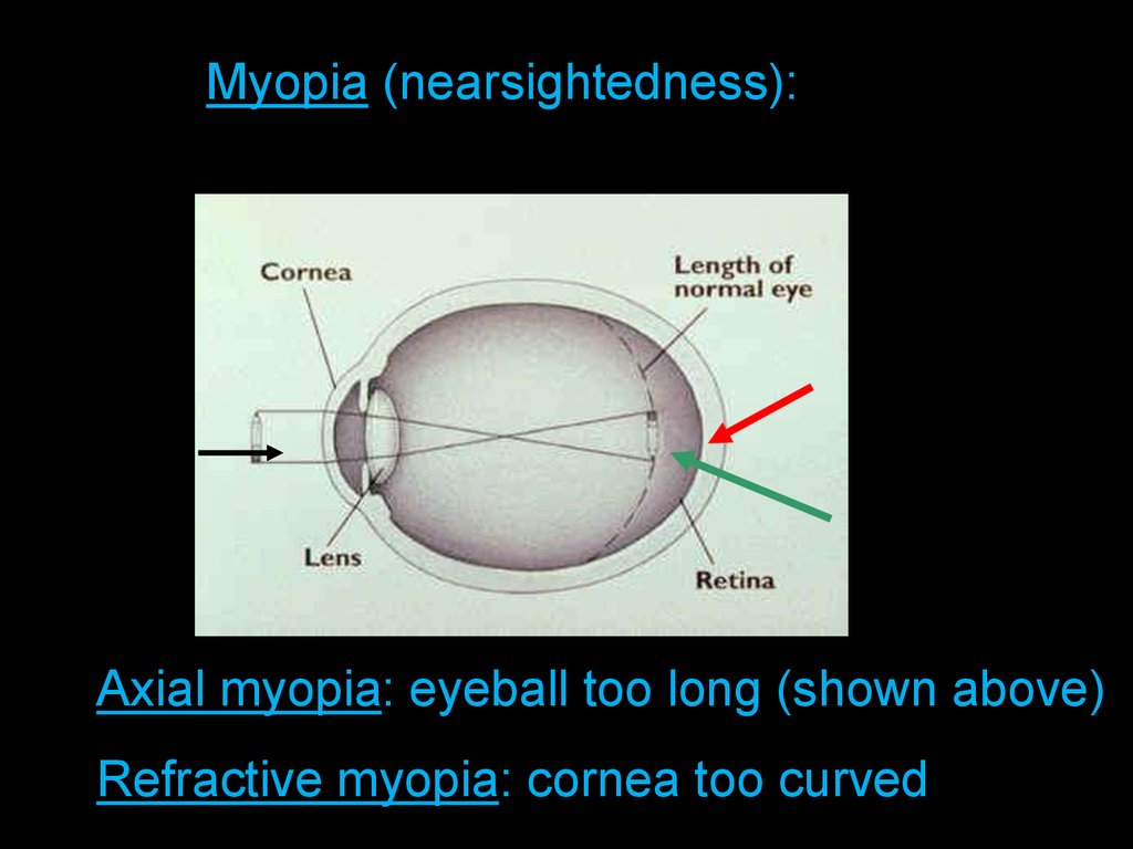
Length of Eyeball + Curvature ofMyopia (nearsightedness):
Cornea
Axial myopia: eyeball too long (shown above)
Refractive myopia: cornea too curved
13.
Myopia Correctionconcave lens: negative diopter; diffraction
laser surgery: remove corneal tissue in
center to reduce curvature
14.
Hyperopia (farsightedness): objects focused behind retinaNormal sight
Farsightedness
15.
Hyperopia Correctionconvex lens: positive diopter; refraction
laser surgery: remove corneal tissue
around sides to increase curvature
16.
AstigmatismNormal sight
Astigmatism
Aspherical cornea:
light at some
orientations is
focused, while light at
others is not
17.
AstigmatismLASER correction
of cornea shape