


 Биология
Похожие презентации:


G-protein-coupled receptors
1. G-protein-coupled receptors
Prepared by: Bayzhigitova A.Akimniyazova A.
Maulenova R.
BT-1602
2.
3.
4.
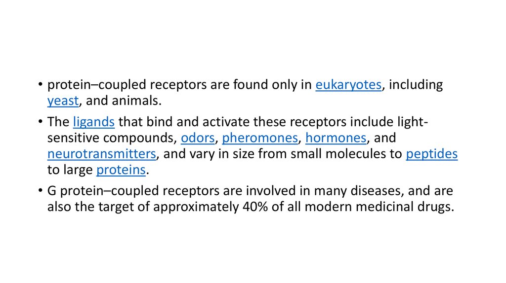
• protein–coupled receptors are found only in eukaryotes, includingyeast, and animals.
• The ligands that bind and activate these receptors include lightsensitive compounds, odors, pheromones, hormones, and
neurotransmitters, and vary in size from small molecules to peptides
to large proteins.
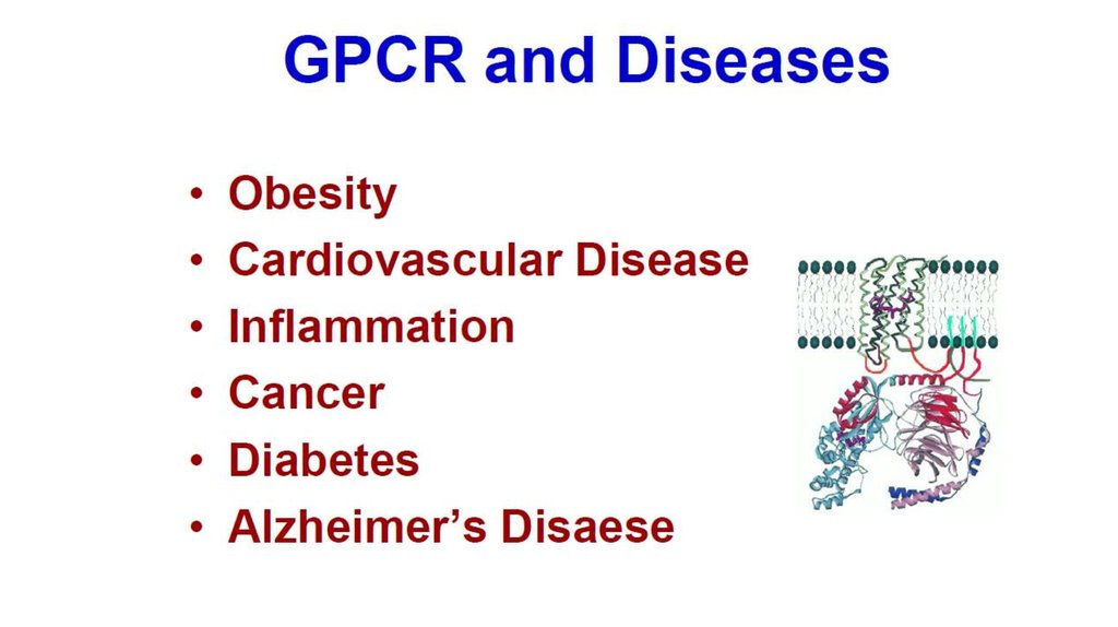
• G protein–coupled receptors are involved in many diseases, and are
also the target of approximately 40% of all modern medicinal drugs.
5.
• There are two principal signal transduction pathways involving the Gprotein–coupled receptors:
• the cAMP signal pathway and
• the phosphatidylinositol signal pathway
6. Physiological roles GPCRs are involved in a wide variety of physiological processes. Some examples of their physiological roles include:
• The visual sense: The opsins use a photoisomerization reaction to translate electromagnetic radiation into cellular signals.Rhodopsin, for example, uses the conversion of 11-cis-retinal to all-trans-retinal for this purpose
• The gustatory sense (taste): GPCRs in taste cells mediate release of gustducin in response to bitter- and sweet-tasting substances.
• The sense of smell: Receptors of the olfactory epithelium bind odorants (olfactory receptors) and pheromones (vomeronasal
receptors)
• Behavioral and mood regulation: Receptors in the mammalian brain bind several different neurotransmitters, including serotonin,
dopamine, GABA, and glutamate
• Regulation of immune system activity and inflammation: Chemokine receptors bind ligands that mediate intercellular
communication between cells of the immune system; receptors such as histamine receptors bind inflammatory mediators and
engage target cell types in the inflammatory response. GPCRs are also involved in immune-modulation and directly involved in
suppression of TLR-induced immune responses from T cells.
• Autonomic nervous system transmission: Both the sympathetic and parasympathetic nervous systems are regulated by GPCR
pathways, responsible for control of many automatic functions of the body such as blood pressure, heart rate, and digestive
processes
• Cell density sensing: A novel GPCR role in regulating cell density sensing.
• Homeostasis modulation (e.g., water balance).
Involved in growth and metastasis of some types of tumors.
7.
8.
9.
Structure - Single protein with 7 transmembrane regionsExtracellular
loops
NH2
N -Terminal chain
Membrane
VII
VI
V
IV
III
II
I
G-Protein
binding region
HO2C
C -Terminal chain
Variable
intracellular loop
Intracellular loops
Transmembrane
helix
10.
G-protein-coupled receptors (7-TM receptors)Ligands
Monoamines e.g. dopamine, histamine, noradrenaline, acetylcholine
(muscarinic)
Nucleotides
Lipids
Hormones
Glutamate
Ca++
11.
G-protein-coupled receptors (7-TM receptors)Ligand binding site - varies depending on receptor type
Ligand
A
B
C
D
A) Monoamines - pocket in TM helices
B) Peptide hormones - top of TM helices + extracellular loops
+ N-terminal chain
C) Hormones - extracellular loops + N-terminal chain
D) Glutamate - N-terminal chain
12.
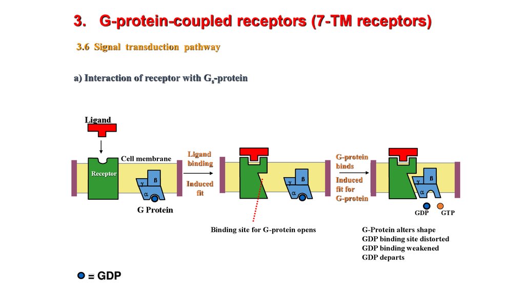
3. G-protein-coupled receptors (7-TM receptors)3.6 Signal transduction pathway
a) Interaction of receptor with Gs-protein
Ligand
Cell membrane
Receptor
ß
g
a
Ligand
binding
Induced
fit
G-protein
binds
ß
g
a
G Protein
ß
g
a
GDP
Binding site for G-protein opens
= GDP
Induced
fit for
G-protein
GTP
G-Protein alters shape
GDP binding site distorted
GDP binding weakened
GDP departs
13.
3. G-protein-coupled receptors (7-TM receptors)3.6 Signal transduction pathway
a) Interaction of receptor with Gs-protein
ß
g
a
GTP binds
Binding site recognises GTP
g
ß
g
a
Fragmentation
and release
Induced fit
G-protein alters shape
Complex destabilised
• Process repeated for as long as ligand bound to receptor
• Signal amplification - several G-proteins activated by one ligand
• as Subunit carries message to next stage
ß
a
14.
3. G-protein-coupled receptors (7-TM receptors)3.6 Signal transduction pathway
GTP
GDP
b) Interaction of as with adenylate cyclase
Binding site
for as subunit
as-subunit
Adenylate cyclase
GTP hydrolysed
to GDP catalysed
by as subunit
Binding
Induced
fit
Active site
(closed)
P
ATP
cyclic AMP
Active site
(open)
Signal
transduction
(con)
as Subunit recombines with b,g dimer
to reform Gs protein
ATP
cyclic AMP
Active site
(closed)
as Subunit changes shape
Weaker binding to enzyme
Departure of subunit
Enzyme reverts to inactive
state
15.
16.
17. Adrenoreceptor
18. Localization and the main effects
• α1- и β1- receptors localized mainly in the postsynaptic membrane and react to the action of noradrenaline released from nerveendings of the postganglionic neurons of the sympathetic division.
• α2- и β2- receptors are extrasynaptic, and are also available on the presynaptic membrane of the same neurons. On the α2receptors act as adrenaline and noradrenaline. β2-receptors are sensitive mainly to adrenaline. Α2-receptors on the presynaptic
membrane noradrenaline acts on the principle of negative feedback - inhibits proper selection .
• α1 — localized in arterioles, stimulation leads to a spasm of arterioles, increasing the pressure, decrease vascular permeability and
a decrease in exudative inflammation.
• α2 — mainly presynaptic receptors are "negative feedback loop" for the adrenergic system and their stimulation leads to lower
blood pressure
• β1 — localized in the heart, the stimulation frequency leads to an increase (positive chronotropic effect) and force of cardiac
contractions (positive inotropic effect) in addition, increases the myocardial oxygen consumption and increase blood pressure. It
is also localized in the kidneys, being receptors juxtaglomerular apparatus.
• β2 — located in the bronchioles, the stimulation causes dilation of the bronchial tubes and the removal of bronchospasm. These
receptors are found on cells of the liver, the effects on them hormone causes glycogenolysis and glucose output in blood.
• β3 — located in the adipose tissue. Stimulation of these receptors enhances lipolysis and leads to the release of energy and to
increase heat production
19.
• Механизм действия адренергических рецепторов. Эпинефрин и норадреналин являются лигандами дляадренергических рецепторов α1, α2 или β. С α1-адренергическим рецептором связывается α-субъединица Gq, что
приводит к повышению внутриклеточной концентрации ионов кальция и, например, к сокращению гладкой
мускулатуры. С α2-адренергическим рецептором α2 связывается α-субъединица Gi, что приводит к снижению
концентрации цАМФ или, например, к сокращению гладкой мускулатуры. С β-рецептором связывается αсубъединица Gs, что приводит к повышению внутриклеточной концентрации цАМФ и, например, к сокращению
сердечной мускулатуры, расслаблению гладкой мускулатуры и гликогенолизу.