


 staging")


 Медицина
 Английский язык
Похожие презентации:


Rectal cancer staging go the full “Distance”. MRI
1. Rectal cancer staging go the full “DISTANCE”
Geertje Noë2. “DISTANCE”
• A mnemonic recently introduced• Simplify reporting rectal cancer staging MRI
3. Overview
• MR imaging sequences• The report for MR rectal cancer staging and
“DISTANCE”
• Primary rectal cancer staging cases
• Post CRT staging and cases
4. We have come such a long way…
Courtesy Dr. Stephen EslerCT tomogram from the 1980’s
5.
• The radiologist plays a central role in themultidisciplinary approach to rectal cancer
• MRI can accurately stage rectal cancer
• Pre-operative staging with MRI important to select
the appropriate therapy
• Rectal cancer staging with MRI remains a challenge
for many radiologists
6. Technique and sequences
• No need for bowel preparation, filling of rectum withcontrast/air
• Antispasmodic agents can be helpful but are not
mandatory
• Only sequence that is required is a T2 –weighted fast spin
echo sequence (high resolution)
• IV contrast is not recommended as it does not improve
diagnostic quality
7.
Additional sequences to consider:• DWI
• T2 fat sat
• T1
8.
Austin protocol:• Three Plane Localiser
• Coronal T2 3D SPACE Whole Pelvis
• Axial T1 Whole Pelvis
• Axial T2 FS Whole Pelvis
• Axial DWI
Modifications Reformat 3D in 3 planes
• Coronal Oblique - Angled parallel to the long axis of the
rectum
• Sagittal
• Axial Oblique – Angled perpendicular to the long axis of
the rectum
9.
Overview• MR imaging sequences
• The report for MR rectal cancer staging and
“DISTANCE”
• Primary rectal cancer staging cases
• Post CRT staging and cases
10.
4 critical questions need to be answered1. Location of the tumor (high, middle, low)
(you can use a specific staging for low rectal tumours describing the
involvement of the sphincters)
2. The T-stage of the tumour
3. Free resection margin for TME (CRM)
4. N-stage
11.
Other things that need to go in the report:• Tumor length, tumor description/morphology
(polypoid, ulcerative etc.)
• Distance of tumour to anal verge (+/- anorectal
junction)
• Circumferential?
• Involvement of pelvic side wall nodes
• Extramural vascular invasion (EMVI)
• Metastasis
12.
• Pedersen et al. reported in 2011 that the reportquality overall could be significantly improved
• There is a need for standardisation of reports and
Taylor et al from Brown’s group created a form based
reporting tool in 2008
• Brown’s group also created the mnemonic
“DISTANCE”
13.
Taylor FG et al. A sytematic approach to the interpretation pre-operative staging MRIfor rectal cancer. Am J Roentgenol. 2008 Dec;191(6):1827-35
14.
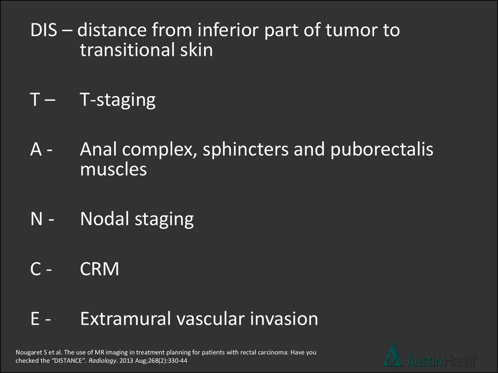
DIS – distance from inferior part of tumor totransitional skin
T–
T-staging
A-
Anal complex, sphincters and puborectalis
muscles
N-
Nodal staging
C-
CRM
E-
Extramural vascular invasion
Nougaret S et al. The use of MR imaging in treatment planning for patients with rectal carcinoma: Have you
checked the “DISTANCE”. Radiology. 2013 Aug;268(2):330-44
15.
Overview• MR imaging sequences
• The report for MR rectal cancer staging and
“DISTANCE”
• Primary rectal cancer staging cases
• Post CRT staging
16.
CASE 1= 7.8 cm
17.
126
18. Report conclusion:
T3 N2 mid rectal tumour with a length ofapproximately 8.6 cm which reaches 7.8 cm
above the anal verge and has a positive CRM.
19.
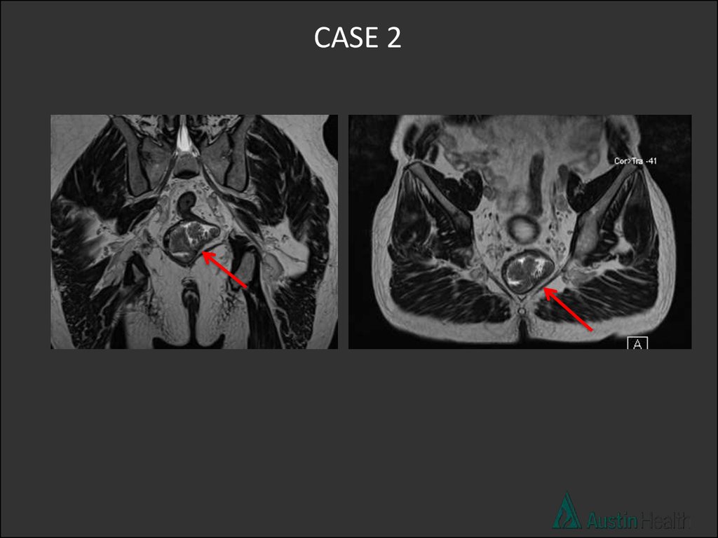
CASE 220.
21. Report conclusion:
T2 N0 low rectal tumour with a length of 5.1 cmand reaches approximately 4.1 cm above the
anal verge.
22.
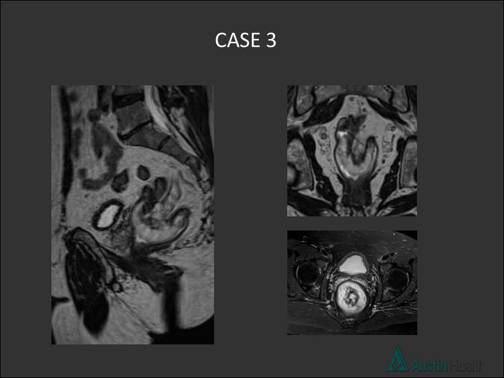
CASE 323.
24.
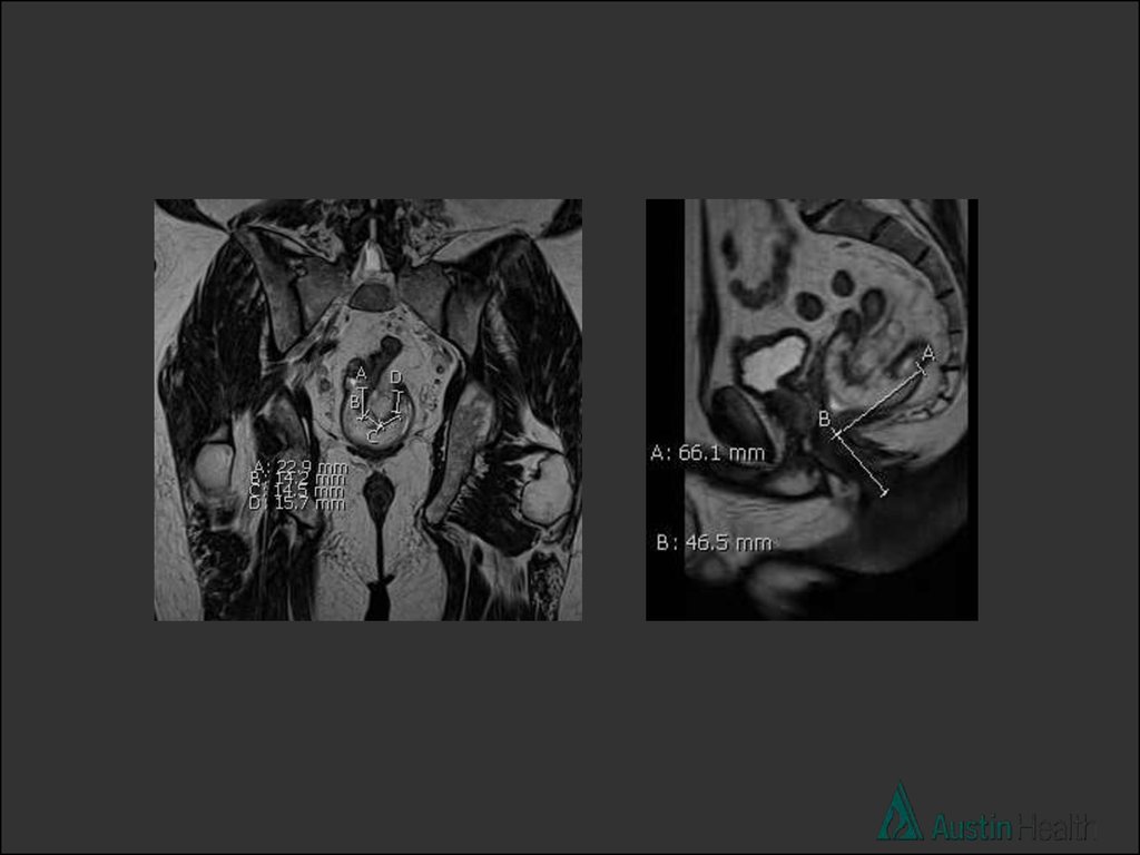
25. Report conclusion:
T3 N1 mid rectal tumour with a length of 6.7 cmwith a distance of 10 cm from the anal verge.
The CRM is negative.
26.
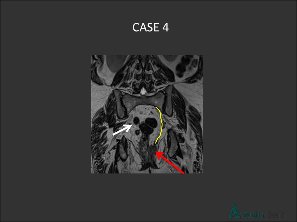
CASE 427.
28.
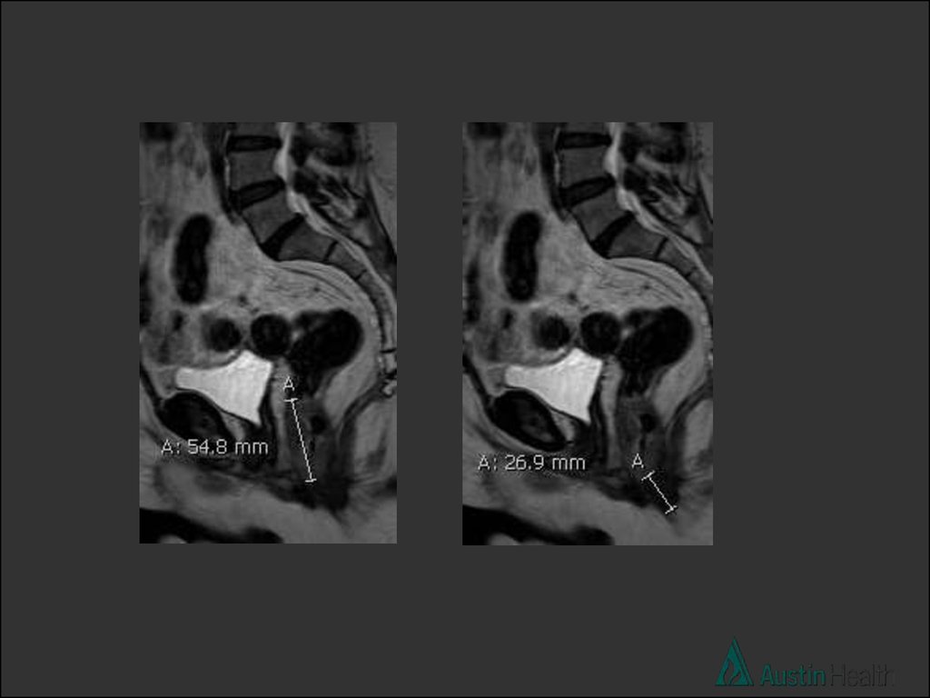
29. Report conclusion:
Low rectal tumour with a length of 5.5 cm withextension to and involvement of the left levator
muscle. It reaches 2.7 cm above the anal verge
and there are 5 abnormal lymph nodes. An
enlarged left pelvic side wall node is present.
Staging in keeping with T4 N2 M1
30.
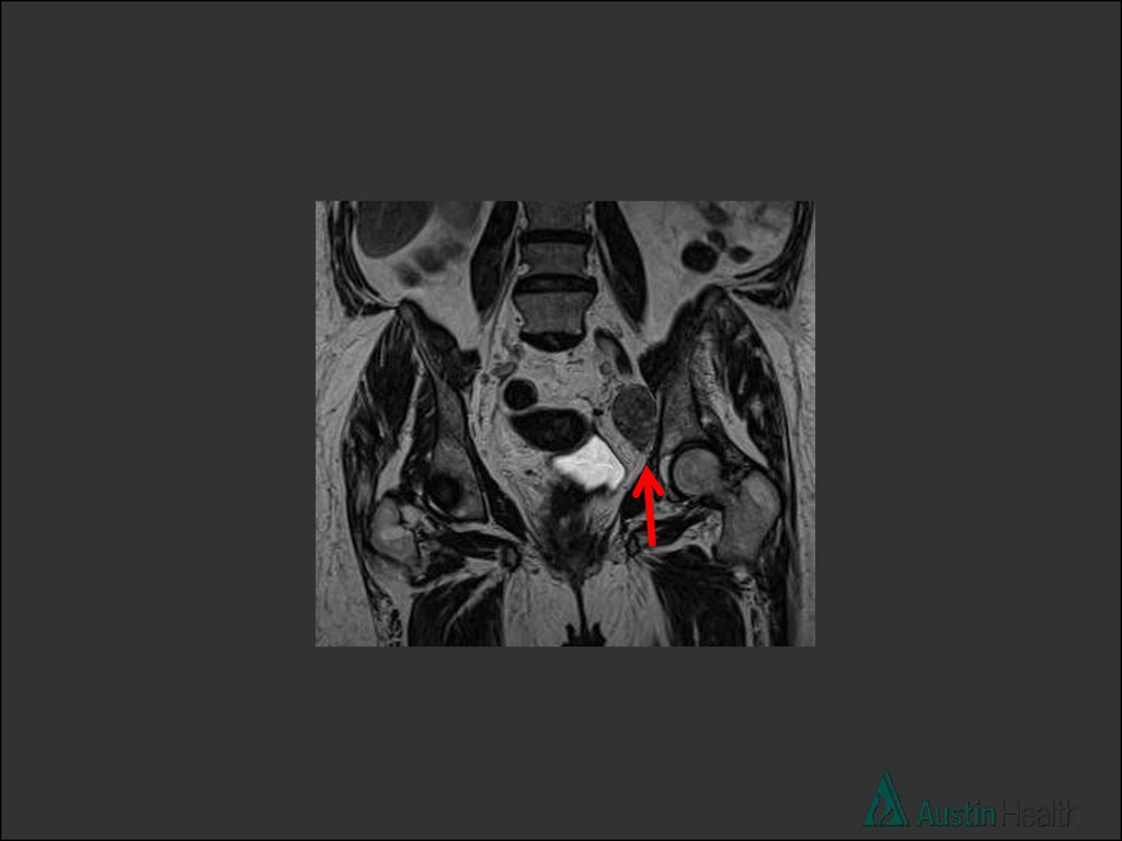
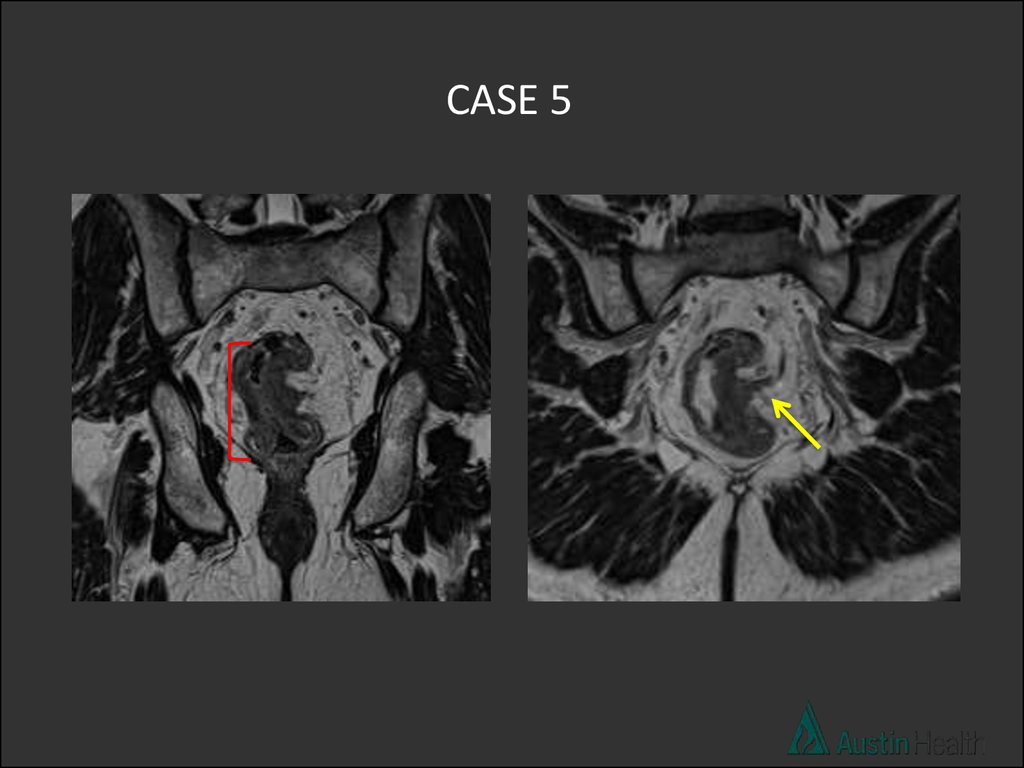
CASE 531.
CASE 632.
33.
Overview• MR imaging sequences
• The report of MR rectal cancer staging and
“DISTANCE”
• Primary rectal cancer staging cases
• Post CRT staging
34. Post chemoradiation therapy (CRT) staging
• Main indications for CRT:– Locally advanced rectal tumor T3 with > 5mm of
extramural spread
– EMVI
– Tumor within 1mm of mesorectal fascia (node,
tumor, EMVI)
– Threatened or involved anal sphincter
– Nodal involvement
35.
• Locally advanced rectal cancer has a poorprognosis
• Benefits of downstaging and downsizing
with neoadjuvant CRT:
1. improves resectability
2. sphincter preservation
3. reduced local recurrence
4. improved overall survival
36.
• MRI is developing a central role in identifyinggood and poor responders
• Can provide a basis to further fine tune
treatment
• In the future MRI may be used to select
patients that will just receive CRT (wait and
see approach)
37.
• Tumour volume reduction of at least 70% predicts disease free survival andgood histologic regression.
Nougaret et al MR volumetric measurement of low rectal cancer helps predict tumour response and outcome after combined
chemotherapy and radiation therapy. Radiology May 2012.
• Post CRT MRI assessment of tumour regression grade correlated
with disease free survival.
Patel et al MRI-detected tumour response for locally advanced rectal cancer predicts survival outcomes JCO 2011
• A pathological complete response following neoadjuvant CRT is associated
with excellent long-term survival, with low rates of local recurrence and
distant failure.
Martin et al. Br J Surg 2012 Systematic review and meta analysis of outcomes following pathological
complete response to neoadjuvant chemoradiotherapy for rectal cancer.
• Tumour volume regression grade of less than 45% is predictive of a poor
tumour outcome.
Yeo et al, Tumour volume reduction rate after preoperative chemoradiotherapy as a prognostic factor in locally advanced rectal
cancer, Int J Radioation Oncolo Biol Phys 2012.
38. Post CRT MRI interpretation
• Predicting the stage prior to CRT ~ 85%, after CRT ~ 50%(fibrosis vs tumour?)
• Need primary rectal cancer staging MRI
• “DISTANCE” comes into play first again (ymr added to the
abbreviations e.g. ymrT)
• Followed by MR Tumour Response Grading (mrTRG)
• Research has shown that ymrT and mrTRG predict the
corresponding histopathological parameters and can identify good
and poor responders to CRT
39.
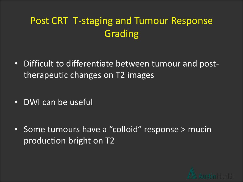
Post CRT T-staging and Tumour ResponseGrading
• Difficult to differentiate between tumour and posttherapeutic changes on T2 images
• DWI can be useful
• Some tumours have a “colloid” response > mucin
production bright on T2
40.
Morphologic descriptions used in T-staging and TumourResponse Grading
• Fibrosis within tumour and rectal wall: low signal.
• Desmoplastic reaction: low intensity spicules.
• Residual tumour: Intermediate signal and nodular margin.
• Mucinous change: mucinous response in non-mucinous
tumours suggests treatment response
1. Uniform mucinous change in tumours exhibiting baseline
mucinous heterogeneity suggests treatment response
2. Persistent heterogeneous mucinous signal unchanged post
treatment no response.
41.
Post CRT changesNougaret S et al. The use of MR imaging in treatment planning for patients with rectal
carcinoma: Have you checked the “DISTANCE”. Radiology. 2013 Aug;268(2):330-44
42.
TRG 1: Complete radiologic response:no evidence of abnormalities
TRG 2: Good response: dense fibrosis
(>75%) no obvious residual tumour
or minimal residual tumour
TRG 3: Moderate response >50% fibrosis or
mucin and visible tumour
TRG 4: Slight response: small areas of
fibrosis or mucin, but mostly
tumour
TRG 5: No response, same appearance as
original tumour
43.
CASE 1 – PRE CRTDWI
ADC
44.
CASE 1 – POST CRTPOST
POST
PRE
PRE
DWI
ADC
45.
mrTRG2Good response with tumour replaced by dense
fibrosis with no obvious tumour left.
46.
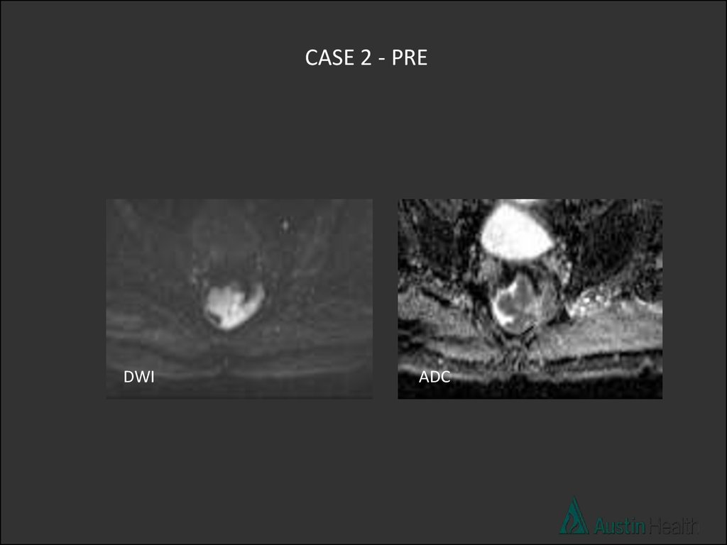
CASE 2 - PREDWI
ADC
47.
Rectal cancers may exhibit restricted or increased diffusion
dependant on tumour cellularity, intra-tumoral oedema, and
presence of cystic/necrotic areas.
Low ADC value is predictive of good treatment response.
Dzik_Jurasz et al DWI-MRI for prediction of response of rectal carcinoma to chemoradiation. Lancet 2002
An early increase in the ADC after commencing treatment is
predictive of better treatment outcome. Hein et al DWI-MRI for monitoring diffusion
changes in rectal carcinoma during combined chemoradiation. EJR 2003
48.
CASE 2-POST CRTPOST
PRE
DWI
ADC
49.
mrTRG 1Complete radiological response
50.
CASE 3 – PRE CRT51.
CASE 3 – POST CRTPOST
POST
PRE
PRE
POST
PRE
52.
mrTRG 4Slight response with some fibrosis but mostly
tumour.
53.
CASE 4 PRE-CRT54.
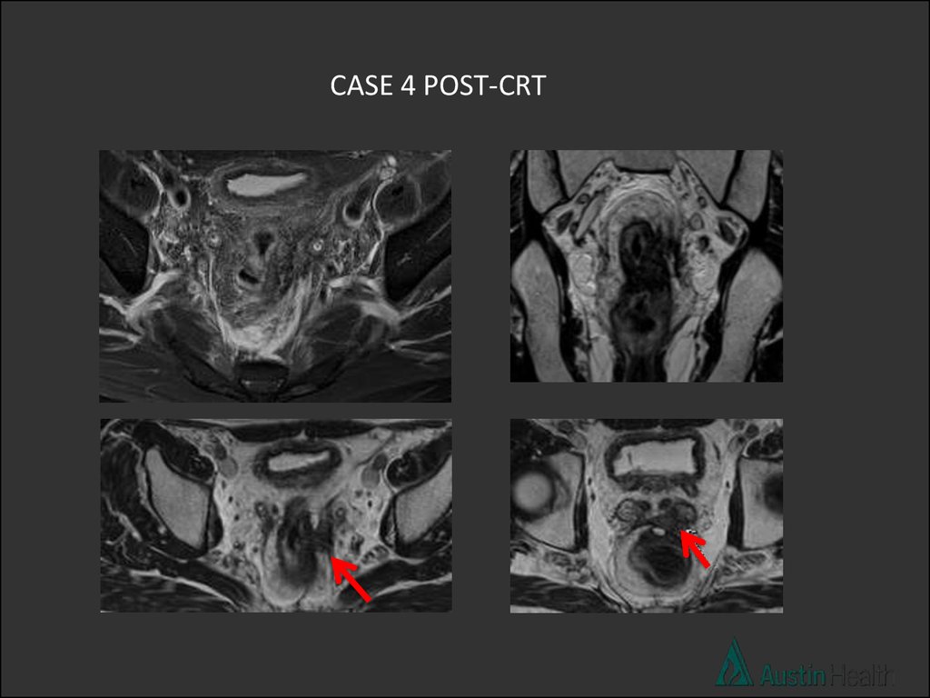
CASE 4 POST-CRT55.
mrTRG 2-3Moderate - good response with > 50% fibrosis
and minimal remaining visible tumour.
T4 stage
56. Summary
• Imaging techniques• DISTANCE easy mnemonic to help us remember
what to report on
• Some example cases and reports of primary
staging
• Brief discussion of post CRT staging and some
cases
57.
Now… challenge yourself to report rectal staging!58. References
Nougaret S, Reinhold C, Mikhael W H et al. The use of MR imaging in treatment planning for
patients with rectal carcinoma: Have you checked the “DISTANCE”. Radiology. 2013
Aug;268(2):330-44
Taylor FG, Swift RI, Blomqvis L et al. A sytematic approach to the interpretation pre-operative
staging MRI for rectal cancer. Am J Roentgenol. 2008 Dec;191(6):1827-35
Pedersen BG, Blomqvist L, Brown G et al. Postgraduate multidisciplinary development
program: impact on the interpretation of pelvic MRI in patients with rectal cancer – a clinical
audit in West Denmark. Dis Colon Rectum 2011:54(3):328-334
Barbaro B, Vitale R, Leccisotti L et al. Restaging locally advanced rectal Cancer with MR
Imaging after chemoradiation therapy. Radiographics 2010;30:699-721
Patel UB, Taylor F, Blomqvist L et al. Magnetic resonance imaging-detected tumor repsonse
for locally advanced rectal cancer predicts survival outcomes: MERCURY experience. J Clin
Oncol 2011; 29 (28):3753-3760
Dzik_Jurasz et al DWI-MRI for prediction of response of rectal carcinoma to chemoradiation.
Lancet 2002
Hein et al DWI-MRI for monitoring diffusion changes in rectal carcinoma during combined
chemoradiation. EJR 2003