


 Медицина
Похожие презентации:

")
")


")
")
Disorders of Skeletal Muscle
1. Disorders of Skeletal Muscle
Made by: Stanislav Aladev,15452.2 group
2. Spinal Muscular Atrophy
3. Spinal Muscular Atrophy
4. Disorders of Neuromuscular Junction
Myasthenia gravis is an autoimmune disease withfluctuating muscle weakness that is caused by
autoantibodies that target the neuromuscular
junction. The most common antigenic target is the
postsynaptic acetylcholine receptor (AChR). Clinically,
myasthenia gravis frequently manifests with ptosis
(drooping eyelids) or diplopia (double vision) because
of weakness in the extraocular muscles. This pattern
of weakness is distinctly different from that of most
primary myopathic diseases, in which there is relative
sparing of facial and extraocular muscles.
5. Disorders of Neuromuscular Junction
Lambert-Eaton syndrome is caused by autoantibodiesthat inhibit the function of presynaptic calcium
channels, thereby reducing the release of
acetylcholine into the synaptic cleft. Patients with
Lambert-Eaton syndrome experience improvement in
weakness with repetitive stimulation, in contrast to
those suffering from myasthenia gravis. Repetitive
stimulation serves to build up sufficient intracellular
calcium to facilitate acetylcholine release. LambertEaton syndrome often arises as a paraneoplastic
disorder, particularly in patients with small cell lung
carcinoma.
6. Miscellaneous Neuromuscular Junction Disorders
Congenital myasthenic syndromes comprise aheterogeneous group of diseases that result from
mutations that disrupt the function of various
neuromuscular junction proteins.
Infections with exotoxin-producing bacteria may be
associated with defects in neural transmission and
muscle contraction. Clostridium tetani and Clostridium
botulinum both release extremely potent neurotoxins
that interfere with neuromuscular transmission.
7. Muscle Fiber Atrophy
Neuropathic changes. Loss of innervation causes atrophy ofmyofibers. The two main morphologic hallmarks of neurogenic
changes, grouped atrophy and fiber type grouping are the result
of multiple rounds of denervation and reinnervation. Loss of an
axon or lower motor neuron results in atrophy of the myofibers
that are part of this motor unit. Atrophic myofibers can be
reinnervated by axonal branches from adjacent motor units,
increasing the size of these motor units and returning trophic
input to the atrophic myofibers. In this setting loss of
innervation will therefore produce large clusters of atrophic
myofibers, grouped atrophy.
Prolonged disuse of muscles from any cause (e.g., prolonged bed
rest in the sick, casting of a broken bone) may cause focal or
generalized muscle atrophy, which tends to affect type II fibers
more than type I fibers.
Glucocorticoid exposure, whether exogenous or endogenous
(e.g., in Cushing syndrome), also may cause muscle atrophy.
Proximal muscles and type II myofibers are affected
preferentially in this setting.
8. Patters of skeletal muscle injury
9. Inherited Disorders of Skeletal Muscle
Muscular dystrophies are associated with progressivemuscle injury in patients who have normal muscle
function at birth.
Congenital muscular dystrophies, by contrast, are
progressive, early-onset diseases. Some are also
associated with malformations of the central nervous
system.
Congenital myopathies typically present in infancy with
muscle defects that tend to be static or to even improve
with time. They are often associated with distinct
structural abnormalities of the muscle.
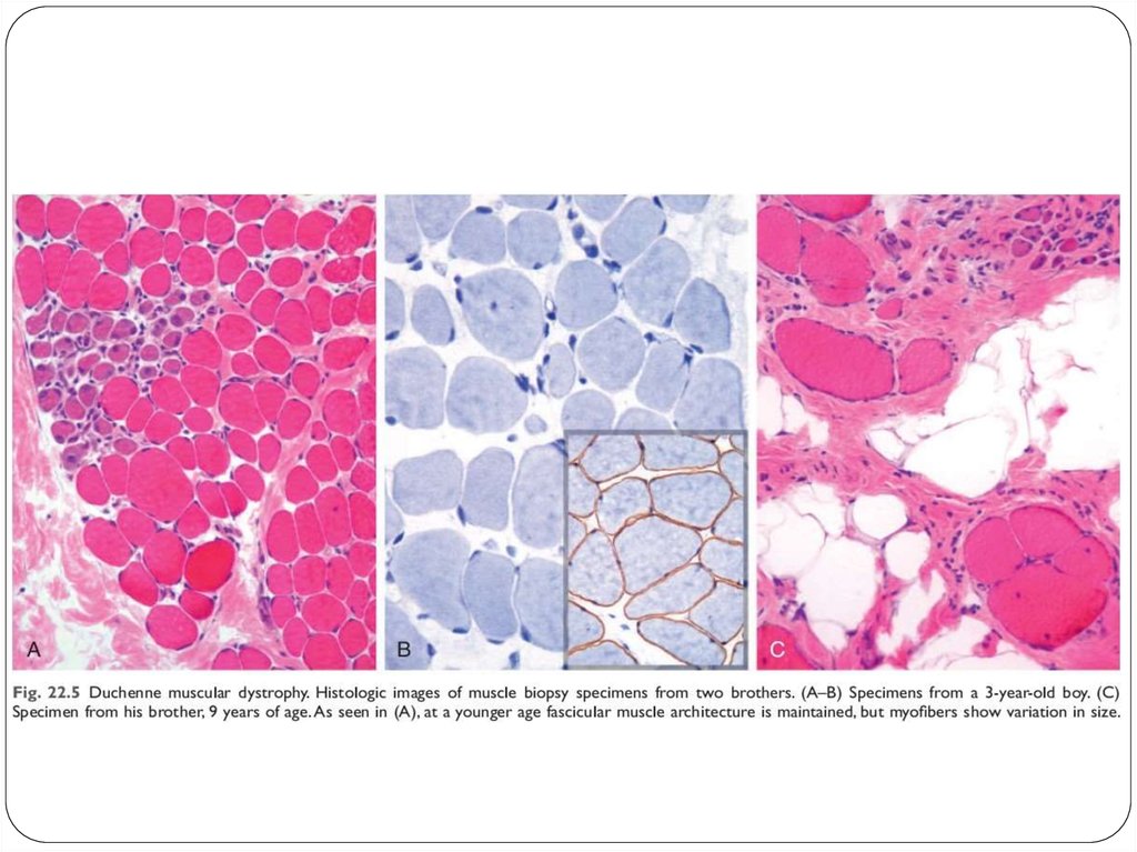
10. Dystrophinopathies: Duchenne and Becker Muscular Dystrophy
The most common muscular dystrophies are X-linkedand are caused by mutations that disrupt the function of
a large structural protein called dystrophin. As a result,
these diseases are referred to as dystrophinopathies Duchenne muscular dystrophy (DMD) and Becker
muscular dystrophy (BMD) are the two most important
diseases in this group.
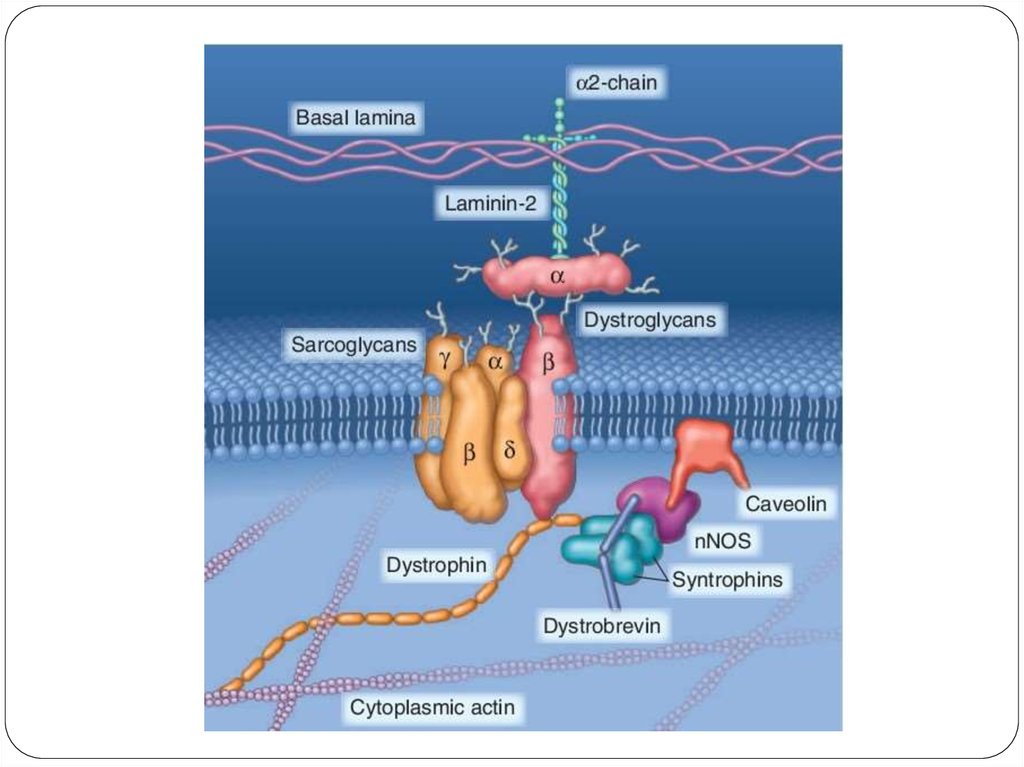
Both DMD and BMD are caused by mutations disrupting
the function of the dystrophin gene located on the short
arm of the X chromosome (Xp21). Dystrophin is a very
large protein (427kD in molecular weight) found in
skeletal and cardiac muscle, brain, and peripheral
nerves; it is part of the dystrophin-glycoprotein complex.
11.
12.
13. Other X-Linked and Autosomal Muscular Dystrophies
Myotonic dystrophy. Myotonia, the sustained involuntarycontraction of a group of muscles, is the cardinal neuromuscular
symptom in myotonic dystrophy. Patients often complain of
stiffness and difficulty in relaxing their grip, for example, after a
handshake. Myotonic dystrophy is a nucleotide repeat
expansion disease that is inherited as an autosomal dominant
trait. More than 95% of patients with myotonic dystrophy have
mutations in the gene that encodes the dystrophia myotonica
protein kinase (DMPK).
Limb-girdle muscular dystrophies. These muscular dystrophies
preferentially affect the proximal musculature of the trunk and
limbs. Their genetic basis is heterogeneous. The growing list
includes at least 7 dominant subtypes and 15 autosomal
recessive subtypes. Some of the responsible mutations affect
components of the dystrophin-glycoprotein complex other than
dystrophin. Others affect proteins involved in vesicle transport
and repair of cell membrane after injury.
14. Other X-Linked and Autosomal Muscular Dystrophies
Emery-Dreifuss muscular dystrophy (EMD) is a geneticallyheterogeneous disorder caused by mutations affecting
structural proteins found in the nucleus. An X-linked form
results from mutations in the gene encoding the protein
emerin, whereas an autosomal dominant form is caused by
mutations in the gene encoding lamin A/C.
Facioscapulohumeral dystrophy is an autosomal dominant
form of muscular dystrophy that is caused by complex
genetic changes that allow expression of the transcription
factor DUX4 that is normally repressed in mature tissues. It
is thought that the disease is caused by over expression of
DUX4 target genes, many of which are involved in the
normal function of skeletal muscles.
15. Channelopathies
Ion channel myopathies are a group of familialdisorders caused by inherited defects in ion
channels that are characterized by myotonia,
relapsing episodes of hypotonic paralysis
associated with abnormal serum potassium
levels, or both. Hyperkalemic periodic paralysis
results from mutations in the gene encoding the
skeletal muscle sodium channel SCN4A, which
regulates sodium entry during contraction.
Malignant hyperthermia is a rare syndrome
characterized by tachycardia, tachypnea, muscle
spasms, and hyperpyrexia.
16. Metabolic Myopathies
Myopathies due to inborn errors of metabolisminclude disorders of glycogen synthesis and
degradation and lipid handling. The latter include
disorders of the carnitine transport system and
deficiencies of the mitochondrial dehydrogenase
enzyme system, both of which can lead to
accumulation of lipid in myocytes (lipid
myopathies).
17. Mitochondrial Myopathies
Mitochondrial myopathies can stem from mutations ineither the mitochondrial or nuclear genomes because
both encode proteins and RNAs that are critical for
mitochondrial function. Mitochondrial myopathies
usually manifest in early adulthood with proximal
muscle weakness and sometimes with severe
involvement of the ocular musculature (external
ophthalmoplegia). There may also be neurologic signs
and symptoms, lactic acidosis, endocrinopathy,
peripheral neuropathy, and cardiomyopathy. Some
mitochondrial diseases are associated with normal
muscle morphology, whereas others show aggregates
of abnormal mitochondria; the latter impart a blotchy
red appearance in special stains - hence the term
ragged red fibers.
18. Acquired Disorders of Skeletal Muscle
19. Inflammatory Myopathies
Polymyositis is an autoimmune disorder associated withincreased expression of MHC class I molecules on
myofibers and predominantly endomysial inflammatory
infiltrates containing CD8+ cytotoxic T-cells. The
autoimmune attack leads to myofiber necrosis and
subsequent regeneration.
Dermatomyositis is the most common inflammatory
myopathy in children, in whom it appears as an isolated
entity. In adults, it often manifests as a paraneoplastic
disorder. In both contexts, it is believed to have an
autoimmune basis. The disease is typically associated with
skin manifestations, as implied by the name, and may also
have systemic manifestations such as interstitial lung
disease.
20. Inflammatory Myopathies
Inclusion body myositis is the most common inflammatorymyopathy in patients older than 60 years of age. It is
grouped with other forms of myositis, but it has yet to be
determined whether inflammation is a cause or an effect
in this disorder. The morphologic hallmark of inclusion
body myositis is the presence of rimmed vacuoles that
contain aggregates of the same proteins that accumulate
in the brains of patients with neurodegenerative diseaseshyperphosphorylated tau, amyloid derived from β-amyloid
precursor protein, and TDP-43-leading some to speculate
that this is a degenerative disorder of aging.
21.
22. Toxic Myopathies
Thyrotoxic myopathy may take the form either of acute orchronic proximal muscle weakness, and it can be the first
indication of thyrotoxicosis. Histologic findings include
myofiber necrosis and regeneration.
Ethanol myopathy occurs after an episode of binge
drinking. The degree of rhabdomyolysis may be severe,
sometimes leading to acute renal failure secondary to
myoglobinuria.
Drug myopathy can be produced by a variety of agents. For
example, myopathy is the most common complication of
statins (e.g., atorvastatin, simvastatin, pravastatin),
occurring in approximately 1.5% of users. Two forms of
statin associated myopathy are recognized: (1) toxicity of
the drug and (2) statin-induced HMG-CoA reductase
autoantibodies causing an immune mediated myopathy.
23. Tumors of Skeletal Muscles
Rhabdomyosarcoma is a malignant mesenchymaltumor with skeletal muscle differentiation. Three main
subtypes are recognized: alveolar (20%), embryonal
(60%), and pleomorphic (20%). Rhabdomyosarcoma
(alveolar and embryonal) is the most common soft
tissue sarcoma of childhood and adolescence, usually
appearing before age 20. Pleomorphic
rhabdomyosarcoma is seen predominantly in adults.
The pediatric forms often arise in the sinuses, head
and neck, and genitourinary tract, locations that do
not normally contain much skeletal muscle.
24.
A – embryonal subtypeB – alveolar subtype
25. Tumors of Skeletal Muscles
Leiomyoma, a benign tumor of smooth muscle, ismost common in the uterus but can arise in any soft
tissue site. Uterine leiomyomas are common and may
cause a variety of symptoms including infertility and
menorrhagia. Leiomyomas also may arise from the
erector pili muscles (pilar leiomyomas) in the skin and
rarely in the deep somatic soft tissues or
gastrointestinal tract.
26. Tumors of Skeletal Muscles
Soft tissue leiomyosarcoma accounts for 10% to 20%of soft tissue sarcomas. They occur in adults and affect
women more frequently than men. Most develop in
the deep soft tissues of the extremities and
retroperitoneum or arise from the great vessels.
Leiomyosarcomas have complex genotypes that stem
from acquired defects that lead to profound genomic
instability.
27. Summary
Skeletal muscle function can be impaired by a primary(inherited or acquired) myopathy or secondarily because
of problems with muscle innervation.
The genetic forms of myopathy fall into several fairly
distinct clinical phenotypes, including muscular dystrophy,
congenital myopathy, and congenital muscular dystrophy.
Dystrophinopathies are X-linked disorders caused by
mutations in the dystrophin gene and disruption of the
dystrophin-glycoprotein complex. Depending on the type
of mutation, the disease may be severe, such as DMD, or
mild (e.g., Becker dystrophy).
Acquired myopathies have diverse causes, including
inflammation and toxic exposures.