


")
")
")
")
")
")

")


 Биология
Похожие презентации:


")
")
")
Pathologic Protozoa (Lesson 1)
1. Lesson 1 Pathologic Protozoa
2. CHARACTERISTICS OF PROTOZOA
1. Unicellular2. Chemoheterotrophs (get their energy by
breaking down organic matter).
3. Most ingest their food; thus, they have
special structures for this.
3. CHARACTERISTICS OF PROTOZOA
4. The vegetative form is the TROPHOZOA (tropho =movement; zoite = animal; they move like an animal).
Trophozoa have special organelles for movement.
5. Capable of reproduction
A. Asexual: fission, budding, or schizogony
(produces a large number of trophozoites)
B. Sexual: conjugation
4. CHARACTERISTICS OF PROTOZOA
6. Some produce cysts.These are not tissue cysts like a human gets under
their skin; protozoa cysts are cellular.
They have a thick cell wall that allows for survival in
harsh environments better than the trophozoite
form.
5. TERMS: Host Types
• The definitive host is the one in which the parasitecompletes its sexual life cycle.
• For instance, in Plasmodium, the definitive host is
the tropical mosquito anopheles.
• The intermediate host is the human.
6. TERMS: Host Types
• Its sexual life cycle also starts in the human, so thatcan be confusing.
• What happens is the sporozoite form enters the
bloodstream when the mosquito bites the human.
• First it begins its asexual reproduction, but if two
mosquitoes inject one male and one female
gametocyte into the human, there can be a sexual
life cycle in the human as well.
7. TERMS
• Trophozoite: any stage in a protozoa’s life cyclewhich can ingest food. In practice it refers to the
motile form (pseudopods, cilia, flagella).
• Cyst: Non-motile form, protected by a membrane.
*infective stage*
• Excystation: process of emergence of the
trophozoite from the cyst.
• Pseudopod: “false foot” temporary cytoplasmic
process at the surface of the trophozoite.
8.
9. Phylum Euglenozoa
10. MASTIGOPHORA DISEASES
• Trypanosomiasis• Leishmaniasis
11. TERMS
Mastigote = flagella
Promastigote: has single flagella
Amastigote: has no flagella
Kinetoplast: round mass of circular DNA
12.
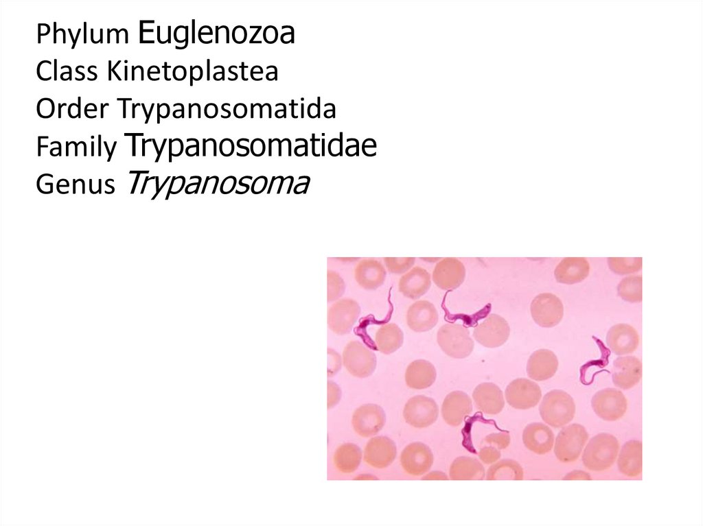
Phylum EuglenozoaClass Kinetoplastea
Order Trypanosomatida
Family Trypanosomatidae
Genus Trypanosoma
13. Trypanosomiasis
• African Trypanosomiasis– (African Sleeping Sickness)
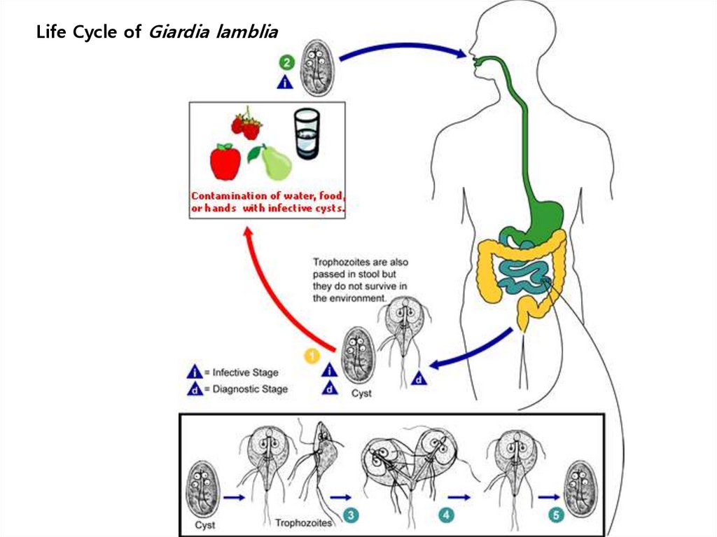
• American Trypanosomiasis
– (Chaga’s Disease)
14. “African Sleeping Sickness”
• Disease: African Tryptanosomiasis• Causal Agents:
– Trypanosoma brucei gambiense
– Trypanosoma brucei rhodesiense
15.
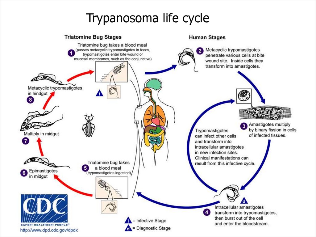
Trypanosoma life cycle16. Geographic Distribution
• T. b. gambiense is found in foci in largeareas of West and Central Africa.
– Humans are the main reservoir for
Trypanosoma brucei gambiense, but this
species can also be found in animals.
• T. b. rhodesiense is found in East and
Southeast Africa.
– Wild game animals are the main reservoir of T.
b. rhodesiense.
17. Trypanosomiasis
• Trypanosomiasis has a biological vector, thetsetse (pronounced “set-see”) fly.
• Wild animals may also be a reservoir
(Zooinotic is when a disease is transmitted to
animals as well as humans.)
18. Trypanosomiasis
• The tsetse fly bites a human and injects thetrypanomastigotes into the skin.
• This causes a chanchre (pronounced
“shanker”), which is an ulcer on the skin.
• Then it enters the lymphatic system.
19. Trypanosomiasis
• It is characterized by Winterbottom’s Sign:swelling of the cervical lymph nodes in the
head and neck area.
• CNS symptoms include a shuffling gait (like a
stroke victim), slurred speech, and malaise
(needing to sleep longer and longer each day).
• They are also restless at night.
20. Trypanosomiasis
• CNS symptoms– Shuffling gait
– Slurred speech
– Malaise (sleeping all day)
• Treatment
– Melarsoprol: which has dangerous side-effects like
chemostherapy. This drug requires administration with a
substance called ethylene glycol, which will break down
regular plastic tubing, so the drug must be administered
with special plastic iv tubing.
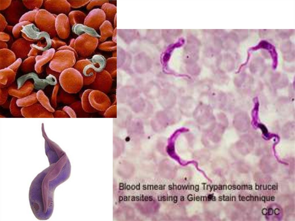
21. Trypanosoma brucei
• Trypomastigote stages are the only form found inpatients.
– Posterior kinetoplast
– Centrally located nucleus
– Undulating membrane
– Anterior flagellum
22. Trypanosoma brucei
23. Trypanosoma brucei gambiense
• trypomastigote24. Trypanosoma
25. Trypanosoma brucei rhodesiense
26.
27. Trypanosoma brucei
UM28. Tsetse Fly
29. “Chaga’s Disease”
• Disease: American TryptanosomiasisA zoonotic disease (can infect animals) that
can be transmitted to humans by bloodsucking bugs.
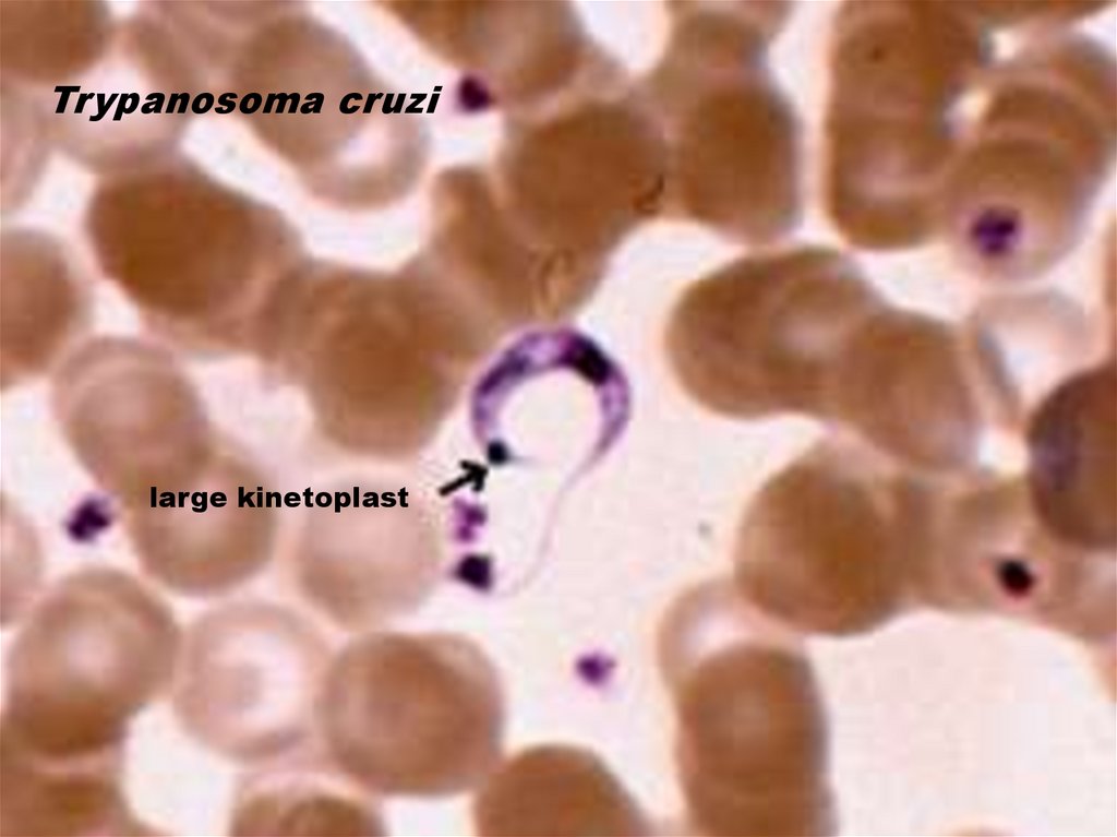
• Causal Agent: Trypanosoma cruzi
– This organism is a little smaller than T. bruceii and
has a pronounced gametoplast.
30. “Chaga’s Disease”
• This disease is NOT found in Africa.• This disease is also zoonotic; it can infect animals as well as
humans.
• The vector is a large bug called the “Kissing Bug”.
• It is found in warm regions and crowded areas, especially in
the cracks of adobe huts.
• It comes out at night and crawls on a human while they sleep.
31. “Chaga’s Disease”
• It prefers the lips because the blood supply is close to thesurface.
• It sucks the blood there, but they don’t transmit the organism
this way.
• When they suck the blood, they also defecate, and the
organism is in the feces.
• When the human wakes up to scratch the itch, feces get into
the tiny wound.
• This is a fecal blood route.
32. “Chaga’s Disease”
• Symptoms include fever, anorexia, swollen lymph nodes,hepatosplenomegally (enlarged liver and spleen), and
myocarditis (inflammation of the heart), which usually causes
death.
• They also have megacolon (large colon) and megaesophagus
(large esophagus).
33.
Trypanosoma life cycle34. Trypanosoma cruzi
• Insect vector is the “kissing” bug. It takes a bloodmeal and releases trypomastigotes in its feces near
the site of the bite wound.
• Trypomastigotes enter the host through the wound
or through intact mucosal membranes, such as the
conjunctiva.
• Trypanosoma cruzi can also be transmitted through
blood transfusions, organ transplantation,
transplacentally, and in laboratory accidents.
35. Trypanosoma cruzi
• Geographic Distribution:The Americas from the southern United States to
southern Argentina. Mostly in poor, rural areas of
Central and South America. Chronic Chagas disease
is a major health problem in many Latin American
countries. With increased population movements,
the possibility of transmission by blood transfusion
has become more substantial in the United States.
36. Trypanosoma cruzi
37. Trypanosoma cruzi
38.
Trypanosoma cruzilarge kinetoplast
39. Trypanosoma cruzi
• Triatomine bug, Trypanosoma cruzi vector,defecating on the wound after taking a blood
meal.
40. Kissing Bug
41. Romana’s sign
• Swollen eye,seen in
Chagra’s
disease.
42. TERMS
• Promastigote: has single flagella• Amastigote: has no flagella
• Kinetoplast: round mass of circular DNA
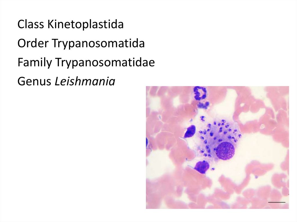
43.
Class KinetoplastidaOrder Trypanosomatida
Family Trypanosomatidae
Genus Leishmania
44. Leishmania donovani
• Disease: Leishmaniasis• Vector-borne disease transmitted by sandflies.
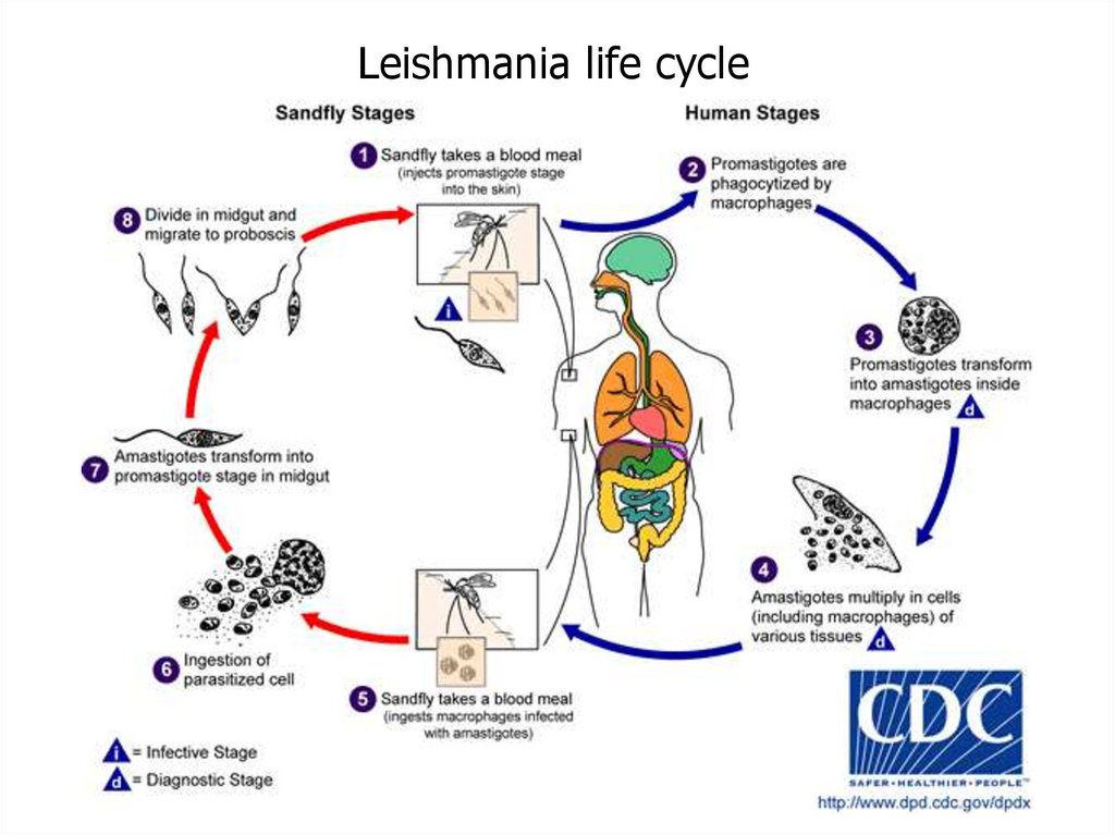
45. Leishmania Life Cycle
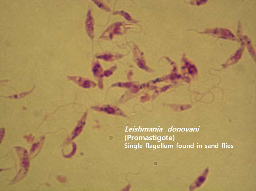
It starts out as a spindleshaped, single flagellatedcell called a promastigote
(mastigote means
flagella).
You can also see the
nucleus and a kinetoplast
(mass of circular DNA).
Kinetoplast
46. Leishmania rosette
• In prepared slidesyou can see
promastigotes align
their nose in a circle,
called a rosette.
47. Leishmaniasis rosette
48. Leishmania Life Cycle
• It reproduces in the gut of a female sandfly,and migrates to her proboscis (mouth part).
• It is introduced into the human by her bite.
• It then enters a macrophage and becomes
intracellular.
• Here, it loses its flagella and is now known as
an amastigote.
49. Leishmaniasis
• These amastigotes multiply in various organsincluding the spleen, liver, and lymph nodes.
• Symptoms include hepatosplenomegaly, lymph
adenopathy, fever, weight loss, and a decrease in all
blood cells: WBC, RBC, and platelets.
• The treatment is almost as bad as the disease
because of the side effects. It is best to catch it early.
50. Leishmania Life Cycle
• The female sandflies inject the infective stage,promastigotes, during blood meals.
• Macrophages phagocytize them and they
transform into amastigotes.
• Other sandflies become infected during blood
meals when they ingest infected macrophages.
• In the sandfly's midgut, the parasites differentiate
into promastigotes, which multiply and migrate to
the proboscis.
51.
Leishmania life cycle52.
Leishmania donovani(Promastigote)
Single flagellum found in sand flies
53. Leishmaniasis
Macrophagerupturing
Amastogotes
Amastogotes with
nucleus and
kinetoplast
54. Leishmania
• Amastigotes55. Sandfly
• This looks like amosquito, except
its body is hairy
and the wings are
feathery.
56. Leishmaniasis
• Geographic Distribution:More than 90 percent of the world's cases of visceral
leishmaniasis are in India, Bangladesh, Nepal, Sudan,
and Brazil.
• Leishmaniasis is also found in Mexico, Central
America, and South America, southern Europe, Asia,
the Middle East, and Africa.
57. Leishmaniasis
• There are three forms of Leishmaniasis:–Cutaneous
–Mucocutaneus
–Visceral
58. Cutaneous Leishmaniasis
• The disease is only at the site of the bite.• This form is seen in Texas, Mexico, Asia, and the Middle East
(our Iraq troops are coming down with this form).
• It manifests as a large, wet sore with raised edges. It looks like
a volcano with weepy serum coming out of the center.
• The wound is not contagious, just the sandfly bite.
• Dogs can get this disease, too.
59. Leishmaniasis (cutaneous)
60. Leishmaniasis (cutaneous)
61. Leishmaniasis (cutaneous)
62. Leishmaniasis (mucocunateous)
• This is when the disease located in themucous membranes of the nose and mouth.
• The most gruesome photos are of this form.
63. Leishmaniasis (mucocunateous)
64. Leishmaniasis (visceral)
• This is the most serious form. It occursespecially in immunocompromised people,
especially HIV patients.
– The amastagotes reproduce inside
macrophages.
– Only T-cells can kill infected macrophages, but
HIV is a disease that infects T-cells.
– This form is known as Kala Azar.
65. Kala Azar
Hepatosplenomegaly66. Kala Azar (duodenum)
67. Phylum Metamonada
68. Phylum Metemonada Order Diplomonada Family Hexamitidae Genus Giardia
ARCHAEZOA DISEASES69. Giardiasis
• Organism: Giardia lamblia• Cysts are resistant forms and are responsible for
transmission of giardiasis.
• Both cysts and trophozoites can be found in the
feces.
• Infection occurs by the ingestion of cysts in
contaminated water, food (includes undercooked
meat), or by the fecal-oral route.
70.
Life Cycle of Giardia lamblia71. Giardia lamblia
• In the small intestine, excystation releasestrophozoites (each cyst produces two trophozoites).
• Trophozoites multiply, remaining in the lumen where
they can be free or attached to the mucosa by a
ventral sucking disk.
• Encystation occurs as the parasites transit toward the
colon. The cyst is the stage found most commonly in
nondiarrheal feces.
• Because the cysts are infectious when passed in the
stool or shortly afterward, person-to-person
transmission is possible.
72. Giardia lamblia
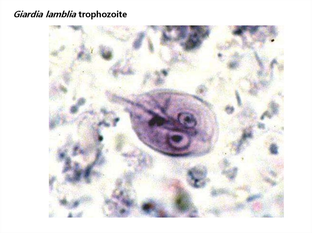
– Trophozoite form: piroform (pear or teardrop shape), lookslike a happy face.
– Discovered by Anton Van Leuwenhoek when he examined
his own feces when he had this infection.
– You won’t see the flagella in lab because you need a special
stain for that.
– Cyst form: oval shaped. Nuclei looks like two eyes.
– Geographic Distribution:
Worldwide, more prevalent in warm climates, and in
children.
73. Giardia lamblia
• Trophozoite74. Giardia lamblia
• Trophozoites75. Giardia lamblia
• Trophozoites76.
Giardia lamblia trophozoite77. Giardia lamblia
• Cysts78. Phylum Metemonada Order Trichomonadida Family Trichomonadidae Genus Trichomonas
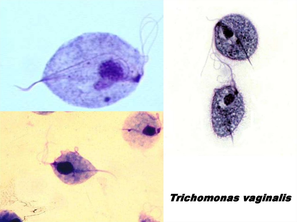
79. Trichomoniasis
• Organism: Trichomonas vaginalis• Trichomonas vaginalis resides in the female lower genital
tract and the male urethra and prostate.
• The parasite is a trophozoite only; it does not have a cyst
form, and does not survive well in the external environment.
• Trichomonas vaginalis is transmitted among humans, its only
known host, primarily by sexual intercourse.
80. Trichomonas vaginalis life cycle
81. Trichomonas vaginalis
Undulatingmembrane
Trophozoite