


")
")
")


")

 of the infant`s brain with congenital virus-associated encephalopathy")
")

")
")


")
")


 Медицина
Похожие презентации:


Development of CNS in embrio. Clinical evaluation of abnormalities
1. Background of the lecture
• Development of CNS in embrio. Clinicalevaluation of abnormalities.
• Features of CNS in fetus and newborn.
• Neurological examination
Complaints&History.
Level of consciousness (LOC)
Mentality
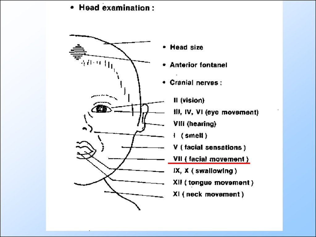
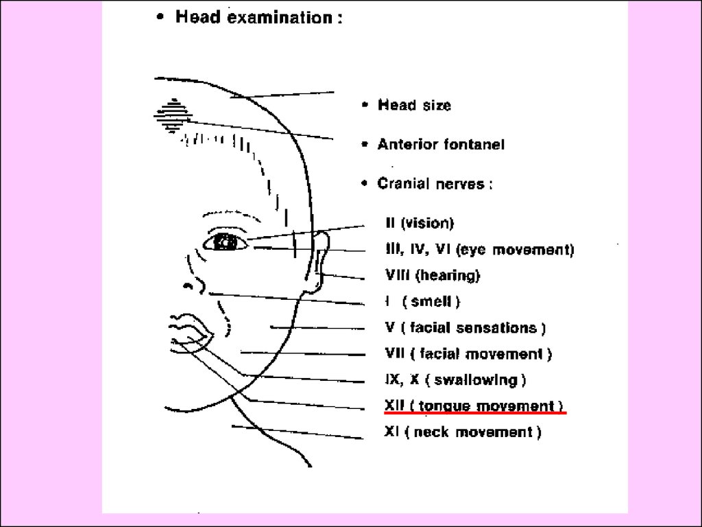
Head examination
Evaluation of motor system
• Main semiotics of CNS disorders. Meningitis.
2.
Anatomo-physiological peculiaritiesof CNS in children and their clinical
importance
3.
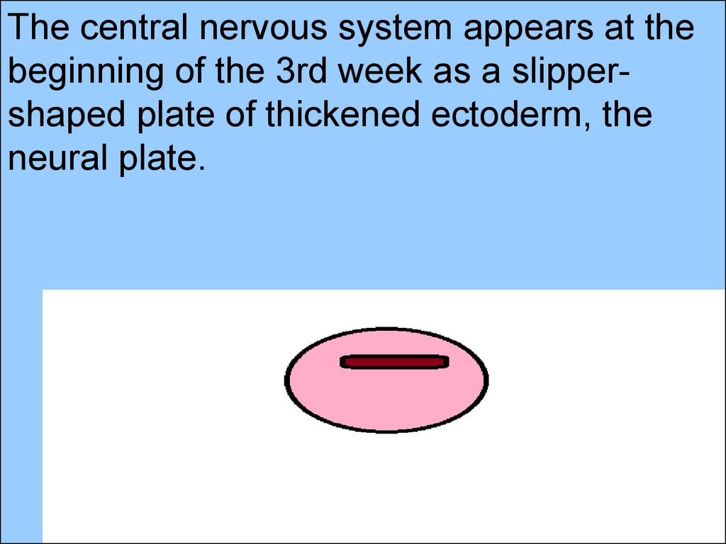
The central nervous system appears at thebeginning of the 3rd week as a slippershaped plate of thickened ectoderm, the
neural plate.
4.
Its lateral edges soon become elevated toform the neural folds. With further
development, the neural folds become more
elevated, approach each other in the
midline, and finally fuse, thus forming the
neural tube.
5.
Neural tube defects account for the mostcongenital anomalies of the CNS and
result from the failure of the neural tube to
close spontaneously between the 3rd and
4th wk of in utero development. Neural
tube defects (NTDs) involve the
meninges, vertebrae, muscles, and skin.
6. Neural tube defects (NTDs)
spina bifida occulta
meningocele
myelomeningocele
encephalocele
anencephal
7. Neural tube defects (NTDs)
• can be diagnosed prenatally byultrasound, and by determination of άfetoprotein (AFP) levels in maternal serum
and amniotic fluid. The cranium or
vertebra can be visualized since 12 weeks
of gestation, and defects can be detected.
• Recent evidence indicates that folic acid
(folate) reduces the incidence of NTDs in
certain populations.
8. Neural tube defects (NTDs)
• Meningocele(Meningoencephalocele) is
herniation of meninges and
brain(medulla) through a defect
in the skull or vertebra split
producing a fluid-filled sac in the
occipital or lumbar region.
9.
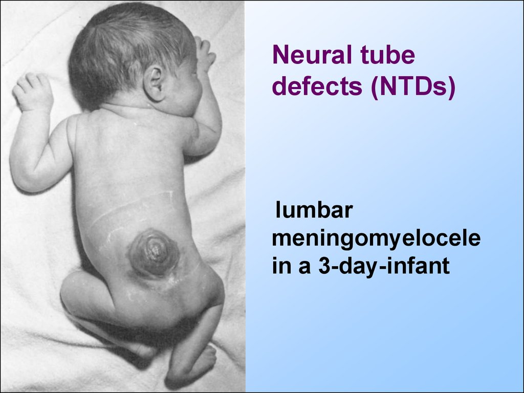
Neural tubedefects (NTDs)
lumbar
meningomyelocele
in a 3-day-infant
10.
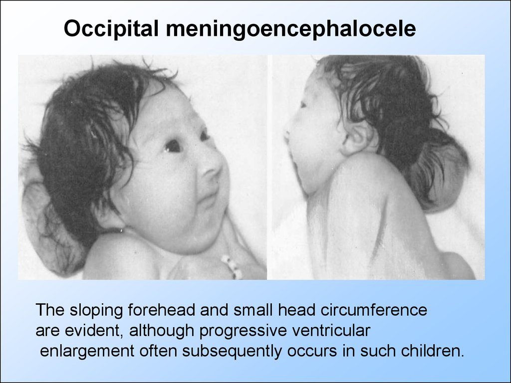
Occipital meningoencephaloceleThe sloping forehead and small head circumference
are evident, although progressive ventricular
enlargement often subsequently occurs in such children.
11.
In embryo at its cephalic end of the neuraltube the brain bladders are forming from
which all parts of the brain are originated
within approximately 2-3 months of in utero
development, including neural parts of ear,
eye and sense of smell.
12.
Hemispheres of the brain are developedfrom the first brain bladder. Errors of
embryogenesis, connected with an action of
a teratogen (the factor inducing
abnormalities) can lead to severe pathology
of the fetus and newborn, for example,
microcephaly and anencephaly. The
cerebral hemispheres and cerebellum are
usually absent, and only a residue of the
brain stem can be identified when
anencephaly presents.
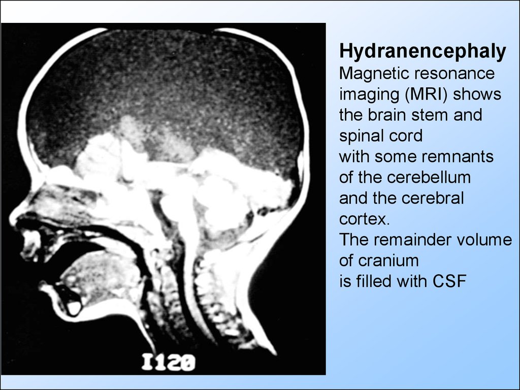
13.
HydranencephalyMagnetic resonance
imaging (MRI) shows
the brain stem and
spinal cord
with some remnants
of the cerebellum
and the cerebral
cortex.
The remainder volume
of cranium
is filled with CSF
14. CSF (cerebral spinal fluid)
• CSF flow results from the pressuregradient that exists between the
ventricular system and venous
channels. The intraventricular pressure
is twice higher than the pressure in the
superior sagittal sinus.
15. CSF
• Hydrocephalus resulting fromCSF accumulation inside the
brain is called internal
hydrocephalus.
16. The cranial computerized tomogram (CT) of the infant`s brain with congenital virus-associated encephalopathy
The cranial computerized tomogram (CT)of the infant`s brain with congenital virusassociated encephalopathy
Cerebral atrophy with
enlarged ventricles and
widened sulsi (internal
hydrocephalus).
17.
• CSF is absorbed primarily by thearachnoid villi through tight junctions
of their endothelium by the pressure
forces.
• Hydrocephalus resulting from
malfunction of the arachnoid villi is
called nonobstructive or
communicating hydrocephalus.
18. External hydrocephalus in the newborn with in utero infection of the brain (MRI)
19. Features of CNS in fetus and newborn
• The brain development is characterizing by gradualformation and maturation of brain structures from
ontologically "old" to "young“. Note the line: the spinal
cord, brain stem, subcortical formations, cerebellum
and at last the cortex are making mature.
• First months of life there is some functional minority of
regulating activity of the cortex in favour to the
subcortical formations with domination of
thalamopallidal and striopallidal areas.
• The child’s brain contains more protein than the brain
of the adult. Cerebral proteins make the tissues of
brain hydrophilic and bent them to cellular edema.
20. Features of CNS in fetus and newborn (continue)
• There is not clear differentiation of thebrain’s layers (grey and white substances
are indistinctly differentiated among
themselves).
• The gyri and sulci of the cortex are not
deep that reduces the absolute and
relative area of the child's cortex in
comparison to adult.
21. Features of CNS in fetus and newborn (continue)
The blood-brain barrier (BBB) of the fetusand newborn
• is normally indiscriminately permeable,
allowing protein and other large and small
molecules to pass freely between the
cerebral vessels and the brain.
• becomes mature only to the ending of the
neonatal period
22. Features of CNS in fetus and newborn
• Central and peripheral neurons formmyelin coating gradually. Myelinization
finally finishes only after the 3-rd year of
life.
• Due to undeveloped myelinization in
children long time the cortex physiology
will be characterizing to be bent to
generalization of irritation and difficulties of
neuronal braking.
23. Features of CNS in fetus and newborn
• The features of the brain vascularsystem of fetus when anastomoses
develop insufficiently make the brain
of premature newborn easily
vulnerable to hypoxia, mechanical,
and thrombotic damages. This can
promote for cerebral ischemia and
hypoxia with form of cerebral palsy.
24.
Neurological examination25. Neurologic evaluation of the child. Complaints&History.
Neurologic evaluation of thechild. Complaints&History.
• Seizures (convulsion) are involuntary, violent
contraction of muscles. Seizures may be:
• tonic or clonic,
• focal or generalized.
Tonic seizures are characterized by increased
tone or rigidity.
Clonic seizures consist of rhythmic muscle
contraction and relaxation, when stereotypic,
wide movements observe in extremities and
other parts of a body.
26.
Opisthotonus in a brain-injured infant.This is the tonic seizure.
27. Objective neurological examination
of the child should include 4 maindiagnostic aspects:
• 1. Level of consciousness (LOC)
• 2. Mentality
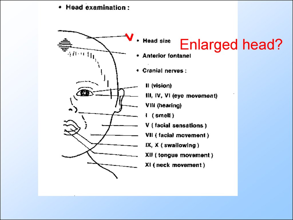
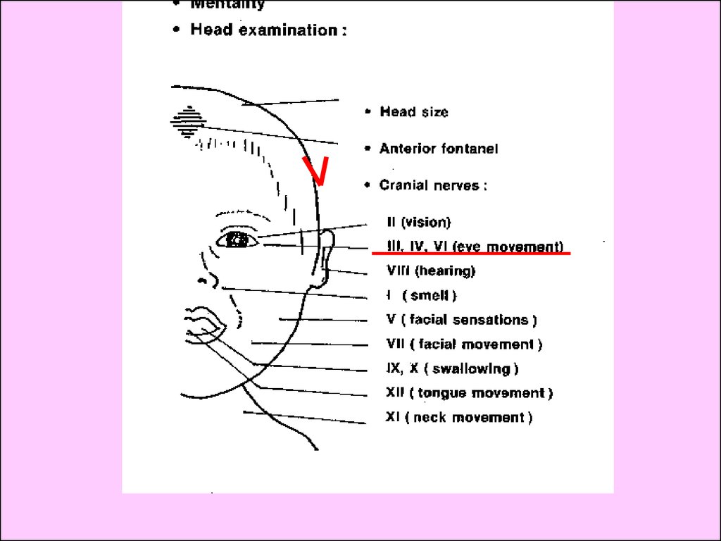
• 3. Head examination
• 4. Evaluation of motor system
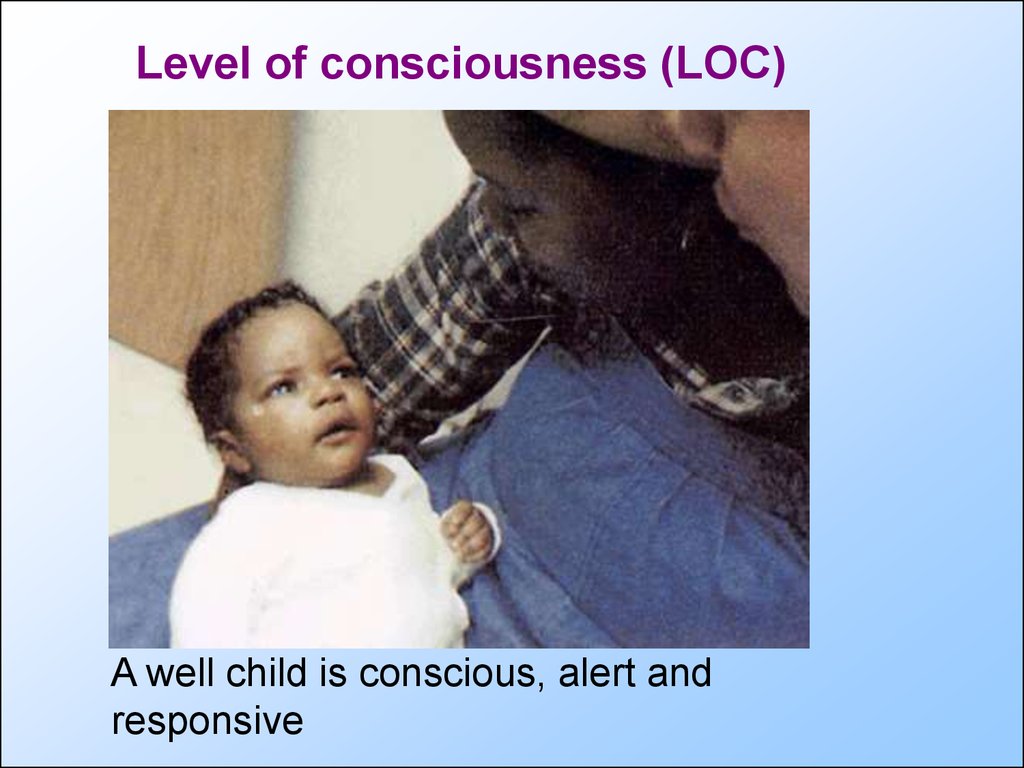
28.
Level of consciousness (LOC)A well child is conscious, alert and
responsive
29. Level of consciousness (LOC)
Lethargy or pathological sleepy
(somnolence) is possible to determine
as an unusual sleep of the patient.
Confusion. The responses of confused
patients demonstrate a failure to
comprehend their surroundings. The
patient is unable to estimate direction or
location, is apt to be disoriented in time
and may misidentify people.
Cоmа is absence of consciousness.
30. Level of consciousness (LOC)
• This is achild with
meningitis.
The child is
somnolent
and can not
arouse. Note
the face of a
gray color.
31. Stages of coma
1.2.
3.
4.
Stupor: The stuporous patient arouses
from sleep only after painful stimuli. Verbal
responses are slow or even absent. The
patient lapses into an unresponsive state
when the stimulus ceases.
Light coma: the patient has response to
painful stimulus.
Deep cоmа: there is no response to
painful stimulus.
Terminal coma: coma with a muscular
relaxation and apnea.
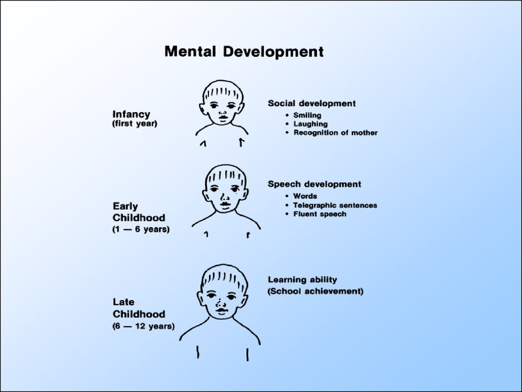
32.
Mental development33.
34.
Head size35.
Enlarged head?36.
AF&PF37.
A fontanelbulging is a
reliable indicator
of increased ICP,
but vigorous
crying can cause
a protuberant
fontanel in a
normal infant.
ICP-intracranial pressure
38.
Cranial nerves39.
40.
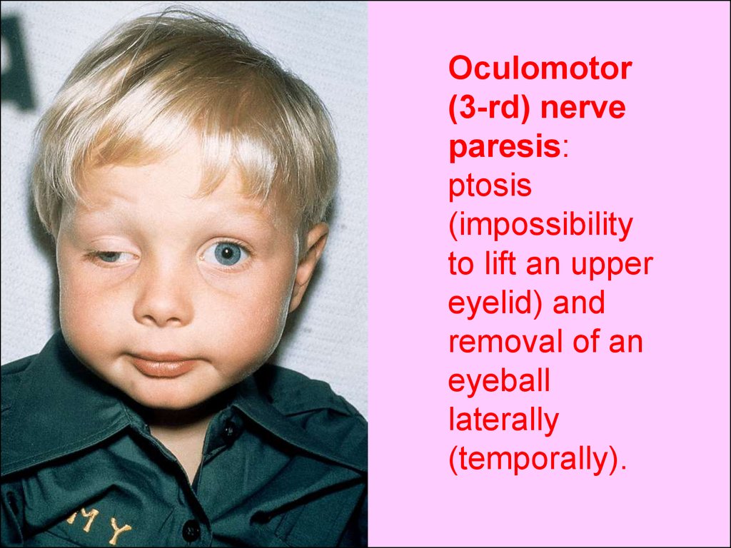
Oculomotor(3-rd) nerve
paresis:
ptosis
(impossibility
to lift an upper
eyelid) and
removal of an
eyeball
laterally
(temporally).
41.
42.
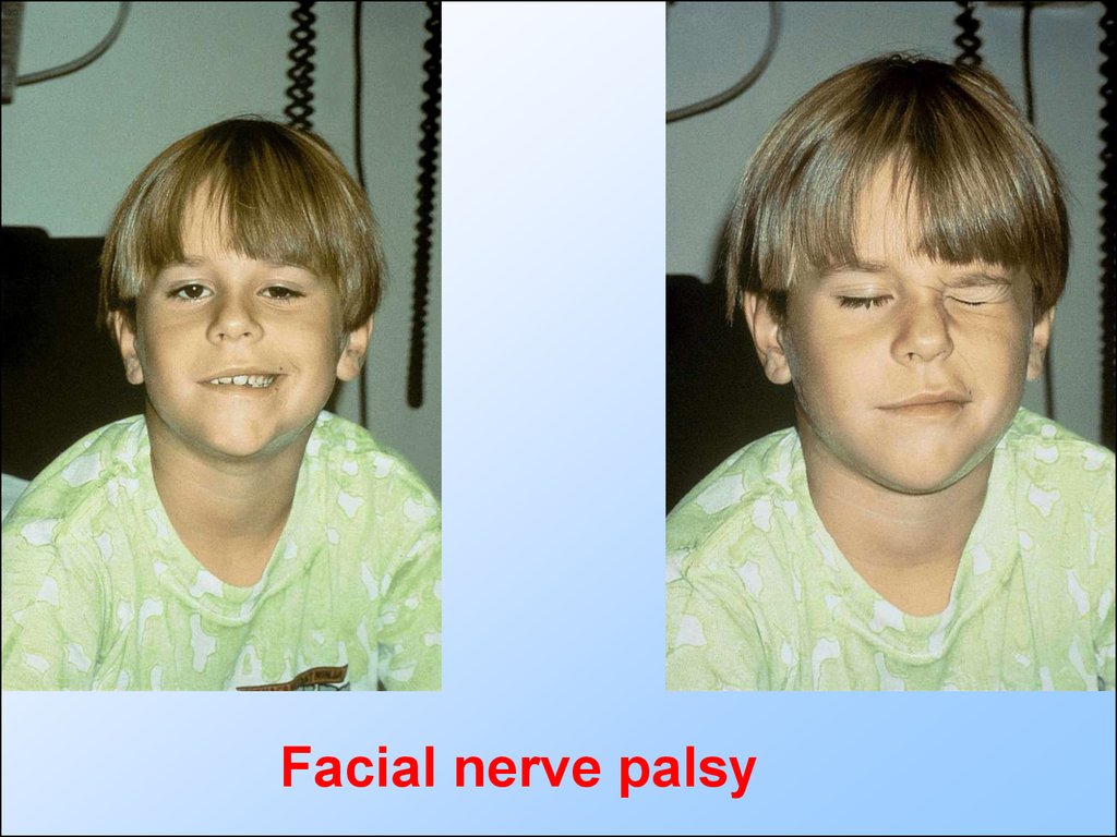
Facial nerve palsy43.
Facial nerve palsy. Notice the loss of the nasolabialfold and the mouth deviated to the left when he
smiles.
44.
A newbornwith right
facial palsy
45.
choking46.
47.
Unilateral(right-side)
hypoglossal
(12th) nerve
paresis.
Tongue
deviation.
48.
Motor examination49.
50. Abnormal gaits
• The spastic gait• Circumduction gait
• Cerebellar ataxia
• waddling gait
• clumsy, tentative gait
51. Movement disorders
• Paralysis (palsy) – the absence of anyvoluntary movements
• Paresis is incomplete paralysis
• Ataxia - gross uncoordination that may
become worse with the eyes closed
• Athetosis - slow, writhing, wormlike,
constant, grossly uncoordinated
movements that increase on voluntary
activity and decrease on relaxation
52.
A newborn withbrachial right
sided paralysis
(palsy). The arm
hangs limp
alongside the body
and internally
rotated, and the
wrist is pronated
hand (hangs limp
downwards).
53. Movement disorders
• Dystonia - slow twisting movements oflimbs or trunk (alternation of a hypotonia
with rigidity, formation of elaborate
postures)
• Tics - involuntary, compulsive,
stereotyped movements of an associated
group of muscles (can be suppressed by
strong-willed effort).
• Tremors - constant small very fast
involuntary movements.
54. Muscles
Examination includes assessment of• muscles’ development: wasting,
pseudohypertrophy
• Tone: hypotonia, hypertonia
• Strength: increase, decrease
55.
Posterioraspect of the
legs of a father
and his 6-yearold son with a
rare autosomal
dominant
muscular
dystrophy.
Hypertrophy
of the calves
resembles
Duchenne
muscular
dystrophy
56.
HypotoniaOn ventral
suspension, the
baby assumes the
position of a rag
doll.
When pulled up
from the supine to
the sitting
position, the head
of the baby lags.
57.
Main semiotics of CNSdisorders. Meningitis.
58.
59.
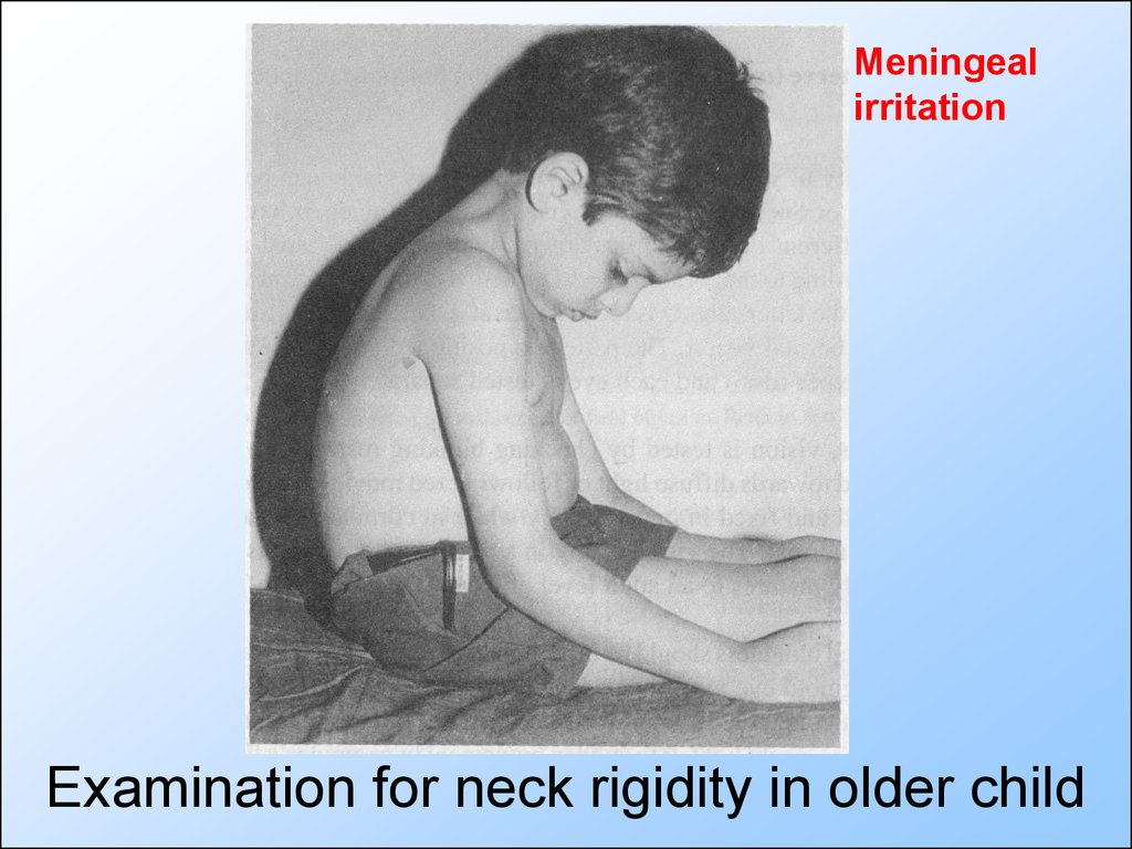
Meningealirritation
Examination for neck rigidity in older child
60.
Meningealirritation
Brudzinski’s sign
61.
Meningealirritation
Kernig’s sign
62. LP
• The lumbar punction confirms themeningitis
63.
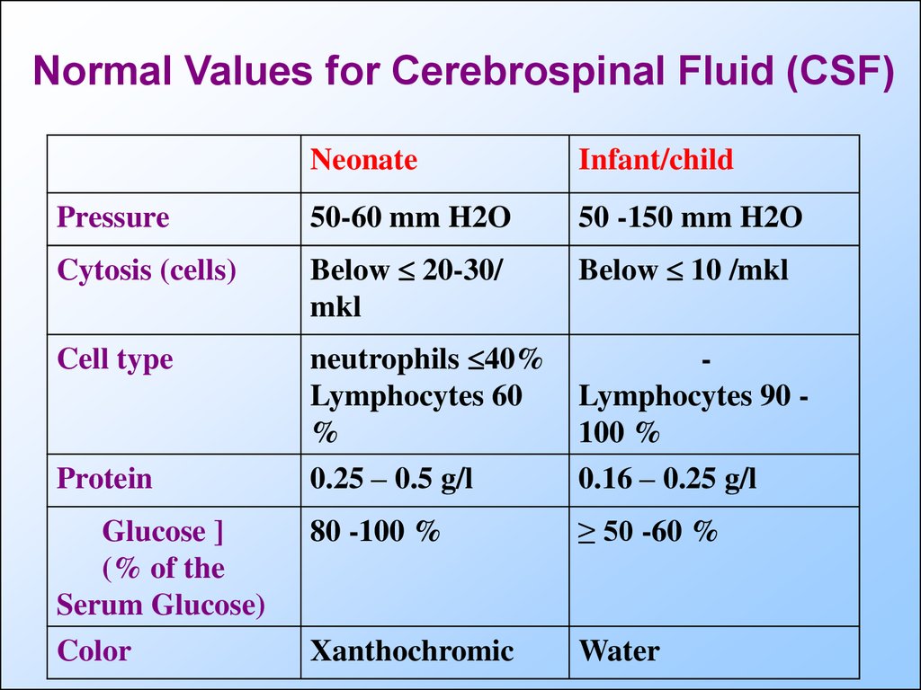
Normal Values for Cerebrospinal Fluid (CSF)Neonate
Infant/child
Pressure
50-60 mm H2O
50 -150 mm H2O
Cytosis (cells)
Below 20-30/
mkl
Below 10 /mkl
Cell type
neutrophils 40%
Lymphocytes 60
%
Lymphocytes 90 100 %
Protein
0.25 – 0.5 g/l
0.16 – 0.25 g/l
Glucose ]
(% of the
Serum Glucose)
80 -100 %
≥ 50 -60 %
Color
Xanthochromic
Water
64. CSF finding in bacterial meningitis
• ICP - increased• White blood cell count, μL - 100 –
10000
• Cell type - neutrophiles 100%
• Protein content - ≥ 40 mg/dl (0.4 g/l)
• Glucose - 40 mg/dl ( 50% blood
glucose)
• Culture - positive
65. Meningizm
• If the analysis of a cerebrospinal fluid findsinflammatory changes, the child has
meningitis even having negative or
doubtful clinical symptoms.
• If there are meningeal irritation symptoms,
but no inflammatory changes in a
cerebrospinal fluid, there is no meningitis.
Such condition is called meningizm, it
means non inflammatory irritation of
meninges in various diseases in children.