


 Медицина
Похожие презентации:


Basic dental instrumentation
1.
Discipline: "Propaedeutics of dental diseases and materials science"Basic dental instrumentation
Department of propaedeutics
dental diseases
2.
Lecture plan:1. Viewing set. The main tools included in the viewing kit. Their
purpose.
2. Tools used in therapeutic dentistry.
3. Tools used in surgical dentistry.
4. Instruments used in orthopedic dentistry.
3.
Dental instrumentsare designed to ensure
technological, efficient and
safe performance of all
medical diagnostic and
preventive dental procedures.
4.
1. Viewing set. The main tools included in the viewingkit. Their purpose.
The examination set of a dentist
includes tools intended for the
initial examination of a patient:
- dental mirror;
- dental probe;
- dental tweezers.
5.
Dental mirrorDental mirror is a round, usually
one-way mirror with a diameter
of 22-24 mm in a metal frame,
screwed into the handle.
Dental mirrors come in two
types: concave, which
magnifies the image, and flat,
which gives a real, undistorted
image. The working part is
made independently of the
handle and is attached to it with
a thread directly in the dental
office.
6.
The dental mirror is intended for the following manipulations:- additional illumination of
darkened areas of the oral cavity;
- inspection of areas inaccessible
to direct observation;
- abduction of the cheeks and
tongue when examining the
patient's oral cavity;
- abduction and protection of soft
tissues of the oral cavity during
preparation and other
manipulations.
7.
Dental probeThe dental probe (pointed probe) is a multifunctional diagnostic tool.
The most commonly used in dentistry is the angled probe. The
working part of this probe is thin, round in cross section, bent at an
angle and ends with a sharp tip.
The handle is small and light for improved tactile sensitivity. Probes
are single-sided or double-sided, may have a different configuration of
the working part.
8.
Appointment of pointed dental probes:- detection of carious cavities,
demineralized dentin, areas of
soreness of hard tissues of the
tooth during mechanical action
(probing);
- assessment of the condition of
the fissures of the teeth, the
presence of areas of carious
lesions in them ("jamming" of the
probe);
- detection of the presence of
communication between the
carious cavity and the tooth
cavity, detection of the mouths of
root canals, perforations, cracks;
9.
Appointment of pointed dental probes:- detection of supragingival dental
deposits;
- introduction into the cavity and
distribution along the bottom and
walls of medical and filling materials
of a fluid consistency;
- the introduction of drugs into the
cavity with the help of a cotton
turunda wound on the working part
of the probe.
- the handle of the probe can be
percussion - tapping on the cutting
edge or chewing surface of the
tooth.
10.
Dental tweezersDental tweezers have
branches (cheeks) curved at
an angle of 115-120°, the
inner side of which can be
smooth or has notches.
Tweezers are made of spring
steel.
branches
11.
Purpose of dental tweezers:- retention and transfer of cotton rolls,
retraction threads and other objects into and
out of the oral cavity;
- the introduction of drugs into the carious
cavity or cavity of the tooth either on a cotton
ball or between the branches of the
tweezers;
- holding and carrying small dental instruments
and pins;
- determination of the degree of tooth mobility;
The tweezer handle can be used as a spatula
to push back cheeks, lips, or percussion
teeth.
Notches on the jaws of the tweezers
contribute to better fixation (capture) of
the material.
12.
2. INSTRUMENTS USED IN THERAPEUTICDENTISTRY
13.
Mixing of filling materials, medical pastes and gaskets iscarried out with a metal or plastic spatula on a special
surface.
14.
Metal spatulaA metal spatula consists of a
handle, at both ends of which
there are elongated straight
spatulas. A metal spatula is
more convenient to use than
a plastic one.
spatula
handle
15.
Plastic spatulasPlastic spatulas are used to
prepare medicinal pastes and
to mix filling materials that
become inactivated or change
color when in contact with
metals (for example, silicate
cements, some composites).
Plastic spatulas are
disposable.
16.
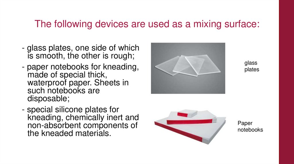
The following devices are used as a mixing surface:- glass plates, one side of which
is smooth, the other is rough;
- paper notebooks for kneading,
made of special thick,
waterproof paper. Sheets in
such notebooks are
disposable;
- special silicone plates for
kneading, chemically inert and
non-absorbent components of
the kneaded materials.
glass
plates
Paper
notebooks
17.
A number of requirements are imposed on the instruments used forfilling cavities:
- The instrument must be ergonomic, easy to clean and sterilize.
- The working part of the tool must be durable and resistant to abrasive,
mechanical and chemical influences.
- The color of the working part of the instrument should contrast against
the background of the filling material, tooth tissues and oral mucosa.
- The working part of the tool should not shine, give glare.
Filling materials should not stick to the surface of the working part of the
instrument.
The most common tools in this group are trowels and pluggers.
18.
Dental trowelA trowel is a dental tool with a
working part in the form of a
short plate of a straight or
curved shape, located at a
certain angle with respect to
the handle. Using a trowel,
paste-like drugs, filling
materials for gaskets,
temporary and permanent
fillings are introduced into the
cavities, and the surface of the
filling is modeled.
Dental trowel
19.
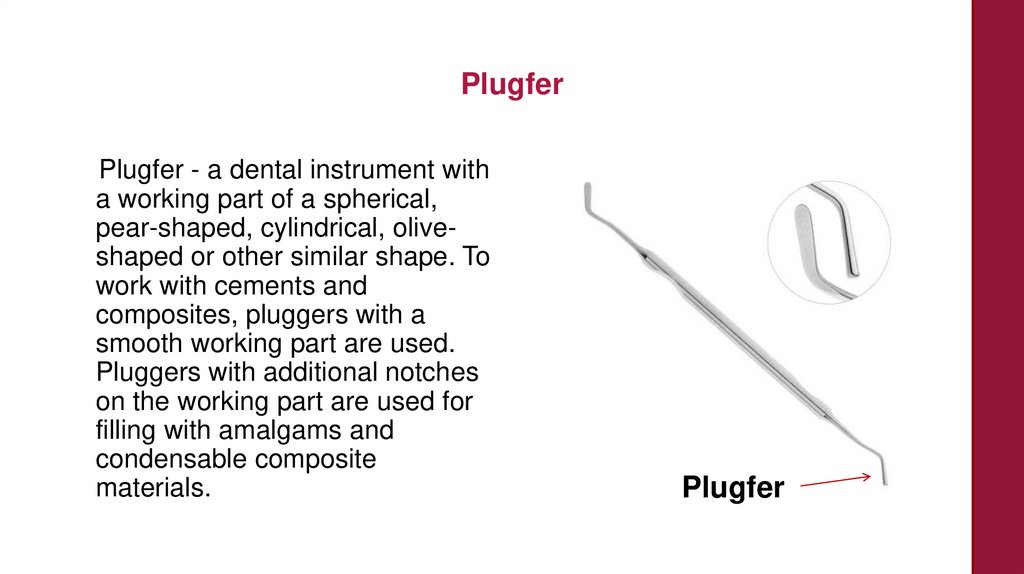
PlugferPlugfer - a dental instrument with
a working part of a spherical,
pear-shaped, cylindrical, oliveshaped or other similar shape. To
work with cements and
composites, pluggers with a
smooth working part are used.
Pluggers with additional notches
on the working part are used for
filling with amalgams and
condensable composite
materials.
Plugfer
20.
Plugfers are conditionally divided into condensingand modeling
Condensing pluggers have the shape of a working part, which
makes it possible to carry out compaction (condensation) of the
material in the cavity with maximum efficiency.
Modeling pluggers have a working part designed to model the
surface layer of the restoration, create the contours of fissures,
masticatory tubercles, marginal ridges, ridges and depressions
on the surface of the tooth crown.
21.
Condensing pluggersModeling pluggers
22.
Pluggers and floaters are available in various sizes, singlesided, double-sided, and also as a combination of a floaterwith a plugger.
23.
Dental excavatorIt is used to remove softened dentin, food debris, temporary
filling material.
24.
There are complete sets of standard instruments for periodontal examination, for example, the set described byMüller H.-P. [2004]. The set consists of six tools.
It includes:
- Flat dental mirror with a diameter of 22 mm;
- Dental tweezers;
- 4 probes:
1. Bilateral "probe-hook" - for the study of carious cavities and the
detection of subgingival dental deposits,
2. WHO probe (with rounded end),
3. Probe for furcations (Nabers probe calibrated to a step depth of 3
mm),
4. Graduated probe (periodontometer - calibration by 1 mm or 3-3-2-3
mm).
25.
Periodontal probeA periodontal probe is a special
probe with a measuring scale.
Using this probe, it is possible
to determine with high
accuracy the depth of the
pocket, the degree of bleeding
of the gums, to assess the root
surfaces of the tooth, the
furcations of the molars and to
identify the presence of
subgingival dental deposits.
26.
3. INSTRUMENTS USED IN SURGICALDENTISTRY
27.
Forceps and elevators for extraction of teethForceps
When removing teeth, the principle of the
lever is used.
In forceps for removing teeth and roots,
there are:
1. Cheeks;
2. Castle
3. Handles;
Some forceps have a transitional part (4)
between the jaws and the lock.
28.
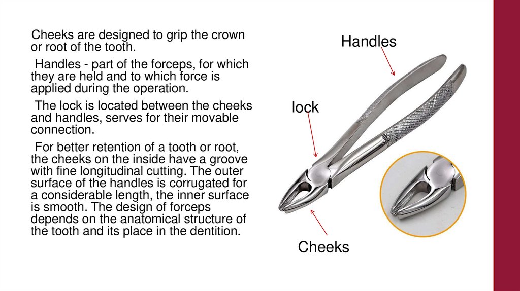
Cheeks are designed to grip the crownor root of the tooth.
Handles - part of the forceps, for which
they are held and to which force is
applied during the operation.
The lock is located between the cheeks
and handles, serves for their movable
connection.
For better retention of a tooth or root,
the cheeks on the inside have a groove
with fine longitudinal cutting. The outer
surface of the handles is corrugated for
a considerable length, the inner surface
is smooth. The design of forceps
depends on the anatomical structure of
the tooth and its place in the dentition.
Handles
lock
Cheeks
29.
There are the following types of forceps:1. Forceps for removing teeth and
roots of the upper and lower jaws.
In forceps for extracting teeth of
the upper jaw, the longitudinal
axis of the cheeks and the axis of
the handles coincide, or are
parallel, or form an obtuse angle
approaching two right angles. In
forceps for extracting teeth of the
lower jaw, the cheeks and
handles are located at a right
angle or at an angle approaching
a right one.
30.
2. Forceps for extracting teethwith a preserved crown
(crown) and for removing
roots (root).
The cheeks of the forceps for
removing teeth with a crown
do not converge when
closing, for removing roots
they converge.
31.
3. Forceps for removingindividual groups of teeth of
the upper and lower jaws.
They differ in the width and
features of the structure of the
cheeks, their location in
relation to the handles, the
shape of the handles.
32.
4. Forceps for removing thefirst and second large molars
of the upper jaw on the right
and left.
The left and right cheeks of
these forceps are arranged
differently.
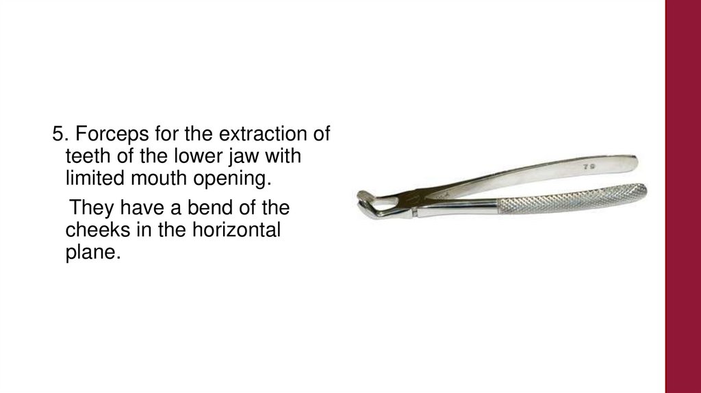
33.
5. Forceps for the extraction ofteeth of the lower jaw with
limited mouth opening.
They have a bend of the
cheeks in the horizontal
plane.
34.
To successfully perform theoperation, forceps should be
used, the design of which
corresponds to the anatomical
features of the tooth being
removed.
35.
The removal of the centralincisor, lateral incisor and
canine of the upper jaw is
carried out with forceps that
have a straight shape - straight
forceps. The longitudinal axes of
the cheeks and handles are in
the same plane and coincide.
Both cheeks are of the same
shape, on the inside they have a
recess (groove), the ends are
rounded. Forceps may have
cheeks of greater or lesser
width.
36.
Removal of small molars ofthe upper jaw is carried out
with forceps having an Sshaped bend. Their cheeks
are located at an obtuse
angle to the handles.
37.
Removal of large molars of the upper jaw is performed with forcepshaving an S-shaped bend and similar in shape to forceps for
removing small molars. However, their cheeks are arranged
differently. They are shorter and wider, the distance between them in
the closed state is greater. Both cheeks on the inside have recesses.
38.
At one cheek, the end is semicircular or flat, at the other it endswith a protrusion (thorn), from which a small ridge extends
along the middle of the inner surface. When a tooth is removed,
the spike enters between the buccal roots, a cheek with a flat
end covers the neck of the tooth from the palatal side. Some
forceps have a cheek with a spike on the right side, others - on
the left. Depending on this, a spike is distinguished for removing
teeth on the right or left side.
39.
Removal of the third large molar ofthe upper jaw is performed with
special forceps. Between the cheeks
and the lock, they have a transitional
part. The longitudinal axis of the
cheeks and the axis of the handles
are parallel. Both cheeks are the
same: wide, with a thin and rounded
end at the edges. On the inside, they
have recesses; when the forceps are
closed, the cheeks do not converge.
The design of the forceps makes it
possible to insert them deep into the
oral cavity, while the lower jaw does
not interfere with the operation.
40.
The roots of the incisors, canineand premolars of the upper jaw
are removed with the same
forceps as the teeth, only with
thinner and narrower cheeks that
converge when closed. To
remove the roots of large molars,
bayonet-shaped forceps are
used. They have a transitional
part, from which long converging
cheeks extend with a thin
semicircular end and a groove
along the entire inner surface.
The longitudinal axis of the
cheeks and the axis of the
handles are parallel.
41.
Removal of the teeth and rootsof the lower jaw is carried out
with forceps, curved along the
edge and having a beak-shaped
shape. The axis of the cheeks
and the axis of the handles form
a right angle or close to it. All
components of the tongs are
located in a vertical plane, the
handles are one above the
other. Depending on the shape
of the crown of the removed
tooth and the number of its
roots, the cheeks of the forceps
have a different structure
42.
To remove the incisors of thelower jaw, the cheeks of the
forceps are narrow with
grooves on the inside, their
end is rounded, and they do
not converge when closed.
The canine and small molars
are removed with the same
forceps, but with wider
cheeks.
43.
Forceps for removing largemolars have wide cheeks that
do not converge when closing.
Each of them ends with a
triangular protrusion (thorn). On
the inside, both cheeks have
recesses. When applied to the
tooth, the protrusions enter the
groove between the anterior and
posterior roots, ensuring good
fixation of the forceps on the
tooth.
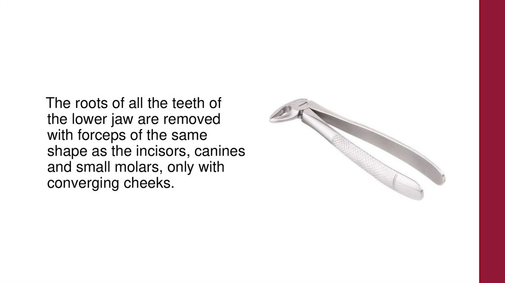
44.
The roots of all the teeth ofthe lower jaw are removed
with forceps of the same
shape as the incisors, canines
and small molars, only with
converging cheeks.
45.
ElevatorsWhen removing teeth with an
elevator, as well as with forceps,
the principle of a lever is used. The
elevator consists of three parts: the
working part, the connecting rod
and the handle.
There are many different designs
of elevators, but three types are
most common:
1. Straight;
2. corner elevator;
3. Bayonet elevator.
Straight elevator
46.
ElevatorsCorner elevator
Bayonet elevator
47.
4. INSTRUMENTS USED IN ORTHOPEDICDENTISTRY
48.
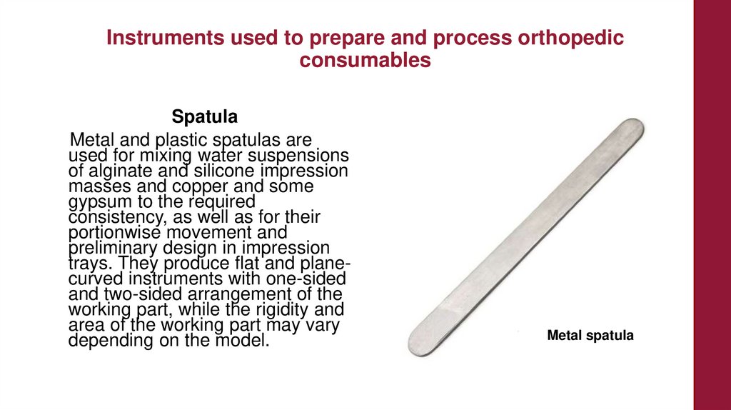
Instruments used to prepare and process orthopedicconsumables
Spatula
Metal and plastic spatulas are
used for mixing water suspensions
of alginate and silicone impression
masses and copper and some
gypsum to the required
consistency, as well as for their
portionwise movement and
preliminary design in impression
trays. They produce flat and planecurved instruments with one-sided
and two-sided arrangement of the
working part, while the rigidity and
area of the working part may vary
depending on the model.
Metal spatula
49.
Knives for making impressions of plaster modelsIn orthopedic dentistry, modeling
knives are used to finish (remove
excess) crystallized gypsum and
design a polymerized impression
mass. Knives for processing
plaster models have a hard blade
and a metal plate on the end part,
designed to open the cuvettes.
50.
Wax knivesFor batch separation of wax, its heat treatment and modeling, wax knives are used,
having a cutting part (blade) and a modeling part (spatula).
The handle of a wax knife is made of a thermally insulating material, since pre-heating
of the tool is necessary to process the wax and give it plastic properties. The working
part of the wax knife can be flat or have a recess for heating the wax over the flame of an
alcohol or gas burner.
51.
Tools used to remove prosthetic structuresForceps
To remove fixed structures fixed on the teeth of the upper jaw,
forceps with an S-shaped bend of the cheeks and handles are
used; perform debonding in the lower jaw area using forceps
curved along the rib or along the plane. To remove cone-shaped
telescopic crowns from the working model at the stage of their
laboratory production, as well as during debonding in the oral
cavity, forceps with divergent necks covered with retention
notches or sintered diamond chips are used.
52.
ElevatorsOrthopedic elevators used to remove prosthetic structures have a
flattened working part placed behind the gingival edge of the crown to
transfer the leverage generated by the rotation of the instrument handle.
The working part, depending on the location of the structure support, can
be oriented longitudinally or perpendicularly to the long axis of the tool.
In universal elevators, the working part has a cruciform shape for
working in the frontal and lateral segments of the jaws.
53.
Crown RemoversCrown removers that directly
transmit manual force are used at
the final stages of debonding or
with a small fixation force of
orthopedic structures. The body
of such instruments, among
which the Treimann crown
remover is the most famous,
consists of a handle with a bend
on the back side, a connecting
rod and a working part that
ensures the retention of the
instrument in the gingival part of
the prosthesis.
54.
55.
Basic dental instrumentationLecturer
Kobzeva Julia Alexandrovna
Associate Professor of the Department of Propaedeutics of Dental Diseases,
Candidate of Medical Sciences
[email protected]