


 Медицина
Похожие презентации:


Abnormalities of bony pelvis
1. ABNORMALITIES OF BONY PELVIS
TEACHER NAME: IRINA KAMILOVASTUDENT NAME: SACHIN PANDEY
GROUP NO. 163-B- LA-1
2. Introduction
INTRODUCTIONDerived from latin word means Basin
Ring of bone:
Two hip bone
Sacro-coccygeal part of vertebral column
The pubic part of hip bone connected by
pubic Symphysis.
Pelvic bone is made up of various
sections:
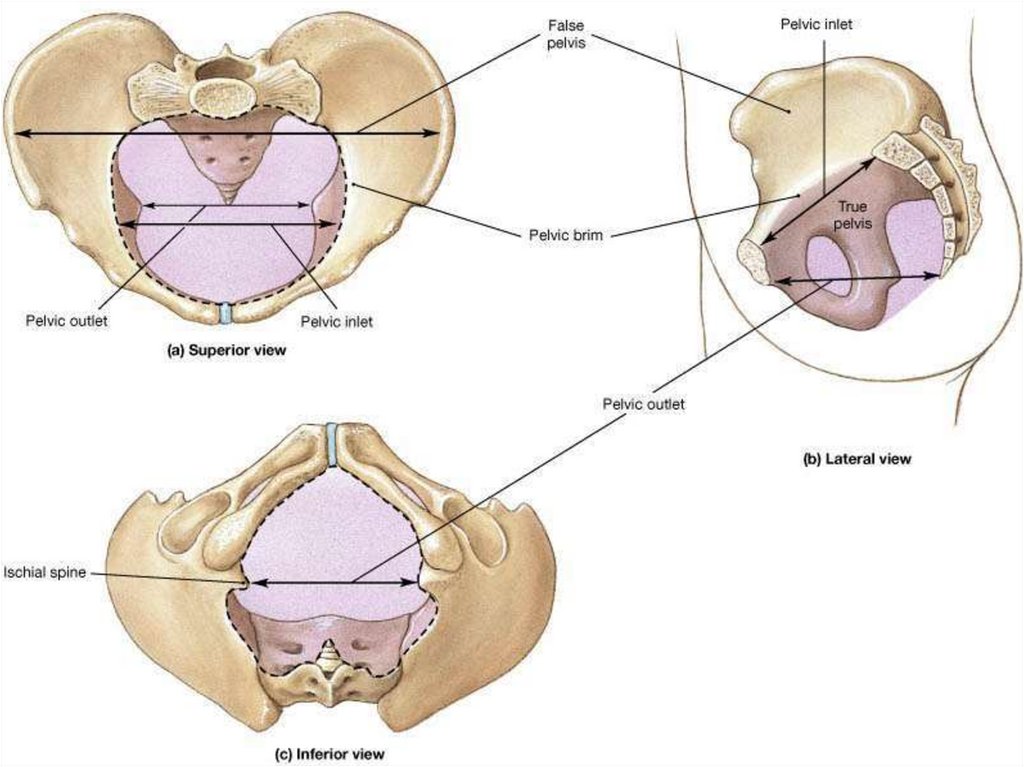
For obstetrical purposes, the pelvis is
divided by the pelvic brim into two parts:
– The False Pelvis
– The True Pelvis
3. False Pelvis
FALSE PELVISThe False Pelvis is that portion
above the pelvic brim. It does not
take part in the mechanism of
delivery and is of no obstetric
interest.
Intercristal diameter [IC ~29 cm]:
widest point on lateral aspect of
iliac crest
Interspinous diameter [IS ~26
cm]: distance between the lateral
tips of the anterior superior iliac
spines
External conjugate [AP] diameter
[EC ~20 cm]: distance between
apex of spine of 5th lumbar
vertebra and centre of the superior
border of symphysis pubis.
4.
5. True Pelvis
TRUE PELVISThe True Pelvis is that portion below the pelvic brim.
It determines the size and shape of the birth canal.
Pelvic Brim or Pelvic
inlet: formed by the
upper margins of pubic
bones, the ilio-pectineal
lines and the anterior
upper margin of the
sacrum.
Cavity: formed by the
pubic bones, ischium,
ilium, and sacrum
Outlet: diamond-shaped
made up of the pubic
bones, ischium, ischial
tuberosities,
sacrotuberous ligament,
and 5th segment of
sacrum
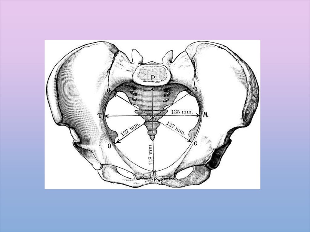
6. Pelvic inlet
PELVIC INLETPelvic inlet is formed from behind forward by
a. Sacral promontory
b. Anterior margins of ala of the sacrum
c. Linea terminalis
d. Upper end of symphysis pubis.
7.
8. Pelvic inlet
PELVIC INLETA-P diameter or anatomical conjugate
Extends from middle of sacral promontory
To the upper margin of symphysis pubis.
Oblique diameter:
Sacroiliac joint of one side to the iliopubic
Eminence of other side.
Transverse diameter:
Widest of all the diameters.
9.
10. Pelvic cavity
PELVIC CAVITYExtends downwards and backwards
from pelvic inlet,intervenes between
inlet and outlet.
Posterior wall of the cavity longer than
anterior wall.
Boundaries
Anteriorly
By symphysis pubis and body of the
pubis with its rami
Posteriorly
Concave pelvic surface of sacrum and
coccyx.
On each side
Quadrangular area formed by pelvic
surface of ilium and ischium.
11. Pelvic cavity
PELVIC CAVITYAnterior posterior diameter:
From middle of the back of symphysis
pubis to the pelvic surface of third sacral
vertebrae.
Oblique diameter:
Lower end of sacroiliac joint to the centre of
obturator membrane.
Transverse diameter:
Across the lateral bony walls of pelvic cavity.
12.
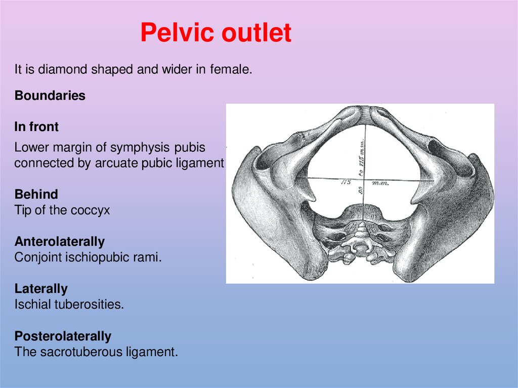
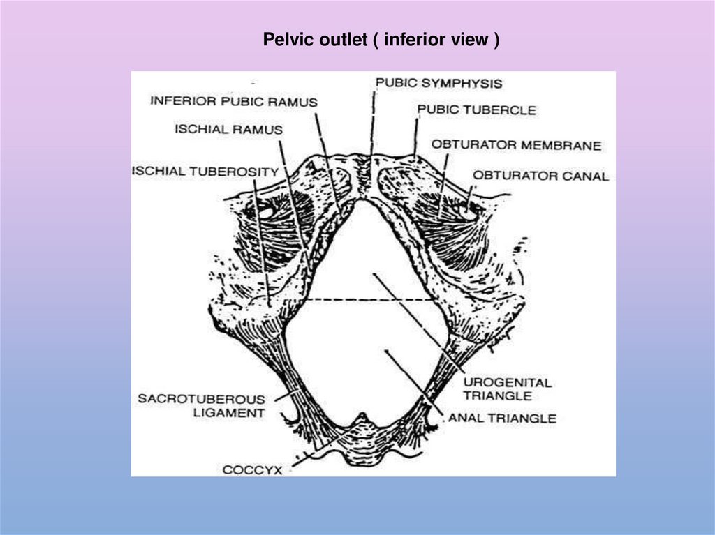
Pelvic outletIt is diamond shaped and wider in female.
Boundaries
In front
.
Lower margin of symphysis pubis
connected by arcuate pubic ligament
Behind
Tip of the coccyx
Anterolaterally
Conjoint ischiopubic rami.
Laterally
Ischial tuberosities.
Posterolaterally
The sacrotuberous ligament.
13.
Pelvic outlet ( inferior view )14. Pelvic outlet
PELVIC OUTLETAnterior –posterior diameter:
from lower border of symphysis pubis
to tip of the coccyx.
Oblique diameter:
Between the junction of ischio-pubic ramus
of one side and middle of the sacrotuberous
ligament of the opposite side.
Transverse diameter:
between the medial surfaces of the lower ends
of ischial tuberosities.
15.
16.
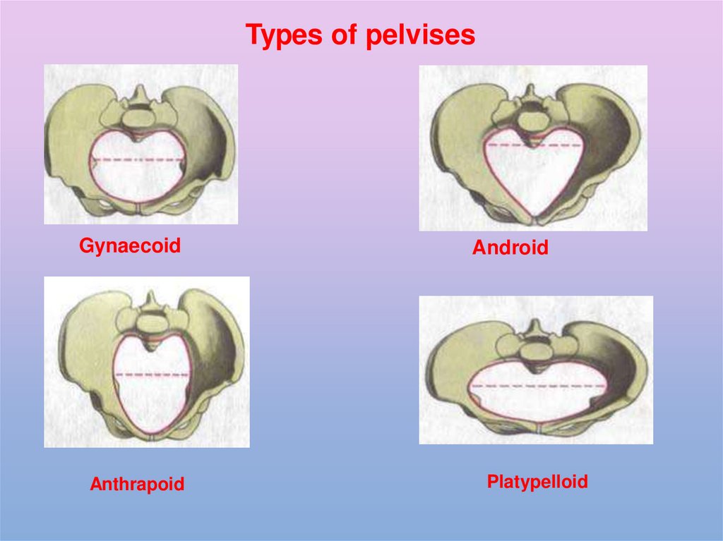
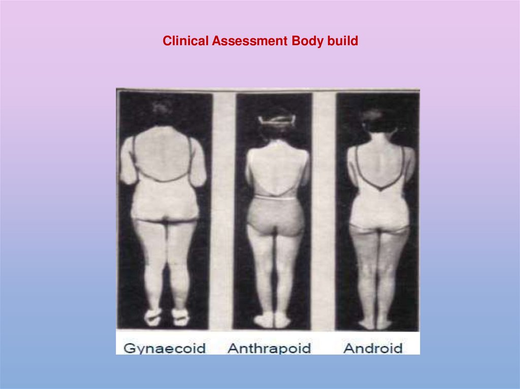
Types of pelvisesGynaecoid
Anthrapoid
Android
Platypelloid
17. Gynaecoid pelvis
GYNAECOID PELVISIdeal pelvis favouring a normal delivery;
50.6% of women
Brim slightly ovaltransversely but almost
Rounded.
Sacrum curved Ischial spines not
prominent
Short-cone pelvis
Obtuse greater sciatic notch
Triangular obturator
foramen
Sub-pubic arch rounded
[Roman arch] angle at least
900
18. Android pelvis
ANDROID PELVISMale-type pelvis favouring OP positions
and apt to cause deep transverse
arrest of head; 22.4% of women.
Brim heart-shaped
Sacrum curved
Ischial spines prominent
Long-cone funnel pelvis
Acute greater sciatic notch
Oval obturator foramen
Sub-pubic arch very narrow
[Gothic arch]
19. Anthrapoid pelvis
ANTHRAPOID PELVISApe-like pelvis favouring OP positions
often requiring operative vaginal
deliveries; 22.7% of women.
Brim AP oval
Sacrum very slightly curved
Ischial spines prominent
Long-cone funnel pelvis
with straight sidewalls
Obtuse greater sciatic notch
Oval obturator foramen
Sub-pubic arch narrow
20. Platypelloid pelvis
PLATYPELLOID PELVISLeads to cephalo-pelvic disproportion;
4.4% of women.
Brim oval transversely
Sacrum very slightly curved
Ischial spines prominent
Short-cone shallow pelvis
Acute greater sciatic notch
Triangular obturator foramen
Wide arch narrow
21.
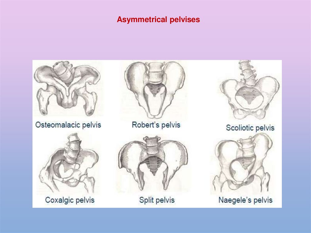
Asymmetrical pelvises22.
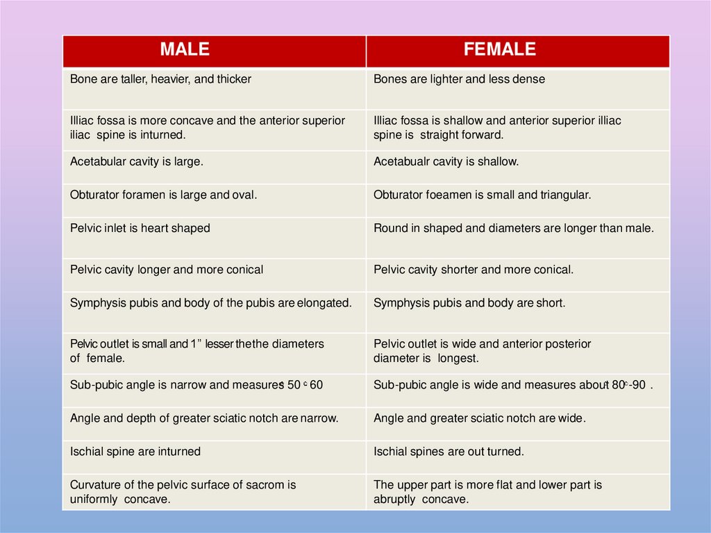
MALEFEMALE
Bone are taller, heavier, and thicker
Bones are lighter and less dense
Illiac fossa is more concave and the anterior superior
iliac spine is inturned.
Illiac fossa is shallow and anterior superior illiac
spine is straight forward.
Acetabular cavity is large.
Acetabualr cavity is shallow.
Obturator foramen is large and oval.
Obturator foeamen is small and triangular.
Pelvic inlet is heart shaped
Round in shaped and diameters are longer than male.
Pelvic cavity longer and more conical
Pelvic cavity shorter and more conical.
Symphysis pubis and body of the pubis are elongated.
Symphysis pubis and body are short.
Pelvic outlet is small and 1” lesser thethe diameters
of female.
Pelvic outlet is wide and anterior posterior
diameter is longest.
Sub-pubic angle is narrow and measures 50 - 60
Sub-pubic angle is wide and measures about 80 -90 .
Angle and depth of greater sciatic notch are narrow.
Angle and greater sciatic notch are wide.
Ischial spine are inturned
Ischial spines are out turned.
Curvature of the pelvic surface of sacrom is
uniformly concave.
The upper part is more flat and lower part is
abruptly concave.
23.
Clinical Assessment Body build24. Clinical Assessment foetal head as pelvimeter
CLINICAL ASSESSMENT FOETAL HEADAS PELVIMETER
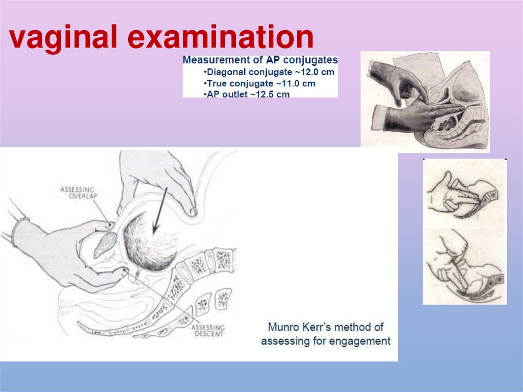
Engagement defined as the point when the engaging
diameter [BPD(biparietal diameter = ~10 cm] goes past the
pelvic brim.
Five fingers = 10 cm.
Fifths palpable above symphysis
pubis
25. Clinical Assessment foetal head as pelvimeter
CLINICAL ASSESSMENT FOETAL HEADAS PELVIMETER
In Gynaecoid & Android pelvis
distance between ischial spine to brim
is ~5 cm.
In Anthropoid pelvis distance is ~7
cm
In Platypelloid pelvis distance is ~3
cm
Station of the head
in
relation to ischial
spines
26.
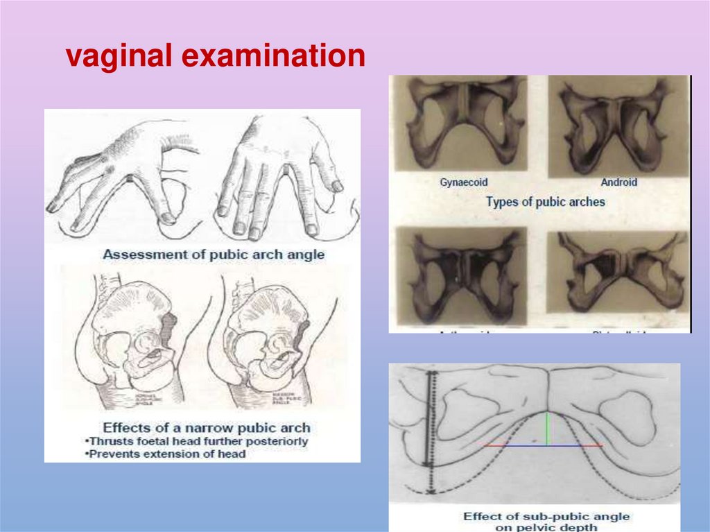
vaginal examination27.
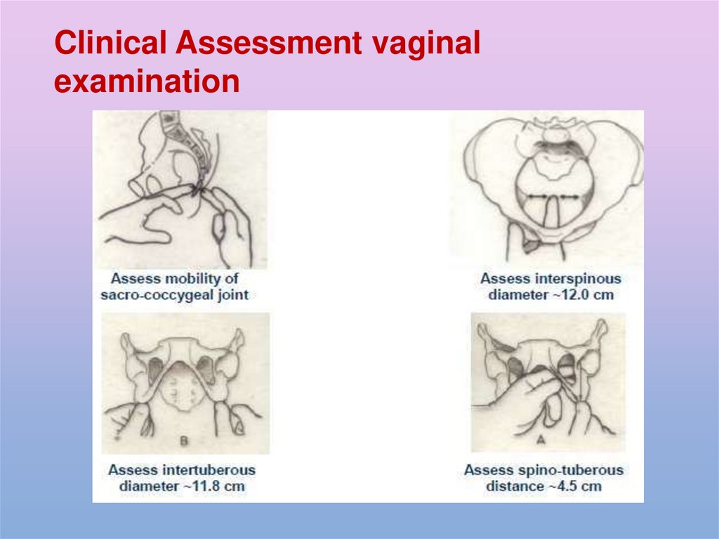
Clinical Assessment vaginalexamination
28.
vaginal examination29.
Presentation of the fetus atthe time of delivery
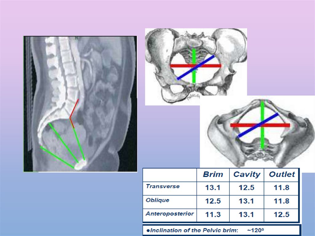
30. Clinical Assessment radiological examination
CLINICAL ASSESSMENT RADIOLOGICALEXAMINATION
1. TRUE AP CONJUGATE
2. OBSTETRIC CONJUGATE
3. MID-CAVITY AP CONJUGATE
4. OUTLET AP CONJUGATE
5. ANGLE GREATER SCIATIC NOTCH
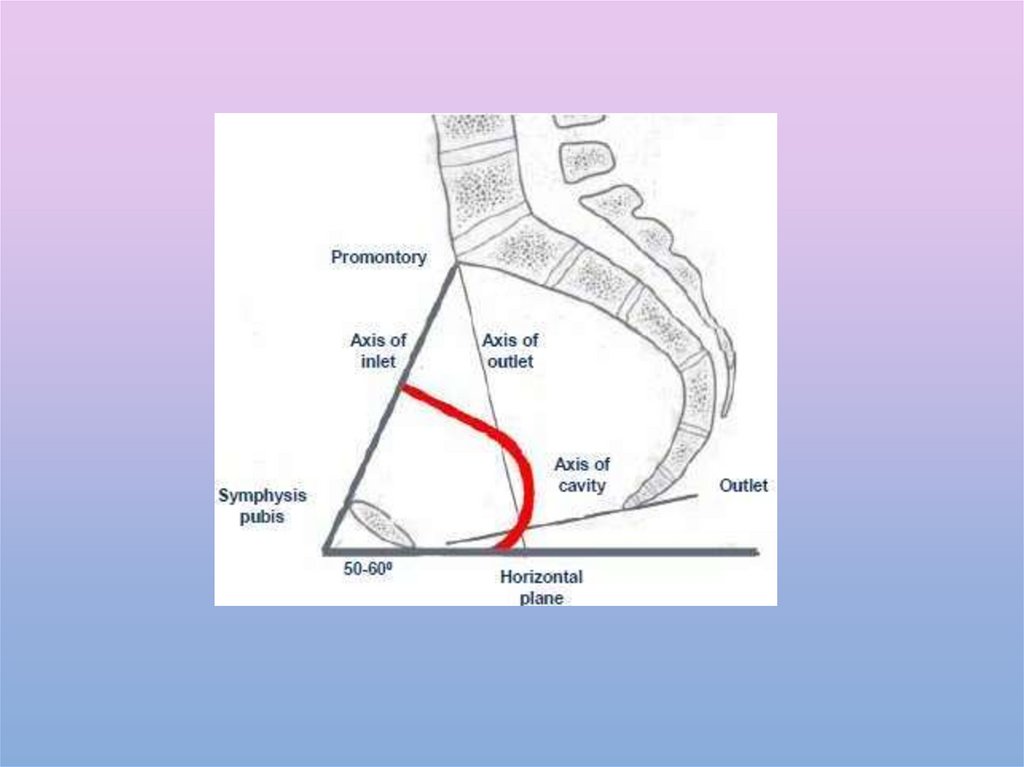
6. ANGLE OF INCLINATION OF PELVIC
BRIM
7. ANGLE OF INCLINATION OF
SACRUM
8. ISCHIAL SPINE
9. ISCHIO-TUBEROUS DISTANCE
10. FOETAL HEAD
• LIE, POSITION, ENGAGEMENT