


")


")


 Биология
Похожие презентации:


General myology
1. General myology
Anastasiia D. Koniaeva, a teaching assistant of anatomydepartment
2.
Myology is the study of the muscular system,including the study of the structure, function
and diseases of muscle
Muscles are the active part of the locomotor
system
3. The Functions of Muscles
• generation of movements• stabilization of the position
of the body
• control of the volume of the
organs
• smooth muscle – sphincters
• motion of the substances in
the body-blood, lymph,
urine, air, food and fluids,
sperm
• generation of body heat
• voluntary and involuntary
contractions of skeletal
striated muscle
4. 3 types of muscle tissue
• Smooth muscle tissue (textus muscularis levis)• Striated muscle tissue (textus muscularis striatus)
• Cardiac striated mucle tissue (textus muscularis striatus cardiacus)
All tipes of muscle tissue convert the chemical energy of ATP into the
mechanical energy of motion.
5.
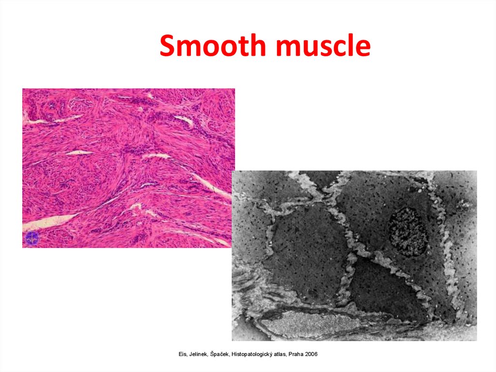
Smooth muscleEis, Jelínek, Špaček, Histopatologický atlas, Praha 2006
6.
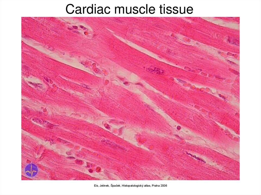
Cardiac muscle tissueEis, Jelínek, Špaček, Histopatologický atlas, Praha 2006
7.
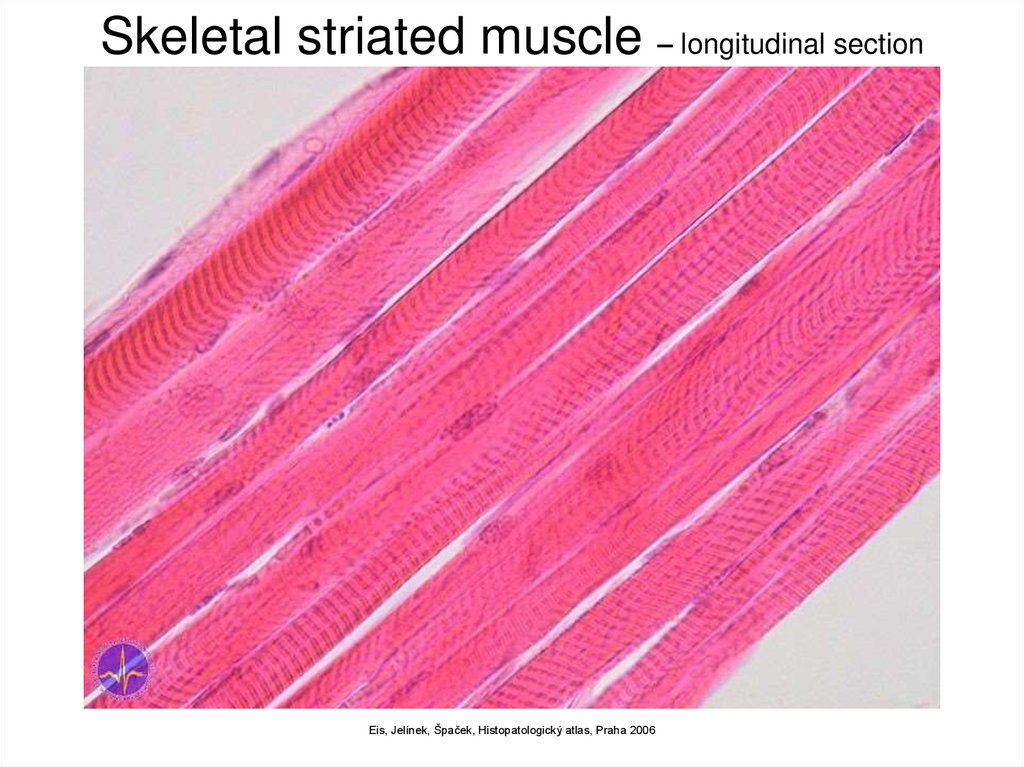
Skeletal striated muscle – longitudinal sectionEis, Jelínek, Špaček, Histopatologický atlas, Praha 2006
8.
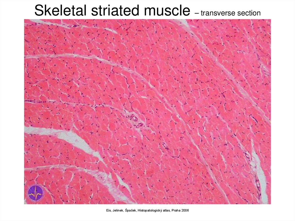
Skeletal striated muscle – transverse sectionEis, Jelínek, Špaček, Histopatologický atlas, Praha 2006
9. Skeletal striated muscle
Myoglobin (pigment causing red colouring)Fibres
• Quick
quickly fatigued
light (white)
in superficial layers
• Slow
more resistant to tiredness
dark (red)
in deeper layer
Inervated by cranial and spinal nerves
without inervation non-functional and atrophies
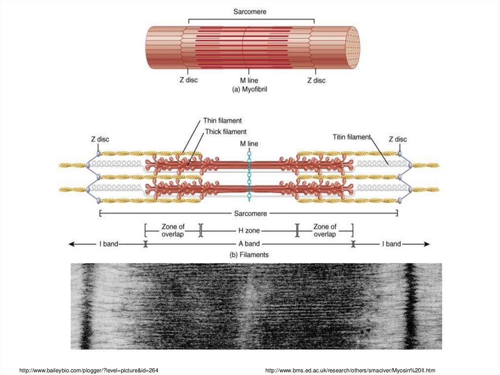
10. Skeletal striated muscle myofibre (myofibra)
Elementary structural unit
Multinucleated
thickness: 10–100 µm
length: mm – cm
origin: merging of elongated mononuclear cells
(myoblasts) → myotubes (nuclei inside, myofibrils at
the surface) → conversion to myofibres (nuclei at the
surface, myofibrils inside)
• sarcolemma on the surface
• striated in the microscope
• lighter and darker sections
11.
http://www.baileybio.com/plogger/?level=picture&id=264http://www.bms.ed.ac.uk/research/others/smaciver/Myosin%20II.htm
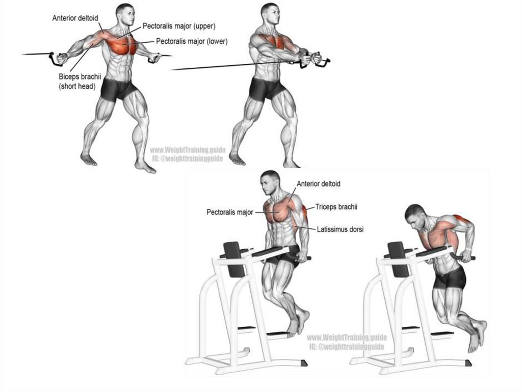
12. Functions of skeletal muscle
1. Movement of animal body2. Control of body openings and passages
"maintain continence“
3. Generate heat by shivering
4. Body support and maintenance of posture
13. Basic muscle structure
• striated muscle fibres• special muscle structures
• primary muscle bundle
– 10-100 fibres connected and covered by
fibrous tissue
• secondary bundles
– connection of primary bundles and covering by
fibrous tissue
• bundles of higher orders
14. Basic muscle structure
• fibrous tissue– endomysium (perimysium internum)
• covers myofibres and bundles
– epimysium (perimysium externum) = fascia
• covers the whole muscle
• tendon (tendo) is a tough band of fibrous connective
tissue that usually connects muscle to bone and is
capable of withstanding tension.
• aponeurosis (aponeurosis)
• myotendinous junction (junctio myotendinea)
– connection of myofibres with first (originating) and
inserting tendon
15. The parts of muscles
• origin (origo)– mobile end (punctum fixum)
• head (caput musculi)
• belly (venter musculi)
• attachment (insertio)
– fixed point (punctum mobile)
origin
belly
insertion
16. Classification of skeletal muscles by form
17. Classification of skeletal muscles by movement
agonists
–
antagonists
–
muscles participating in one movement (working together)
main (principal) muscle
–
counteracting muscles
synergists
–
in the same direction acting muscles
one out of the group of synergists
auxiliary (accesory) muscles
–
they act together with the principal muscle
18. Classification of skeletal muscles by number of joints
• one-jointed muscles– they´re causing the movement only in 1 joint
• double-jointed muscles
• multiple-jointed muscls
-they act mainly in the joint closest to the insertion
19. Classification of skeletal muscles by the direction of movement
• flexor (m. flexor)– makes the angle in the joint
more acute
• extensor (m. extensor)
– makes the angle in the joint
more obtuse
• adductor (m. adductor)
– moves the bone medially
• abductor (m. abductor)
– moves the bone laterally
• rotator (m. rotator)
– turns the bone around its
long axis
• levator (m. levator)
– lifts up a part of the body
• depressor (m. depressor)
– drops down a part of the body
• pronator (m. pronator)
– helps with pronation
• supinator (m. supinator)
– helps with supination
• opponens (m. opponens)
– places the thumb against other
fingers
• sphincter (m. sphincter)
• dilator (m. dilatator)
20. The work of muscles
Dynamic-work in which musclesmove parts of a person’s
body, and the body moves in
relation to a support, earth or
water surface.
• Holding;
• Overcoming;
• Yield.
Static work is observed while
maintaining the positions of
parts of the body. At the
same time, there are no
noticeable movements in the
joints, there is no external
mechanical effect.
• Holding items
• Holding the posture
21.
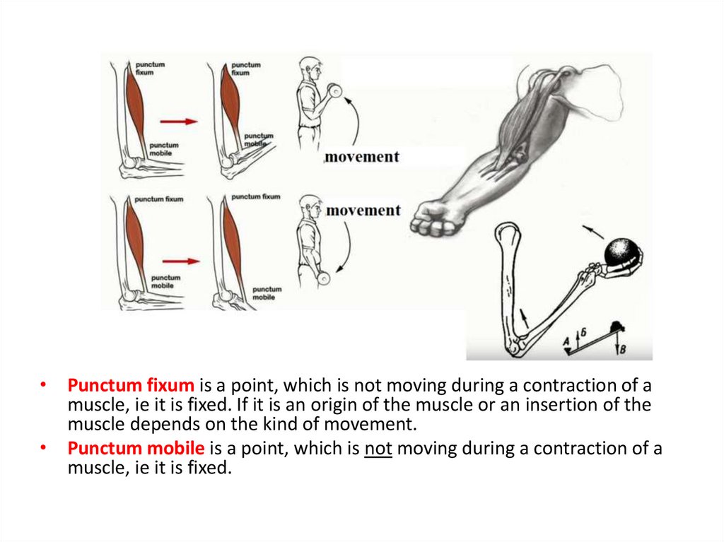
• Punctum fixum is a point, which is not moving during a contraction of amuscle, ie it is fixed. If it is an origin of the muscle or an insertion of the
muscle depends on the kind of movement.
• Punctum mobile is a point, which is not moving during a contraction of a
muscle, ie it is fixed.
22. Special muscle structures
• fascia (= perimysium externum)– fibrous envelope of muscle or muscle group
– barrier for spreading of inflammation in that
specific area
• osteofascial septum (= septum
osteofasciale)
– fascial divider from the superficial fascia to
the periosteum
– separates the space for muscle groups –
compartment (compartimentum)
23. Fasciotomy
http://lifeinthefastlane.com/ortho-library/compartment-syndrome/24. Special muscle structures
• tendon (tendo)– strip of tough fibrous connective tissue composed of bundles of collagenous
fibrils
– connects the muscle to the bone
– peritenonium internum (covers the bundles)
– peritenonium externum (consistent envelope on the surface of the tendon)
• aponeurosis (aponeurosis)
– flat tendon
– mutually crossing bundles in layers
• tendinous sheath (vagina tendinum)
– space along the tendon lined by synovial membrane
– vagina fibrosa: surrounds the vagina synovialis, holds the tendons to the
bone
– vagina synovialis
• epitenonium: inner layer (covers the tendon)
• peritenonium: outer layer
• mesotenonium: mutual switching of both previous things
http://www.zoology.ubc.ca/~biomania/tutorial/bonejt/jt01ac03.htm
25. Special muscle structures
• bursae mucosae– pouches in the vincinity of the joints, tendons and muscles
– lined by synovial membrane, filled up with synovial fluid
– reduction of rubbing in places exposed to considerable movement and
pressure
• retinacula
– strengthened stripes of the superficial fascia
– tie the inserting tendons to the bone
– together with the skeleton create osteofibrous canal
• vincula
– mesotenonium of tendinous sheaths of the flexors of the hand
– vascular supply for corresponding tendons run through them
– vincula brevia et longa
http://medical-dictionary.thefreedictionary.com/bursa
http://www.blackburnfeet.org.uk/hyperbook/trauma/ankleFx/ankleFxBasic1.htm
http://www.msdlatinamerica.com/ebooks/SurgicalExposuresinOrthopaedicsTheAnatomicApproach/sid139333.html
26.
27.
28.
29.
30.
31.
32.
33.
34.
35. EMG (electromyography)
• detection of the superficial muscle or the intramuscularactivity
• detects the change of electrical potential
• diagnostics for various muscle and neural malfunctions
http://biomech.ftvs.cuni.cz/pbpk/kompendium/biomechanika/experiment_metody_emg.php
http://www.fsps.muni.cz/inovace-SEBS-ASEBS/elearning/biomechanika/vyzkumne-metody-v-biomechanice
36. Functional muscle test
• informs us about the muscle strength• helps to assess the extent and location of the impairment
• analysis and examination of performance for the whole
movement
• assessment – 6 grades
• 0 – no sign of contraction
• 1 – twich (not enough to do the move)
• 2 – very weak (movement in the whole extent, doesn´t overcome
the resistance of the tested part of the body)
• 3 – weak (overcomes the gravity)
• 4 – good (overcomes medium-sized outer resistance)
• 5 – normal (very good function)
37.
Abnormal contraction• spasm – involuntary contraction of one muscle
• cramp – painful spasm
• tetanus – multiple spasms of skeletal muscles
• tic – involuntary twiches of muscles, usually under
voluntary control
• tremor – rhythmical, involuntary contractions of opposite
groups of muscles
• fasciculations – involuntary, short twiches on motor unit
visible under the skin
• fibrilace – spontaneous contractions of fibres of one
muscle that aren´t visible under the skin
38. Homework
• #Brightanatomy• @ssmutomsk
• @salome_mee