


:")


 Медицина
Похожие презентации:


")


ЭКГ при нарушениях ритма и проводимости
1. ЭКГ при нарушениях ритма и проводимости
ФГОУ ВО КубГМУ МЗ РоссииЭКГ при нарушениях
ритма и проводимости
Кафедра пропедевтики внутренних
болезней
2.
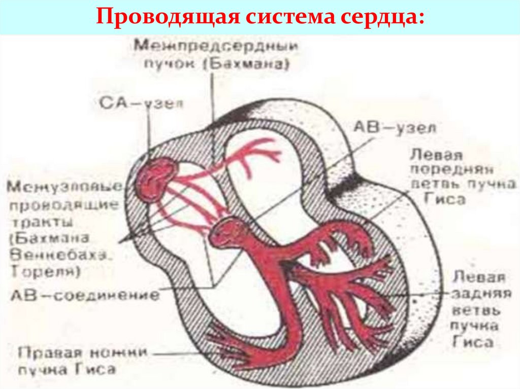
Проводящая система сердца:3.
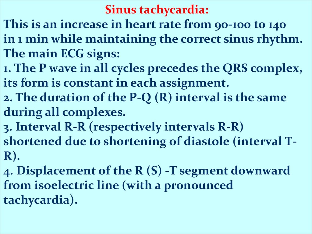
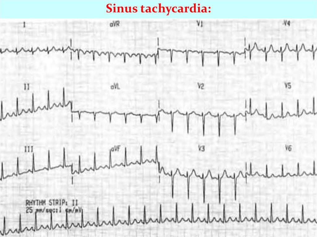
Sinus tachycardia:This is an increase in heart rate from 90-100 to 140
in 1 min while maintaining the correct sinus rhythm.
The main ECG signs:
1. The P wave in all cycles precedes the QRS complex,
its form is constant in each assignment.
2. The duration of the P-Q (R) interval is the same
during all complexes.
3. Interval R-R (respectively intervals R-R)
shortened due to shortening of diastole (interval TR).
4. Displacement of the R (S) -T segment downward
from isoelectric line (with a pronounced
tachycardia).
4.
Sinus tachycardia:5.
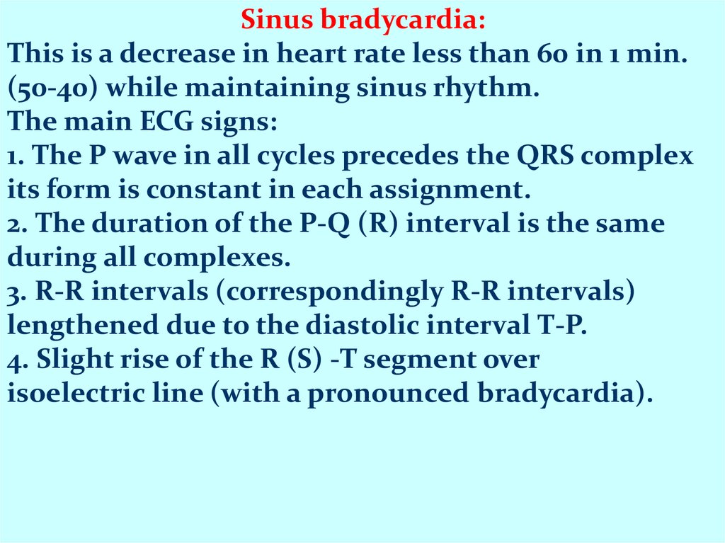
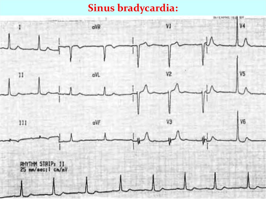
Sinus bradycardia:This is a decrease in heart rate less than 60 in 1 min.
(50-40) while maintaining sinus rhythm.
The main ECG signs:
1. The P wave in all cycles precedes the QRS complex
its form is constant in each assignment.
2. The duration of the P-Q (R) interval is the same
during all complexes.
3. R-R intervals (correspondingly R-R intervals)
lengthened due to the diastolic interval T-P.
4. Slight rise of the R (S) -T segment over
isoelectric line (with a pronounced bradycardia).
6.
Sinus bradycardia:7.
Sinus arrhythmia:This is the alternation of periods of increased heart rate with
periods of its slowdown. Distinguish between respiratory and
non associated with breathing. With respiratory sinus
arrhythmias there is an increase in cardiac activity on
inhalation and slowing down on exhalation (physiological
phenomenon).
The main ECG signs:
1. The P wave in all cardiac cycles precedes complex QRS, its
form is constant in each lead.
2. The duration of the interval P-Q (R) is the same in all
complexes.
3. Different intervals R-R (P-P), and this the difference is
greater than 10% of the average R-R distance (R-R), usually
about 12-0.15 C and more.
8.
Sinus arrhythmia:9.
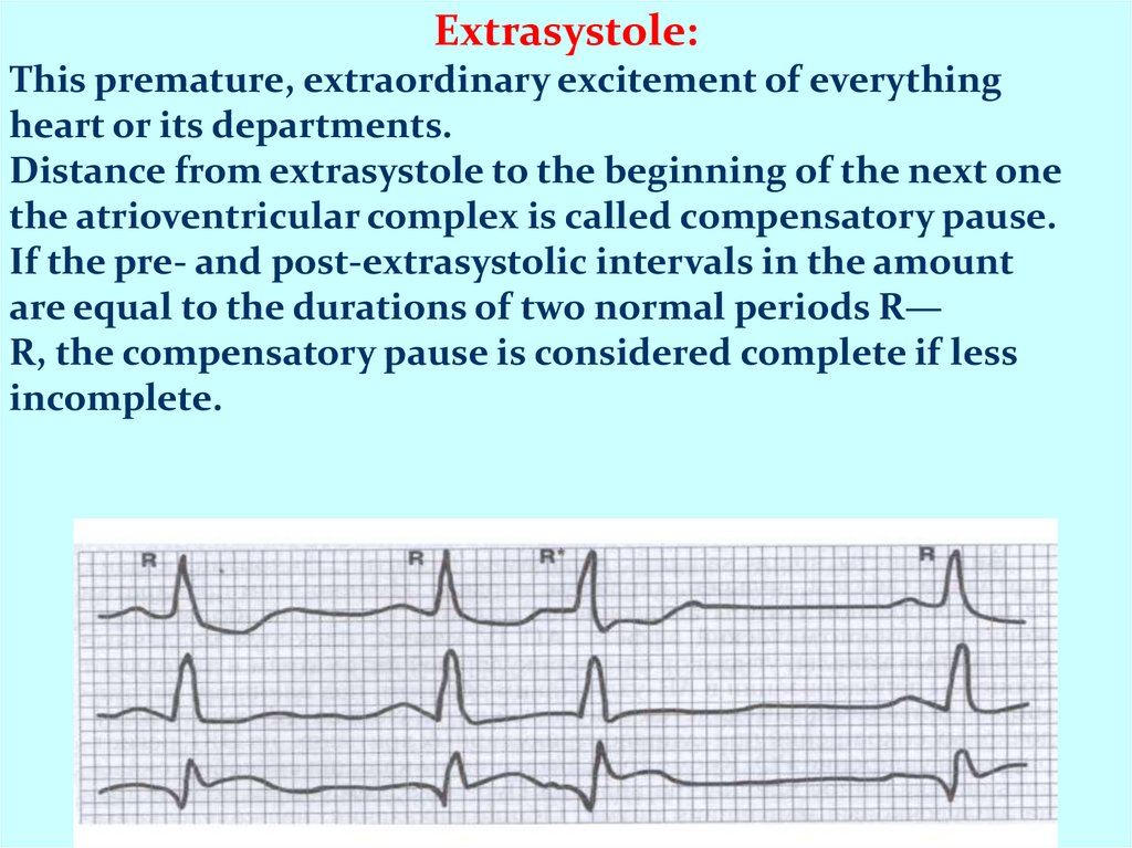
Extrasystole:This premature, extraordinary excitement of everything
heart or its departments.
Distance from extrasystole to the beginning of the next one
the atrioventricular complex is called compensatory pause.
If the pre- and post-extrasystolic intervals in the amount
are equal to the durations of two normal periods R—
R, the compensatory pause is considered complete if less
incomplete.
10.
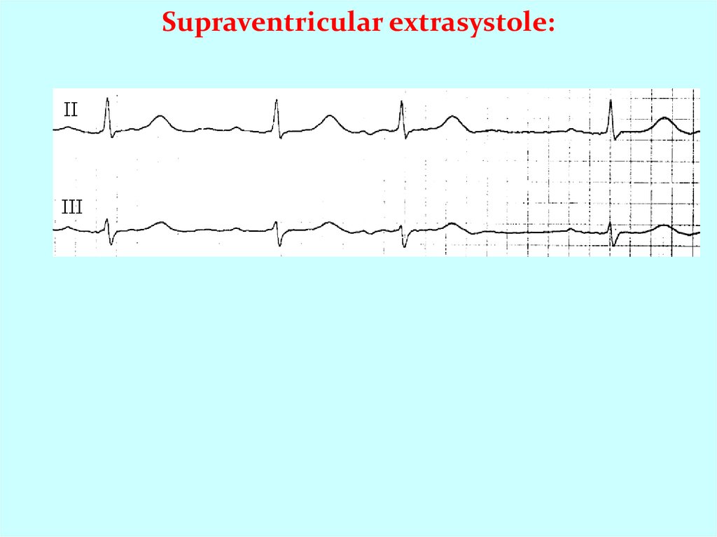
Supraventricular extrasystole:This is the premature appearance of an excitatory
impulse within the atria of the AV node.
The main ECG signs:
1. Premature contraction
2. The presence of a P wave in front of a QRS complex other
than sinus.
A) P positive - extra-atrial extrasystole.
B) P negative - lower atrial extrasystole.
B) P negative immediately precedes QRS,
not defined, follows it - extrasystole from AB connections.
3. The QRS complex of the supraventricular extrasystole is
usually not changed, differs little from that of sinus
reduction.
4. Incomplete compensatory pause.
11.
Supraventricular extrasystole:12.
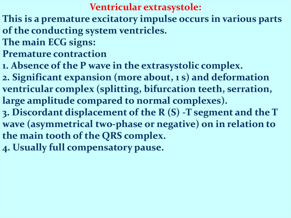
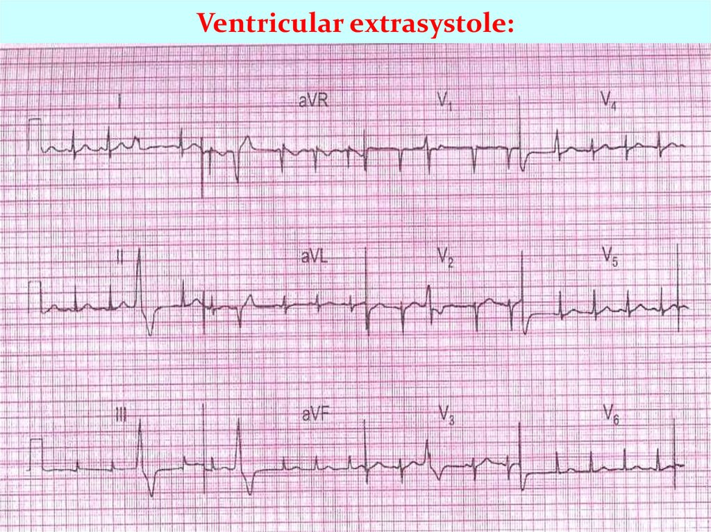
Ventricular extrasystole:This is a premature excitatory impulse occurs in various parts
of the conducting system ventricles.
The main ECG signs:
Premature contraction
1. Absence of the P wave in the extrasystolic complex.
2. Significant expansion (more about, 1 s) and deformation
ventricular complex (splitting, bifurcation teeth, serration,
large amplitude compared to normal complexes).
3. Discordant displacement of the R (S) -T segment and the T
wave (asymmetrical two-phase or negative) on in relation to
the main tooth of the QRS complex.
4. Usually full compensatory pausе.
13.
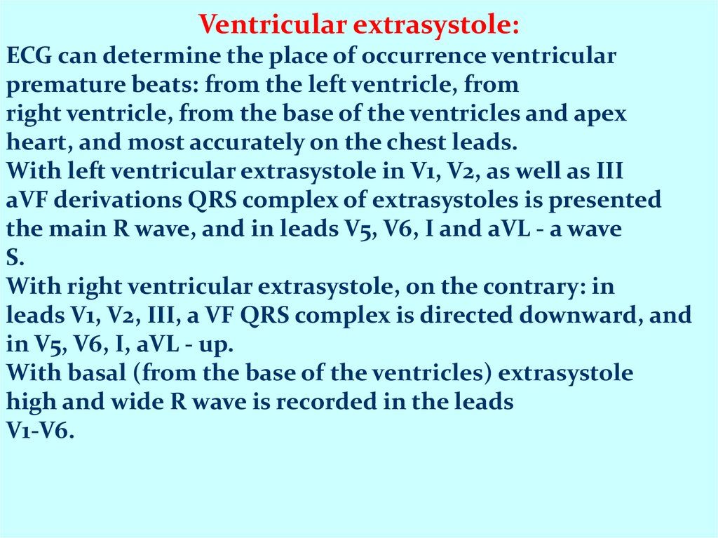
Ventricular extrasystole:ECG can determine the place of occurrence ventricular
premature beats: from the left ventricle, from
right ventricle, from the base of the ventricles and apex
heart, and most accurately on the chest leads.
With left ventricular extrasystole in V1, V2, as well as III
aVF derivations QRS complex of extrasystoles is presented
the main R wave, and in leads V5, V6, I and aVL - a wave
S.
With right ventricular extrasystole, on the contrary: in
leads V1, V2, III, a VF QRS complex is directed downward, and
in V5, V6, I, aVL - up.
With basal (from the base of the ventricles) extrasystole
high and wide R wave is recorded in the leads
V1-V6.
14.
Ventricular extrasystole:15.
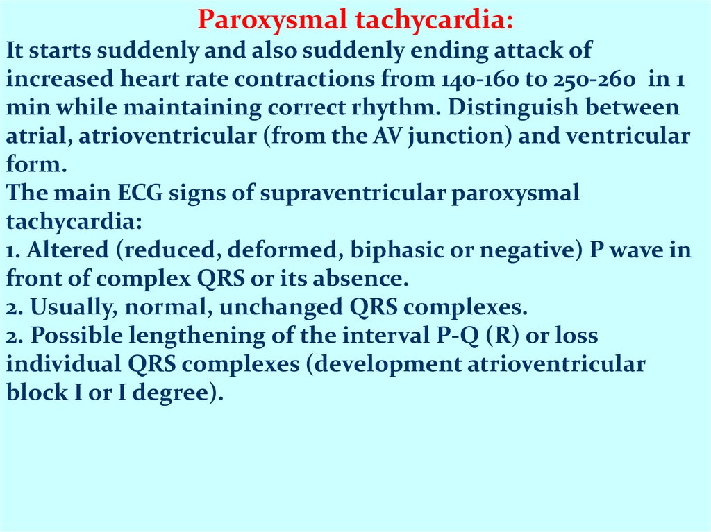
Paroxysmal tachycardia:It starts suddenly and also suddenly ending attack of
increased heart rate contractions from 140-160 to 250-260 in 1
min while maintaining correct rhythm. Distinguish between
atrial, atrioventricular (from the AV junction) and ventricular
form.
The main ECG signs of supraventricular paroxysmal
tachycardia:
1. Altered (reduced, deformed, biphasic or negative) P wave in
front of complex QRS or its absence.
2. Usually, normal, unchanged QRS complexes.
2. Possible lengthening of the interval P-Q (R) or loss
individual QRS complexes (development atrioventricular
block I or I degree).
16.
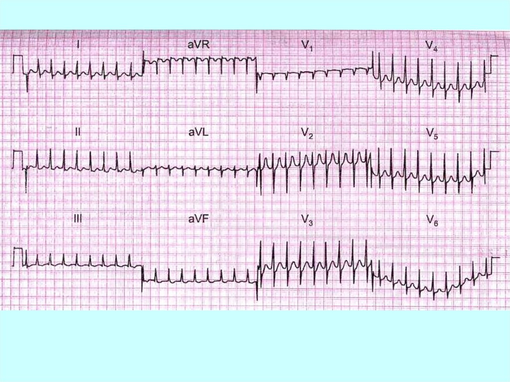
17. Paroxysmal ventricular tachycardia:
1. Paroxysmal ventricular tachycardia ≤ 120 bpm.2. P-waves are absent.
3. QRS-complexes are wide and irregular.
18.
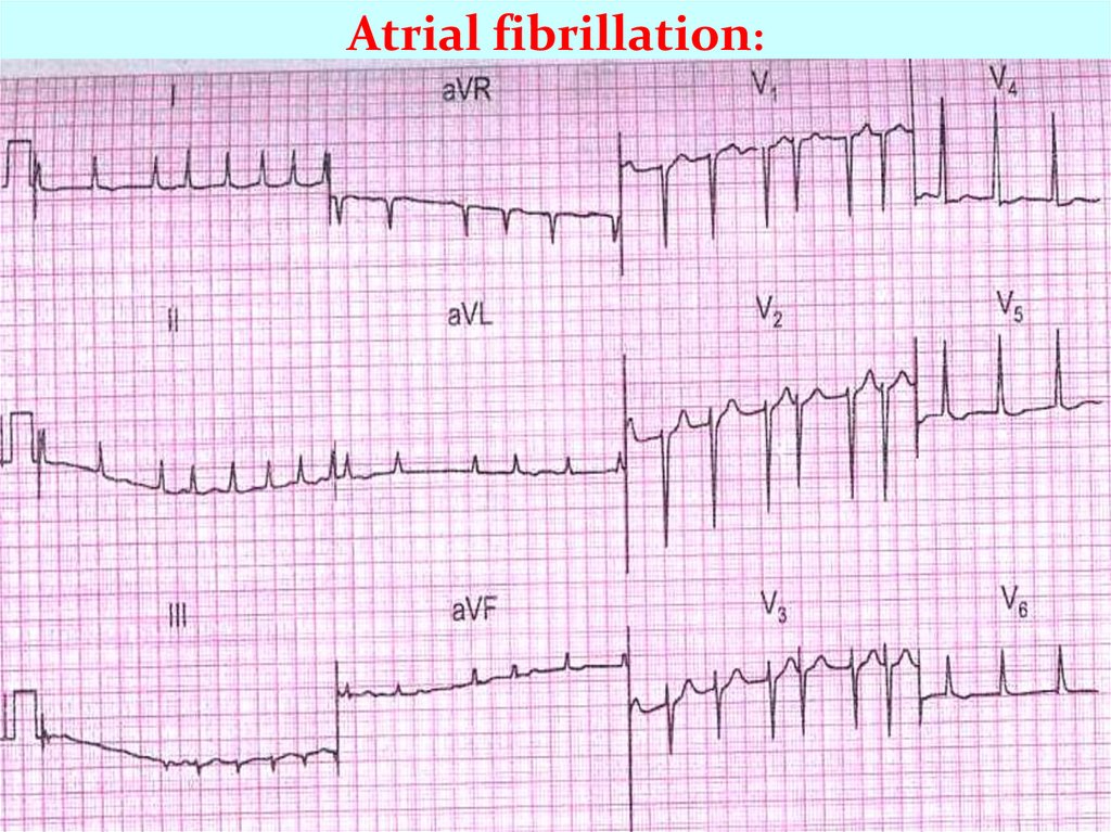
Фибрилляция предсердий:Основные ЭКГ-признаки:
1. Отсутствие зубца Р во всех отведениях ЭКГ.
2. Разные расстояния R - R
3. Наличие на протяжении всего сердечного цикла беспорядочных,
различных по величине, форме и продолжительности предсердных
волн (f-волн), которые лучше регистрируются в III, и часто II, aVF, V1
V2 отведениях.
4. Разная амплитудазубцов R.
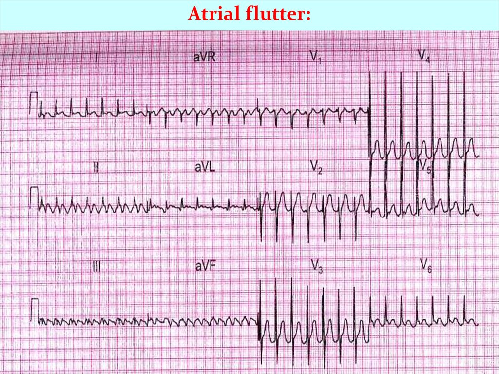
19. Atrial fibrillation and flutter:
1. Atrial fibrillation - more than 400 P-waves per min,QRS-frequency of 150-180 bpm, f-waves
2. Atrial flutter atrial frequency is about 300 bpm,
sawtooth-like P-waves
20.
Atrial fibrillation:21.
Atrial flutter:22. Ventricular fibrillation:
1. Ventricular fibrillation irregular ventricular rate is200-600 twitches/min.
2. The heart does not pump blood.
3. It leads to unconsciousness within 5 seconds.
4. The trigger is anoxia.
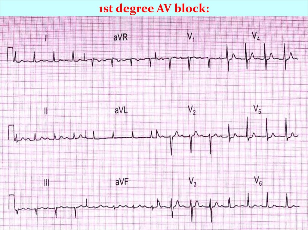
23. Atrioventricular block:
1. Atrioventricular block is the blockage of theconduction from the atria to the AV-node. Three
degrees of AV block are known.
2. 1st degree AV block: PQ - above 0.2 s
24. Atrioventricular block:
1. 2nd degree AV block- some of the P-waves are not followedby QRS-complexes
2. Mobitz type I - PQ-interval is increased progressively until a
P-wave is not followed by a QRS-complex. (Wenchebach
block).
3. Mobitz type II block - the ventricles drop some beats
25. Atrioventricular block:
1. 3rd degree AV block (complete AV-block) is a totalblock of the conduction between the SN and the
ventricles.
2. Atriums are regulated by SA node, ventricles by AV
node
P
P
P
P
P
P
P
P
26.
1st degree AV block:27. Mobitz type I:
III
V1
p
p
p
p
p
p
p
V2
III
V3
AVR
V4
AVL
V5
AVF
V6
28. 3rd degree AV block (complete AV-block):
IV1
p
II
V2
III
V3
AVR
V4
AVL
V5
AVF
p
p
V6
Независимое сокращение предсердий и желудочков
p
p
29.
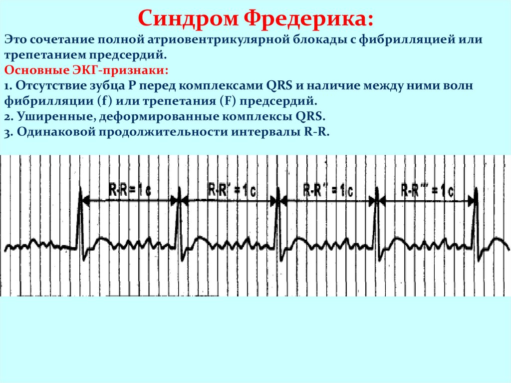
Синдром Фредерика:Это сочетание полной атриовентрикулярной блокады с фибрилляцией или
трепетанием предсердий.
Основные ЭКГ-признаки:
1. Отсутствие зубца Р перед комплексами QRS и наличие между ними волн
фибрилляции (f) или трепетания (F) предсердий.
2. Уширенные, деформированные комплексы QRS.
3. Одинаковой продолжительности интервалы R-R.
30.
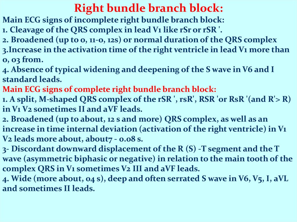
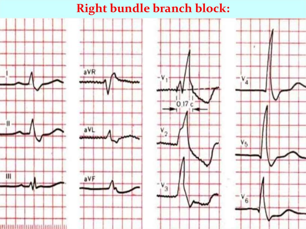
Right bundle branch block:Main ECG signs of incomplete right bundle branch block:
1. Cleavage of the QRS complex in lead V1 like rSr or rSR '.
2. Broadened (up to o, 11-o, 12s) or normal duration of the QRS complex
3.Increase in the activation time of the right ventricle in lead V1 more than
o, o3 from.
4. Absence of typical widening and deepening of the S wave in V6 and I
standard leads.
Main ECG signs of complete right bundle branch block:
1. A split, M-shaped QRS complex of the rSR ', rsR', RSR 'or RsR '(and R'> R)
in V1 V2 sometimes II and aVF leads.
2. Broadened (up to about, 12 s and more) QRS complex, as well as an
increase in time internal deviation (activation of the right ventricle) in V1
V2 leads more about, about7 - 0.08 s.
3- Discordant downward displacement of the R (S) -T segment and the T
wave (asymmetric biphasic or negative) in relation to the main tooth of the
complex QRS in V1 sometimes V2 III and aVF leads.
4. Wide (more about, 04 s), deep and often serrated S wave in V6, V5, I, aVL
and sometimes II leads.
31.
Right bundle branch block:32.
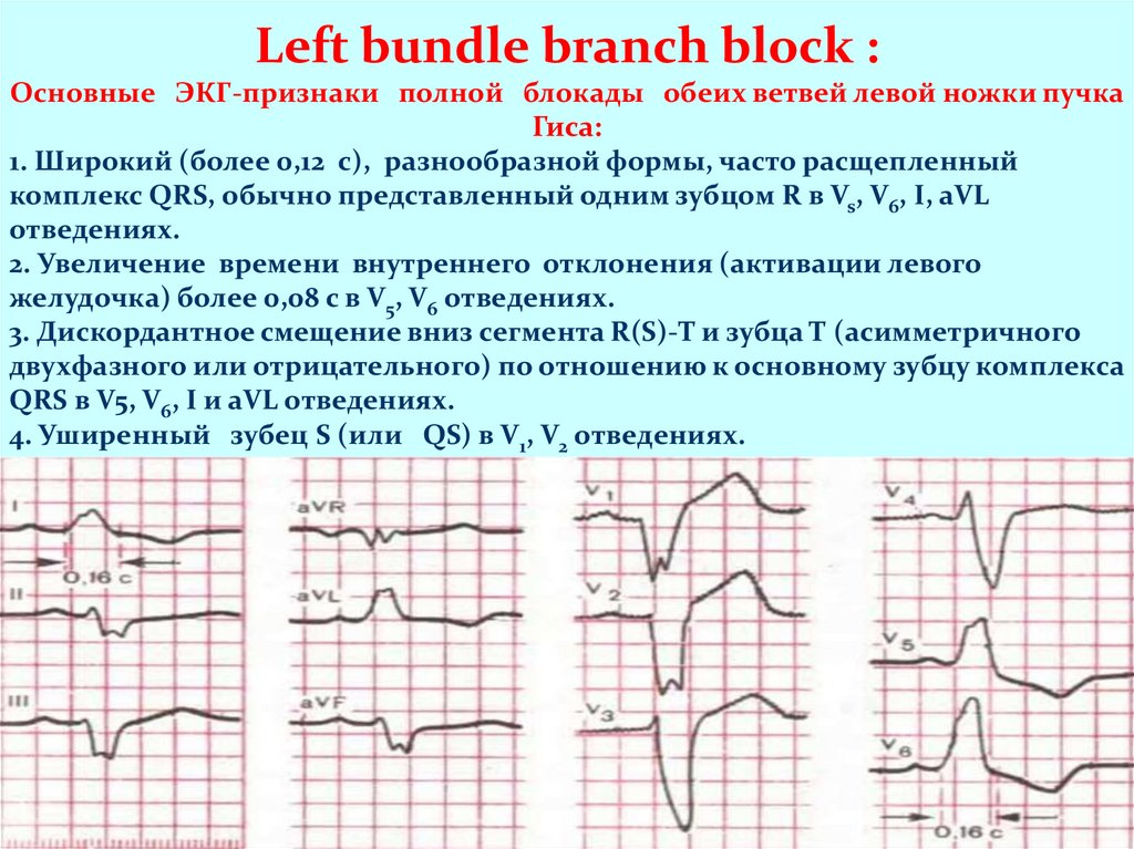
Left bundle branch block :Основные ЭКГ-признаки полной блокады обеих ветвей левой ножки пучка
Гиса:
1. Широкий (более 0,12 с), разнообразной формы, часто расщепленный
комплекс QRS, обычно представленный одним зубцом R в Vs, V6, I, aVL
отведениях.
2. Увеличение времени внутреннего отклонения (активации левого
желудочка) более 0,08 с в V5, V6 отведениях.
3. Дискордантное смещение вниз сегмента R(S)-T и зубца Т (асимметричного
двухфазного или отрицательного) по отношению к основному зубцу комплекса
QRS в V5, V6, I и aVL отведениях.
4. Уширенный зубец S (или QS) в V1, V2 отведениях.
33.
Основные ЭКГ-признаки неполной блокады передней ветви левой ножкипучка Гиса:
1. Выраженное отклонение ЭОС влево (угол альфа превышает -30°). В I и aVL
отведениях комплекс QRS типа qR, а во II, III и aVF отведениях — типа rS.
2. Увеличение амплитуды зубца S в V5, V6 отведениях.
3. Нормальный или несколько уширенный (до 0,10-0,11 с) комплекс QRS.
34.
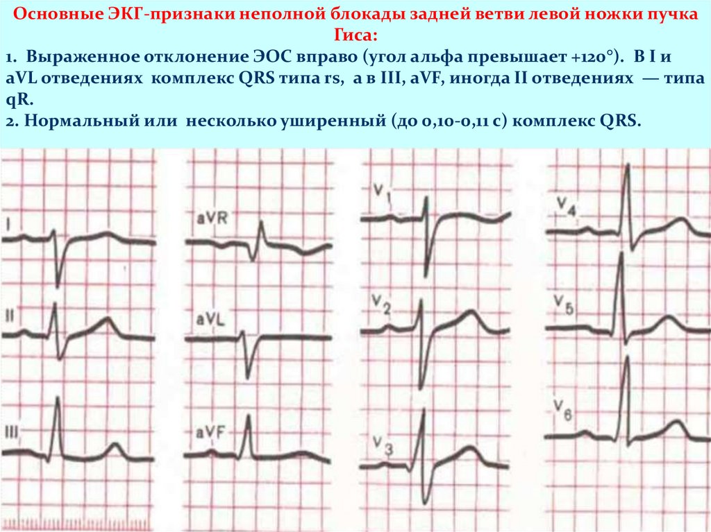
Основные ЭКГ-признаки неполной блокады задней ветви левой ножки пучкаГиса:
1. Выраженное отклонение ЭОС вправо (угол альфа превышает +120°). В I и
aVL отведениях комплекс QRS типа rs, а в III, aVF, иногда II отведениях — типа
qR.
2. Нормальный или несколько уширенный (до 0,10-0,11 с) комплекс QRS.
35.
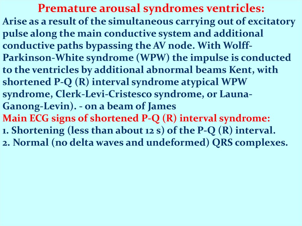
Premature arousal syndromes ventricles:Arise as a result of the simultaneous carrying out of excitatory
pulse along the main conductive system and additional
conductive paths bypassing the AV node. With WolffParkinson-White syndrome (WPW) the impulse is conducted
to the ventricles by additional abnormal beams Kent, with
shortened P-Q (R) interval syndrome atypical WPW
syndrome, Clerk-Levi-Cristesco syndrome, or LaunaGanong-Levin). - on a beam of James
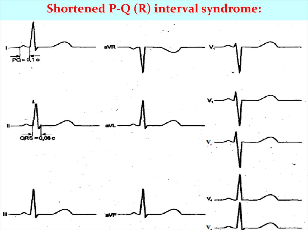
Main ECG signs of shortened P-Q (R) interval syndrome:
1. Shortening (less than about 12 s) of the P-Q (R) interval.
2. Normal (no delta waves and undeformed) QRS complexes.
36.
Shortened P-Q (R) interval syndrome:37.
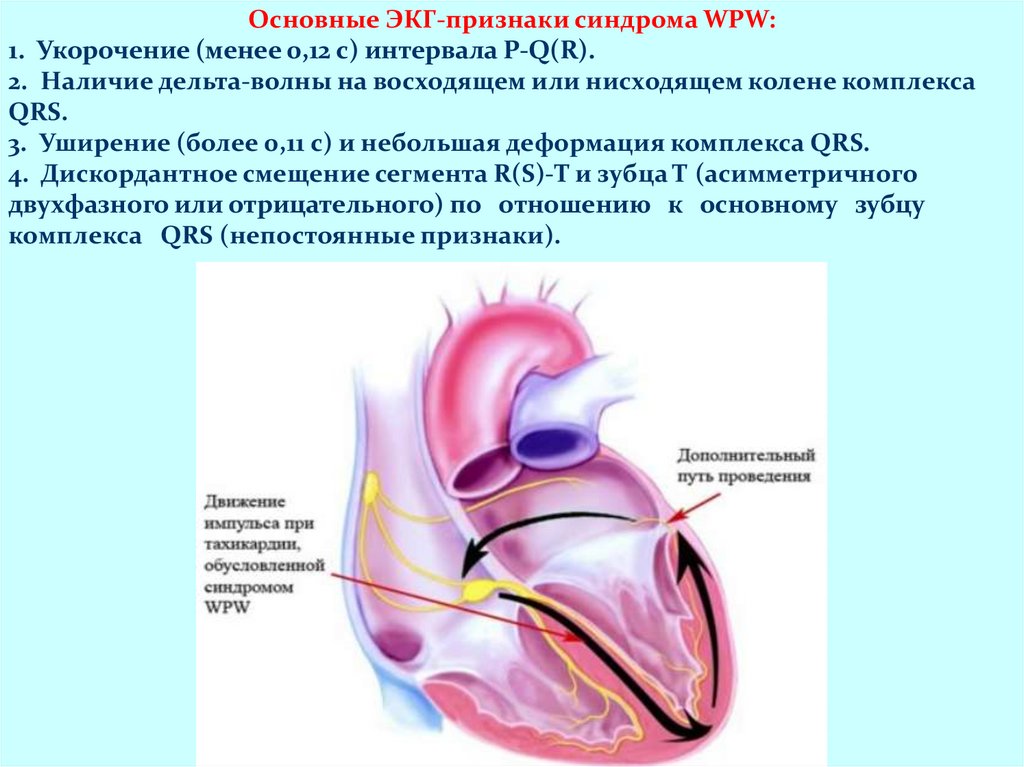
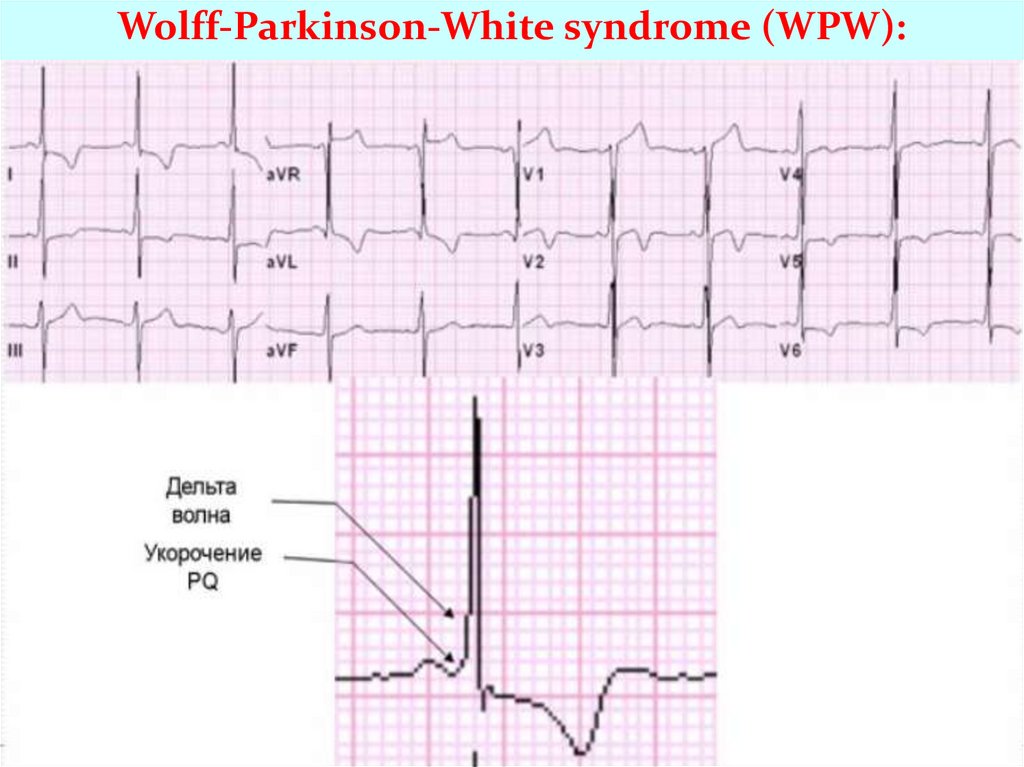
Основные ЭКГ-признаки синдрома WPW:1. Укорочение (менее 0,12 с) интервала P-Q(R).
2. Наличие дельта-волны на восходящем или нисходящем колене комплекса
QRS.
3. Уширение (более 0,11 с) и небольшая деформация комплекса QRS.
4. Дискордантное смещение сегмента R(S)-T и зубца Т (асимметричного
двухфазного или отрицательного) по отношению к основному зубцу
комплекса QRS (непостоянные признаки).
38.
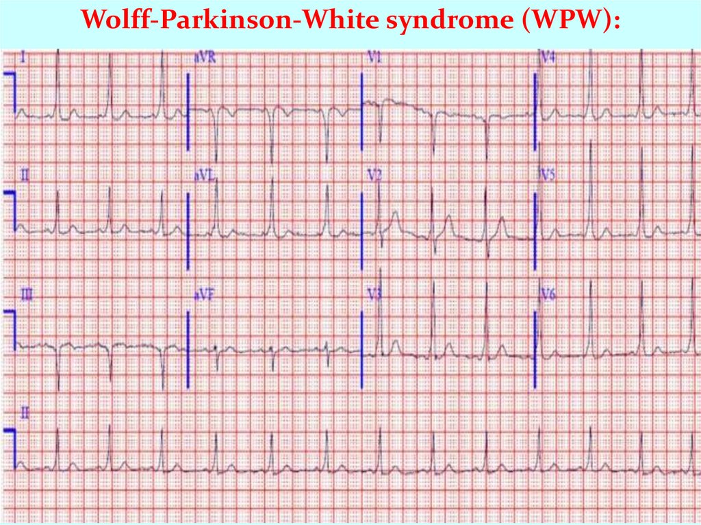
Wolff-Parkinson-White syndrome (WPW):39.
Wolff-Parkinson-White syndrome (WPW):40.
Благодарю завнимание!