


 Медицина
Похожие презентации:


Operative gynecology
1.
OPERATIVEGYNECOLOGY
2.
Hysterectomy• Hysterectomy is the surgical removal of the
uterus.
• It may also involve removal of the cervix,
ovaries, fallopian tubes and other surrounding
structures.
3.
Hysterectomy• Hysterectomy may be total (removing the body,
fundus, and cervix of the uterus; often called
"complete") or partial (removal of the uterine
body while leaving the cervix intact; also called
"supracervical"). It is the most commonly
performed gynecological surgical procedure
• Oophorectomy (removal of ovaries) is frequently
done together with hysterectomy to decrease the
risk of ovarian cancer
4.
Incidence• In the UK, 1 in 5 women are likely to have a
hysterectomy by the age of 60
• Ovaries are removed in about 20% of
hysterectomies
5.
Indications• Certain types of reproductive system cancers or tumors,
including uterine fibroids that do not respond to more
conservative treatment options
• Severe endometriosis and/or adenomyosis, after
pharmaceutical or other surgical options have been
exhausted
• Chronic pelvic pain, after pharmaceutical or other surgical
options have been exhausted
• Postpartum to remove either a severe case of placenta
praevia (a placenta that has either formed over or inside
the birth canal) or placenta accreta (a placenta that has
grown into and through the wall of the uterus to attach
itself to other organs)
6.
Indications• Several forms of vaginal prolapse
• Prophylaxis against certain reproductive system
cancers, especially if there is a strong family history of
reproductive system cancers (especially breast cancer
in conjunction with BRCA1 or BRCA2 mutation), or as
part of recovery from such cancers
• Part of overall gender transition for trans men
• Severe developmental disabilities, though this
treatment is controversial at best, and specific cases of
sterilization due to developmental disabilities
• And others
7.
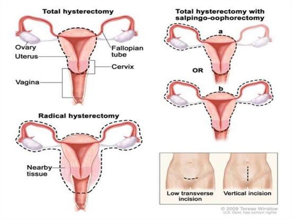
Types• Radical hysterectomy: complete removal of
the uterus, cervix, upper vagina, and
parametrium. Indicated for cancer. Lymph
nodes, ovaries and fallopian tubes are also
usually removed in this situation, such as in
Wertheim's hysterectomy.
• Total hysterectomy: complete removal of the
uterus and cervix, with or without
oophorectomy.
8.
Types• Subtotal hysterectomy: removal of the uterus,
leaving the cervix in situ.
• Supracervical (subtotal) hysterectomy does
not eliminate the possibility of having cervical
cancer since the cervix itself is left intact and
may be contraindicated in women with
increased risk of this cancer.
9.
10.
Abdominal hysterectomy• Is done via laparotomy (abdominal incision, not to be
confused with laparoscopy).
• The recovery time for an open hysterectomy is 4–6
weeks and sometimes longer due to the need to cut
through the abdominal wall.
• Historically, the biggest problem with this technique
were infections, but infection rates are well-controlled
and not a major concern in modern medical practice.
• An open hysterectomy provides the most effective way
to explore the abdominal cavity and perform
complicated surgeries.
11.
Vaginal hysterectomy• Vaginal hysterectomy is performed entirely
through the vaginal canal and has clear
advantages over abdominal surgery such as fewer
complications, shorter hospital stays and shorter
healing time.
• Abdominal hysterectomy, the most common
method, is used in cases such as after caesarean
delivery, when the indication is cancer, when
complications are expected or surgical
exploration is required.
12.
Laparoscopic-assistedvaginal hysterectomy
• With the development of the laparoscopic techniques in
the 1970-1980s, the "laparoscopic-assisted vaginal
hysterectomy" (LAVH) has gained great popularity among
gynecologists because compared with the abdominal
procedure it is less invasive and the post-operative recovery
is much faster.
• It also allows better exploration and slightly more
complicated surgeries than the vaginal procedure. LAVH
begins with laparoscopy and is completed such that the
final removal of the uterus (with or without removing the
ovaries) is via the vaginal canal.
• Thus, LAVH is also a total hysterectomy, the cervix must be
removed with the uterus.
13.
Laparoscopic-assistedsupracervical hysterectomy
• The "laparoscopic-assisted supracervical
hysterectomy" (LASH) was later developed to
remove the uterus without removing the
cervix using a morcellator which cuts the
uterus into small pieces that can be removed
from the abdominal cavity via the
laparoscopic ports
14.
Total laparoscopichysterectomy
• TLH is performed solely through the
laparoscopes in the abdomen, starting at the
top of the uterus, typically with a uterine
manipulator.
• The entire uterus is disconnected from its
attachments using long thin instruments
through the "ports". Then all tissue to be
removed is passed through the small
abdominal incisions.
15.
Advantages and disadvantages of different hysterectomy techniquesTechnique
Abdominal hysterectomy
Vaginal hysterectomy
Laparoscopic supracervical
hysterectomy
Benefits
No limitation by the size of the
uterus
Longest duration of hospital
treatment
Combination with reduction and
incontinence surgery possible
Highest rate of complications
Longest recovery period
Shortest operation time
Short recovery period
Limitation by the size of the uterus
and previous surgery
Combination with reduction
operations are possible
Highest blood loss
Limited ability to evaluate the
fallopian tubes and ovaries
10-17% of patients continue to have
minimal menstrual bleeding
Low risk of complication
Less blood loss
Short inpatient treatment duration
Laparoscopic-assisted vaginal
hysterectomy
Total laparoscopic
hysterectomy
Disadvantages
Possible even with larger uterus and
after previous surgery
Combination with reduction
operations are possible
Less blood loss
Short inpatient treatment duration
Long operation time
High instrumental costs by changing
the access path
None to date
16.
Adverse effects andComplications
• Hysterectomy has like any other surgery certain risks
and side effects.
• Risk of general anesthesia, DVT, and pulmonary
embolism.
• Mortality and surgical risks Short term mortality
(within 40 days of surgery) is usually reported in the
range of 1–6 cases per 1000 when performed for
benign causes.
• The mortality rate is several times higher when
performed in patients that are pregnant, have cancer
or other complications.
17.
Injury to adjacent organs• Bladder injury.
• Bowel injury.
• Ureteral injury is not uncommon and can range
from 2.2% to 3% depending on whether the
modality is abdominal, laparoscopic, or vaginal.
The injury usually occurs in the distal ureter close
to the infundibulopelvic ligament or as a ureter
crosses below the uterine artery, often from blind
clamping and ligature placement to control
hemorrhage.
18.
Convalescence• Hospital stay is 3 to 5 days or more for the
abdominal procedure and between 2 to 3 days
for vaginal or laparoscopically assisted vaginal
procedures.
• Time for full recovery is very long and largely
independent on the procedure that was used.
Depending on the definition of "full recovery“
3 to 12 months have been reported. Serious
limitations in everyday activities are expected
for a minimum of 4 months.
19.
Effects on sexual life andpelvic pain
• After hysterectomy for benign indications the
majority of women report improvement in
sexual life and pelvic pain.
• A smaller share of women report worsening of
sexual life and other problems.
20.
Premature menopauseand its effects
• Estrogen levels fall sharply when the ovaries are
removed, removing the protective effects of estrogen
on the cardiovascular and skeletal systems.
• This condition is often referred to as "surgical
menopause", although it is substantially different from
a naturally occurring menopausal state; the former is a
sudden hormonal shock to the body that causes rapid
onset of menopausal symptoms such as hot flashes,
while the latter is a gradually occurring decrease of
hormonal levels over a period of years with uterus
intact and ovaries able to produce hormones even
after the cessation of menstrual periods.
21.
• Concequences of this is cardiovasculardisease, osteoporosis (decrease in bone
density) and increased risk of bone fractures
are associated with hysterectomies.
• This has been attributed to the modulatory
effect of estrogen on calcium metabolism and
the drop in serum estrogen levels after
menopause can cause excessive loss of
calcium leading to bone wasting.
22.
Urinary incontinence andvaginal prolapse
• Urinary incontinence and vaginal prolapse are
well known adverse effects that develop with
high frequency a very long time after the
surgery. Typically, those complications develop
10–20 years after the surgery.
• Vault prolapse complicate 1% of total
hysterectomy.
23.
Adhesion formation andbowel obstruction
• The formation of postoperative adhesions is a
particular risk after hysterectomy because of
the extent of dissection involved as well the
fact the hysterectomy wound is in the most
gravity-dependent part of the pelvis into
which a loop of bowel may easily fall.
24.
Uterine myomectomy• Myomectomy, sometimes also fibroidectomy,
refers to the surgical removal of uterine
leiomyomas, also known as fibroids. In
contrast to a hysterectomy the uterus remains
preserved and the woman retains her
reproductive potential.
25.
Indications• The presence of a fibroid does not mean that
it needs to be removed. Removal is necessary
when the fibroid causes pain or pressure,
abnormal bleeding, or interferes with
reproduction. The fibroids needed to be
removed are typically large in size, or growing
at certain locations such as bulging into the
endometrial cavity causing significant cavity
distortion.
26.
Procedure• A myomectomy can be performed in a
number of ways, depending on the location
and number of lesions and the experience and
preference of the surgeon. Either a general or
a spinal anesthesia is administered.
27.
Laparotomy• Traditionally a myomectomy is performed via a
laparotomy with a full abdominal incision, either
vertically or horizontally. Once the peritoneal
cavity is opened, the uterus is incised, and the
lesion(s) removed. The open approach is often
preferred for larger lesions. One or more incisions
may be set into the uterine muscle and are
repaired once the fibroid has been removed.
Recovery after surgery takes six to eight weeks.
28.
Laparoscopy• Using the laparoscopic approach the uterus is
visualized and its fibroids located and removed.
Morcellators are available to shred larger fibroids so
that they can be removed through the small port holes
of laparoscopy.
• Studies have suggested that laparoscopic
myomectomy leads to lower morbidity rates and faster
recovery than does laparotomic myomectomy.
• As with hysteroscopic myomectomy, laparoscopic
myomectomy is not generally used on very large
fibroids (3-10cm).
29.
Hysteroscopy• A fibroid that is located in a submucous
position (that is, protruding into the
endometrial cavity) may be accessible to
hysteroscopic removal.
• This may apply primarily to smaller lesions not
greater than 5 cm.
30.
Complications and risksComplications of the surgery include:
• the possibility of significant blood loss leading
to a blood transfusion
• the risk of adhesion or scar formation around
the uterus or within its cavity
• the possible need later to deliver via cesarean
section
31.
Complications and risks• It may not be possible to remove all lesions, nor will
the operation prevent new lesions from growing.
Development of new fibroids will be seen in 42-55% of
patients undergoing a myomectomy.
• It is well known that myomectomy surgery is
associated with a higher risk of uterine rupture in later
pregnancy. Thus, women who have had myomectomy
(with the exception of small submucosal myoma
removal via hysteroscopy, or largely pedunculated
myoma removal) should get Cesarean delivery to avoid
the risk of uterine rupture that is commonly fatal to the
fetus.
32.
Cervical polypectomy• Cervical polypectomy is a procedure to remove small
tumors (polyps), often growing on a stalk, from the
opening of the cervix or inside the cervical canal
(endocervix). The polyps are generally noncancerous
(benign).
Cervical polyps are caused by an overgrowth of normal
tissue. They are relatively common and most do not
cause symptoms. Cervical polyps are frequently the
result of infection, and may be linked to chronic
inflammation, an abnormal response to higher levels of
estrogen, or local congestion of cervical blood vessels.
33.
Reason for procedure• Cervical polyps do not usually cause symptoms.
Some individuals may experience light bleeding
or spotting caused by irritation from a tampon or
sexual intercourse (postcoital bleeding).
• Polyps are generally removed because of this
bleeding, or to prevent additional future irritation
and bleeding. Although most polyps are benign,
all should be removed and examined because
cancerous (malignant) changes may develop;
some cervical cancers first appear as polyps.
34.
How procedure isperformed
• Polypectomy is usually an outpatient procedure performed
in the physician's office. It is generally painless, so no
anesthesia is required. The woman lies on the exam table
with her legs in the stirrups (lithotomy position); a
speculum is then inserted into the vagina to hold it open to
visualize the cervix. The cervix is cleansed using a vaginal
swab soaked in an antiseptic solution. The polyp is grasped
with a surgical clamp (hemostat), twisted several times, and
pulled until it is freed. The polyp is sent for microscopic
examination (pathology) to rule out cancer. The base of the
polyp is then removed by scraping it off with a sharp
surgical instrument (curettage), or by using heat, cold, or
chemicals to destroy the tissue (cauterization).
35.
How procedure isperformed
• If the polyp is large, or if it is attached by a broad base
rather than a stalk, it may need to be cut off and the
wound stitched (sutured) closed. This procedure may
be done under local anesthesia in the hospital because
of the possible risk of excessive bleeding (hemorrhage).
• If the cervix is soft, distended, or partially opened, and
the polyp is large or not clearly visible, dilation and
curettage (D&C) will be done. The cervical opening will
be widened (dilated) so that the cervical canal and
uterus may be examined for other polyps. All removed
polyps will be biopsied for evidence of cancer.
36.
Complications of cervicalpolypectomy
• Complications following cervical polypectomy
are rare; however, hemorrhage and infection
can occur.
37.
Cone biopsy (conization) forabnormal cervical cell changes
• A cone biopsy is an extensive form of a cervical
biopsy. It is called a cone biopsy because a coneshaped wedge of tissue is removed from the
cervix and examined under a microscope. A cone
biopsy removes abnormal tissue that is high in
the cervical canal. A small amount of normal
tissue around the cone-shaped wedge of
abnormal tissue is also removed so that a margin
free of abnormal cells is left in the cervix.
38.
39.
A sample of tissue can beremoved for a cone biopsy using:
• A surgical knife (scalpel)
• A carbon dioxide (CO2) laser
• Loop electrosurgical excision procedure (LEEP)
40.
A cone biopsy is a surgicaltreatment with some risks.
• A few women may have serious bleeding that
requires further treatment.
• Narrowing of the cervix (cervical stenosis) that
causes infertility may occur (rare).
• Inability of the cervix to stay closed during
pregnancy (incompetent cervix) may occur.
Women who have had a cone biopsy may
have an increased risk of miscarriage or
preterm delivery
41.
Cervical cerclage• Cervical cerclage (tracheloplasty), also known as
a cervical stitch, is used for the treatment of
cervical incompetence (or insufficiency), a
condition where the cervix has become slightly
open and there is a risk of miscarriage because it
may not remain closed throughout pregnancy.
Usually this treatment would be done, in the
second trimester of pregnancy, for a woman who
had either suffered from one or more
miscarriages in the past, or is carrying multiples.
42.
Cervical cerclage• The treatment consists of a strong suture
being inserted into and around the cervix
early in the pregnancy, usually between weeks
12 to 14, and then removed towards the end
of the pregnancy when the greatest risk of
miscarriage has passed.
43.
Types• A McDonald cerclage, is essentially a pursestring stitch; the cervix
stitching involves a band of suture at the upper part of the cervix
while the lower part has already started to efface. This cerclage is
usually placed between 12 weeks and 14 weeks of pregnancy. The
stitch is generally removed around the 37th week of gestation.
• A Shirodkar cerclage is very similar, but the sutures pass through
the walls of the cervix so they're not exposed. This type of cerclage
is less common and technically more difficult than a McDonald, and
is thought (though not proven) to reduce the risk of infection.
• An abdominal cerclage, the least common type, is permanent and
involves stitching at the very top of the cervix, inside the abdomen.
This is usually only done if the cervix is too short to attempt a
standard cerclage, or if a vaginal cerclage has failed or is not
possible.
44.
45.
Risks of cerclageWhile cerclage is generally a safe procedure, there are a number of
potential complications that may arise during or after surgery.
These include:
risks associated with regional or general anesthesia
premature labor
premature rupture of membranes
infection of the cervix
infection of the amniotic sac (chorioamnionitis)
cervical rupture (may occur if the stitch is not removed before onset
of labor)
injury to the cervix or bladder
bleeding
Cervical Dystocia with failure to dilate requiring Cesarean Section
46.
Postoperativemanagement
• In the rehabilitation program for patients after surgical
interventions all over the world, elements of the socalled “Fast track surgery” - rapid recovery surgery and
Enhanced Recovery After Surgery (ERAS) - accelerated
recovery after surgery are being actively introduced.
• This technique is a comprehensive approach used for a
wide variety of surgical interventions.
• The protocol is divided into 3 main parts.
47.
Postoperativemanagement
Before surgery:
• preoperative risk assessment (presence of
concomitant diseases)
• refusal of premedication with narcotic drugs,
which accelerates the restoration of intestinal
function
• minimizing fasting, amounting to no more than 6
hours, and for patients with diabetes 3-4 hours
before surgery, liquid nutrition
• prevention of thrombo-embolic complications
48.
Postoperativemanagement
During the operation:
• intraoperative regional anesthesia and analgesia
• refusal of a nasogastric tube
• short-acting anesthetics, including narcotic ones
• refusal of massive infusion therapy
• refusal to use drainage
• minimally invasive (laparoscopic) approach
• refusal of intraoperative blood transfusion,
except in cases of extreme necessity
49.
Postoperativemanagement
After operation:
• early use of laxatives after surgery
• early mobilization on the day of surgery (standing
up, walking around the ward)
• removal of the urinary catheter on the day of
surgery
• prevention and treatment of nausea and
vomiting
• prescribing a light diet 6 hours after surgery
• refusal of narcotic analgesics
50.
Antibiotic regimens forgynecological surgery
• Cefotetan, cefazolin, cefoxitin, cefuroxime or
ampicillin/sulbactam
• For beta-lactam allergy: clindamycin + gentamicin
or clindamycin + ciprofloxacin or clindamycin +
aztreonam
• Metronidazole + gentamicin or metronidazole +
ciprofloxacin
• Vancomycin + aminoglycosides or vancomycin +
aztreonam or vancomycin + quinolone
51.
Caprini risk assessment model52.
Caprini risk assessment modelfor venous thromboembolism