
 Биология
Похожие презентации:


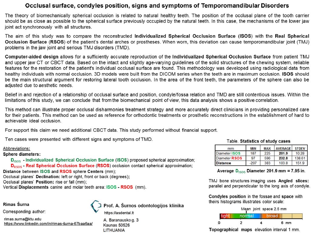
Occlusal surface
1.
https://asdental.lt[email protected]
https://www.linkedin.com/in/rimas-šurna-67baa6aa/
2.
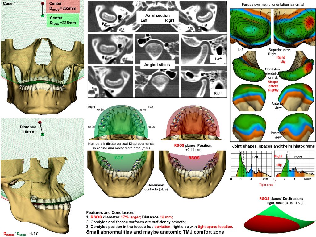
Case 1Center
DRSOS =262mm
Fossae symmetric, orientation is normal
Axial section
Left
Right
Center
DISOS =225mm
Left
Superior view
Right
Right
slip
Left
Angled slices
Condyles
orientation
normal,
Shape
differs
slightly
Right
Anterior
view
Distance
19mm
Posterior
view
Numbers indicate vertical Displacements
in canine and molar teeth area (mm)
ISOS
RSOS planes’ Position:
+0.44 mm
Joint shapes, spaces and theirs histograms
RSOS
Left
Right
Right
slip
Occlusion
contacts (blue)
RSOS planes’ Declination:
right, back (0.04, 0.89)o
Features and Conclusion:
1. RSOS diameter 17% larger; Distance 19 mm;
2. Condyles and fossae surfaces are sufficiently smooth;
3. Condyles position in the fossae has deviation, right side with tight space location.
DRSOS / DISOS = 1.17
Small abnormalities and maybe anatomic TMJ comfort zone
3.
Case 2Fossae asymmetric,
their orientation, rotation and shape are different
Center
DRSOS =237mm
Axial section
Center
DISOS =204mm
Left
Right
Left
Superior view
Condyles
asymmetric,
shapes
and
orientation are
sizeable
differ!
Left
Angled slices
Right
Anterior view
Posterior view
Distance
18mm
Joint shapes, spaces and theirs histograms
Left
Numbers indicate vertical Displacements
in canine and molar teeth area (mm)
RSOS planes’ Position:
-0.10 mm
ISOS
RSOS
Occlusion
contacts (blue)
Asymmetry
Right
RSOS planes’ Declination:
left, front (0.62, 3.59)o
Features and Conclusion:
1. RSOS diameter 16% larger; Distance 18 mm;
2. Condyles and fossae surfaces are sufficiently smooth;
3. Condyles position looks like in normal anatomic comfort zone.
DRSOS / DISOS = 1.16
Right
Asymmetric joints, small abnormalities, maybe anatomic TMJ comfort zone
4.
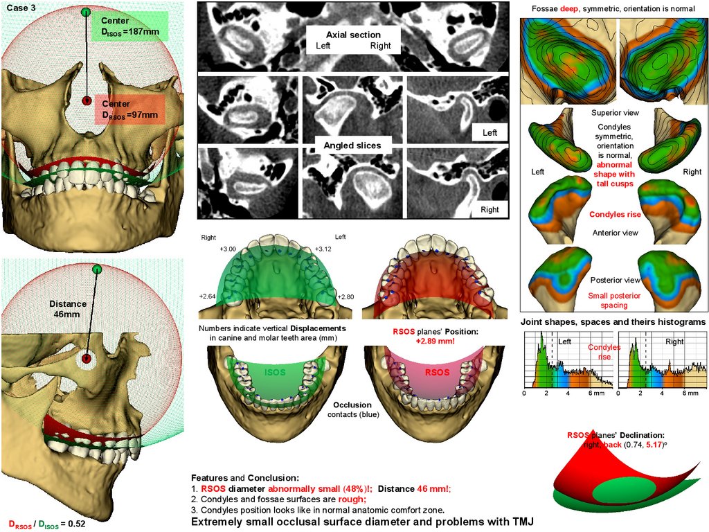
Case 3Fossae deep, symmetric, orientation is normal
Center
DISOS =187mm
Axial section
Left
Right
Center
DRSOS =97mm
Superior view
Condyles
symmetric,
orientation
is normal,
abnormal
shape with
tall cusps
Left
Angled slices
Left
Right
Right
Condyles rise
Anterior view
Posterior view
Small posterior
spacing
Distance
46mm
Numbers indicate vertical Displacements
in canine and molar teeth area (mm)
ISOS
Joint shapes, spaces and theirs histograms
RSOS planes’ Position:
+2.89 mm!
Left
Condyles
rise
Right
RSOS
Occlusion
contacts (blue)
RSOS planes’ Declination:
right, back (0.74, 5.17)o
Features and Conclusion:
1. RSOS diameter abnormally small (48%)!; Distance 46 mm!;
2. Condyles and fossae surfaces are rough;
3. Condyles position looks like in normal anatomic comfort zone.
DRSOS / DISOS = 0.52
Extremely small occlusal surface diameter and problems with TMJ
5.
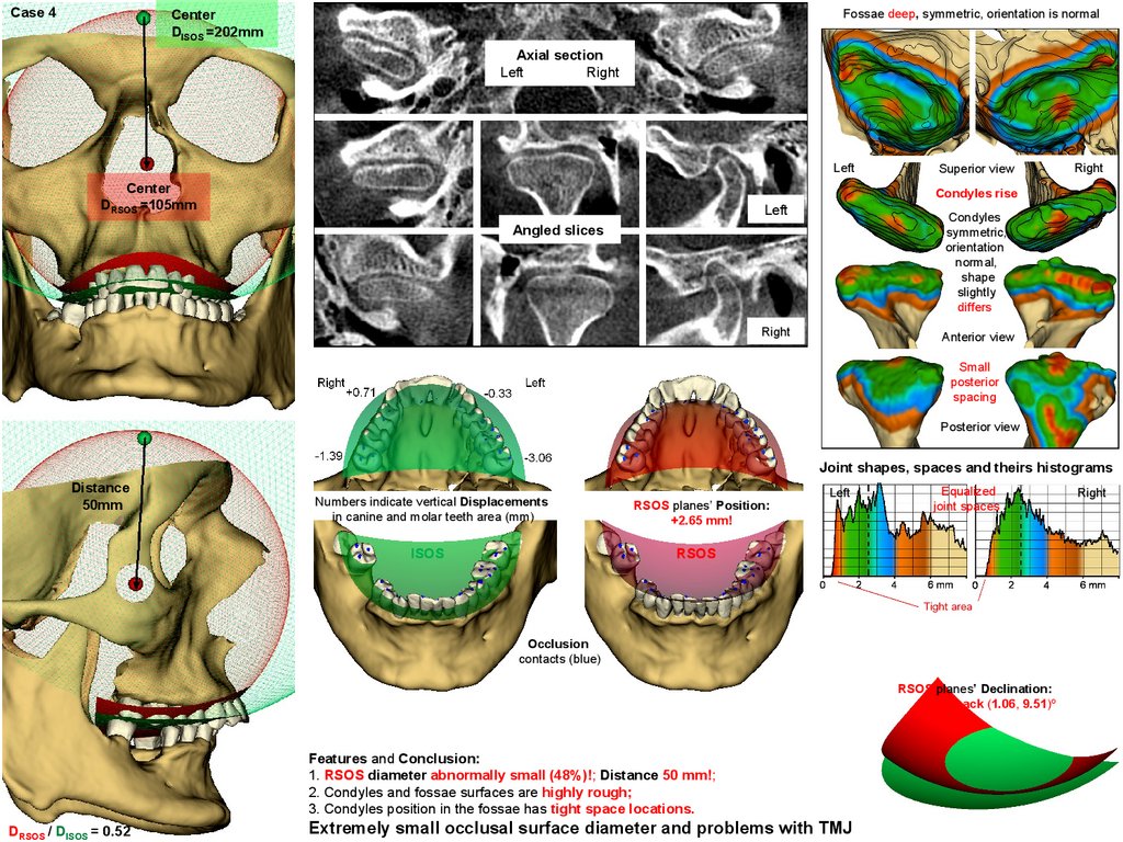
Case 4Fossae deep, symmetric, orientation is normal
Center
DISOS =202mm
Axial section
Left
Right
Left
Center
DRSOS =105mm
Superior view
Right
Condyles rise
Left
Condyles
symmetric,
orientation
normal,
shape
slightly
differs
Angled slices
Right
Anterior view
Small
posterior
spacing
Posterior view
Joint shapes, spaces and theirs histograms
Distance
50mm
Numbers indicate vertical Displacements
in canine and molar teeth area (mm)
ISOS
RSOS planes’ Position:
+2.65 mm!
Left
Equalized
joint spaces
RSOS
Occlusion
contacts (blue)
RSOS planes’ Declination:
left, back (1.06, 9.51)o
Features and Conclusion:
1. RSOS diameter abnormally small (48%)!; Distance 50 mm!;
2. Condyles and fossae surfaces are highly rough;
3. Condyles position in the fossae has tight space locations.
DRSOS / DISOS = 0.52
Extremely small occlusal surface diameter and problems with TMJ
Right
6.
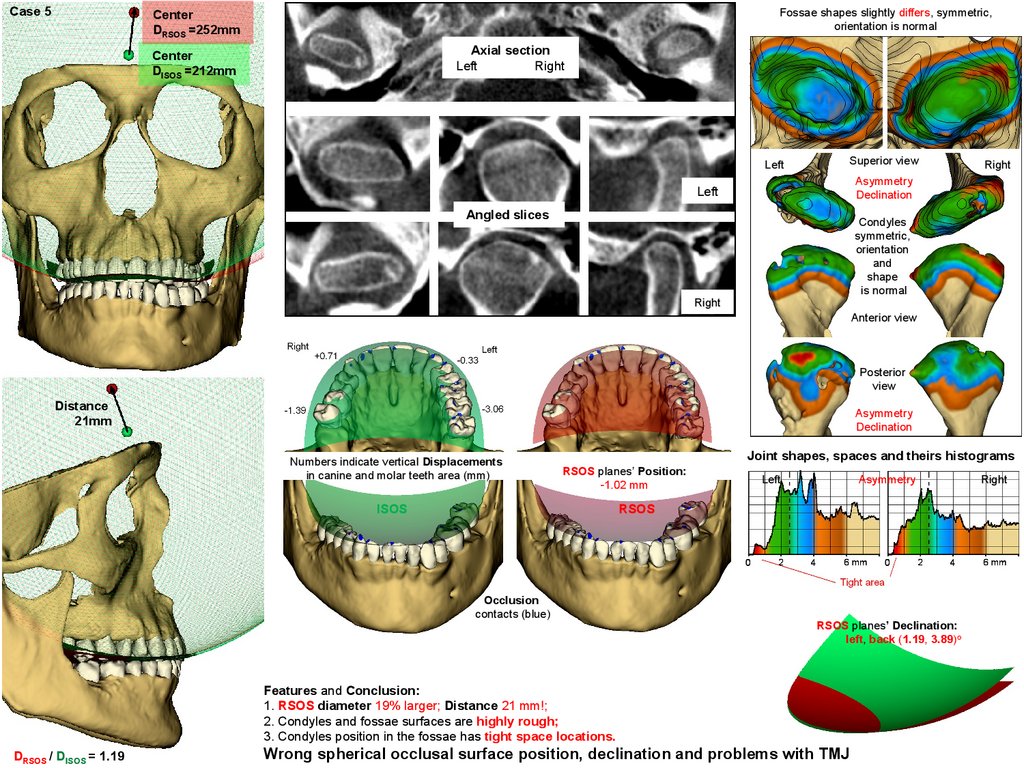
Case 5Fossae shapes slightly differs, symmetric,
orientation is normal
Center
DRSOS =252mm
Axial section
Left
Right
Center
DISOS =212mm
Left
Superior view
Right
Asymmetry
Declination
Left
Angled slices
Condyles
symmetric,
orientation
and
shape
is normal
Right
Anterior view
Posterior
view
Distance
21mm
Asymmetry
Declination
Numbers indicate vertical Displacements
in canine and molar teeth area (mm)
Joint shapes, spaces and theirs histograms
RSOS planes’ Position:
-1.02 mm
ISOS
Left
Asymmetry
RSOS
Occlusion
contacts (blue)
RSOS planes’ Declination:
left, back (1.19, 3.89)o
Features and Conclusion:
1. RSOS diameter 19% larger; Distance 21 mm!;
2. Condyles and fossae surfaces are highly rough;
3. Condyles position in the fossae has tight space locations.
DRSOS / DISOS = 1.19
Wrong spherical occlusal surface position, declination and problems with TMJ
Right
7.
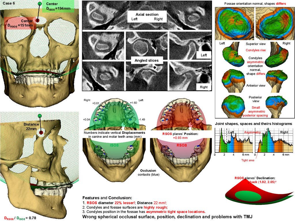
Case 6Fossae orientation normal, shapes differs
Center
DISOS =194mm
Axial section
Left
Right
Center
DRSOS =151mm
Left
Superior view
Right
Condyles rise
Left
Condyles
asymmetric,
orientation
normal,
shape differs
Angled slices
Anterior view
Right
Posterior
view
Small
asymmetric
posterior spacing
Joint shapes, spaces and theirs histograms
Distance
22mm
Numbers indicate vertical Displacements
in canine and molar teeth area (mm)
RSOS planes’ Position:
+0.93 mm
ISOS
Left
Asymmetry
RSOS
Occlusion
contacts (blue)
RSOS planes’ Declination:
right, back (1.92, 2.05)o
Features and Conclusion:
1. RSOS diameter 22% lesser!; Distance 22 mm!;
2. Condyles and fossae surfaces are highly rough;
3. Condyles position in the fossae has asymmetric tight space locations.
DRSOS / DISOS = 0.78
Wrong spherical occlusal surface, position, declination and problems with TMJ
Right
8.
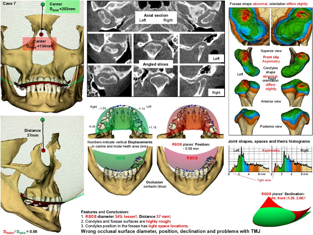
Case 7Fossae shape abnormal, orientation differs slightly
Center
DISOS =203mm
Axial section
Left
Right
Center
DRSOS =134mm
Superior view
Front slip
Asymmetry
Left
Angled slices
Condyles
shape
abnormal,
Right
orientation
differs
slightly
Left
Right
Anterior view
Posterior view
Distance
37mm
Numbers indicate vertical Displacements
in canine and molar teeth area (mm)
ISOS
RSOS planes’ Position:
- 0.59 mm
Joint shapes, spaces and theirs histograms
Left
Asymmetry
Right
RSOS
Occlusion
contacts (blue)
RSOS planes’ Declination:
right, front (1.29, 2.06)o
Features and Conclusion:
1. RSOS diameter 34% lesser!; Distance 37 mm!;
2. Condyles and fossae surfaces are highly rough;
3. Condyles position in the fossae has tight space locations.
DRSOS / DISOS = 0.66
Wrong occlusal surface diameter, position, declination and problems with TMJ
9.
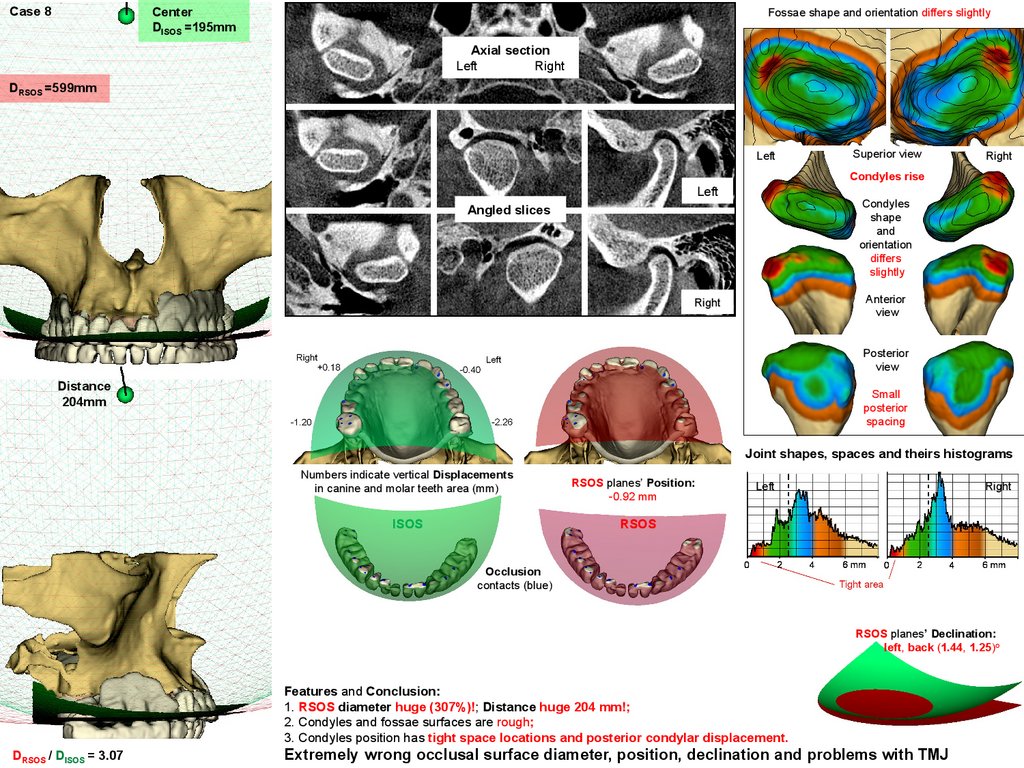
Case 8Center
DISOS =195mm
Fossae shape and orientation differs slightly
Axial section
Left
Right
DRSOS =599mm
Left
Superior view
Right
Condyles rise
Left
Condyles
shape
and
orientation
differs
slightly
Angled slices
Anterior
view
Right
Posterior
view
Distance
204mm
Small
posterior
spacing
Joint shapes, spaces and theirs histograms
Numbers indicate vertical Displacements
in canine and molar teeth area (mm)
ISOS
RSOS planes’ Position:
-0.92 mm
Left
Right
RSOS
Occlusion
contacts (blue)
RSOS planes’ Declination:
left, back (1.44, 1.25)o
Features and Conclusion:
1. RSOS diameter huge (307%)!; Distance huge 204 mm!;
2. Condyles and fossae surfaces are rough;
3. Condyles position has tight space locations and posterior condylar displacement.
DRSOS / DISOS = 3.07
Extremely wrong occlusal surface diameter, position, declination and problems with TMJ
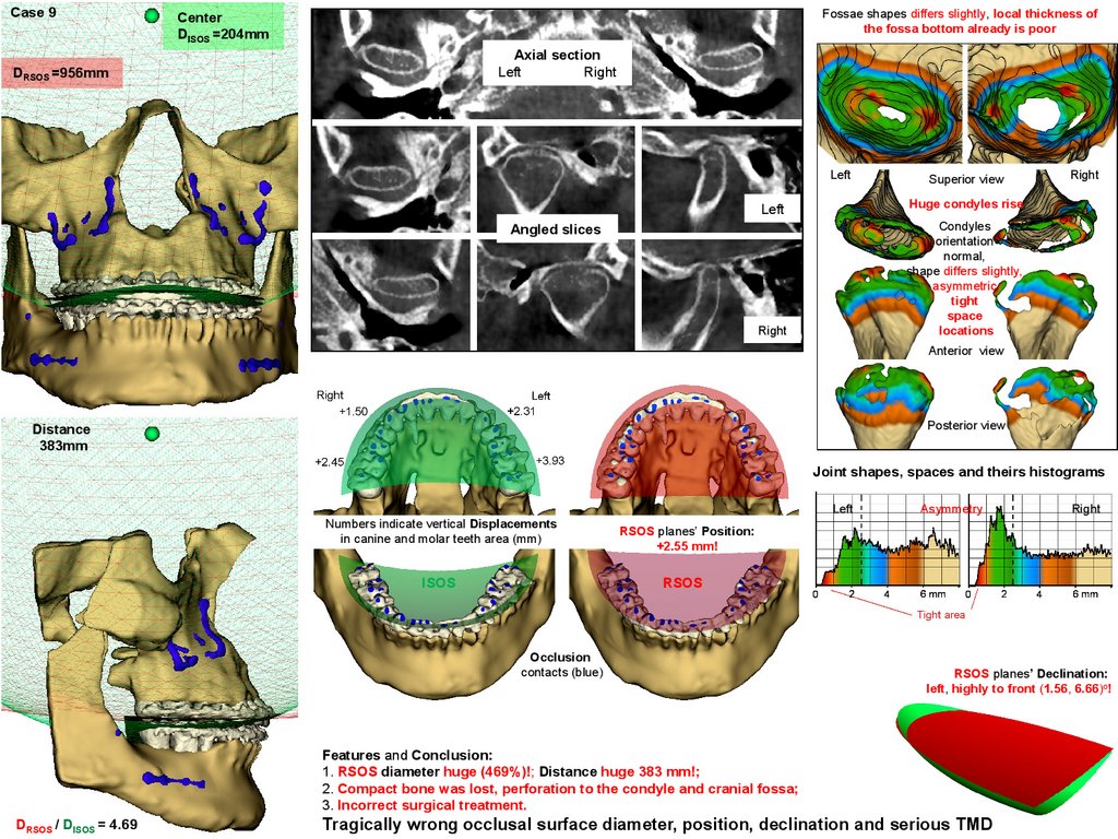
10.
Case 9Fossae shapes differs slightly, local thickness of
the fossa bottom already is poor
Center
DISOS =204mm
Axial section
Left
Right
DRSOS =956mm
Left
Superior view
Right
Huge condyles rise
Left
Condyles
orientation
normal,
shape differs slightly,
asymmetric
tight
space
locations
Angled slices
Right
Anterior view
Posterior view
Distance
383mm
Joint shapes, spaces and theirs histograms
Left
Numbers indicate vertical Displacements
in canine and molar teeth area (mm)
ISOS
Asymmetry
RSOS planes’ Position:
+2.55 mm!
RSOS
Occlusion
contacts (blue)
RSOS planes’ Declination:
left, highly to front (1.56, 6.66)o!
Features and Conclusion:
1. RSOS diameter huge (469%)!; Distance huge 383 mm!;
2. Compact bone was lost, perforation to the condyle and cranial fossa;
3. Incorrect surgical treatment.
DRSOS / DISOS = 4.69
Right
Tragically wrong occlusal surface diameter, position, declination and serious TMD
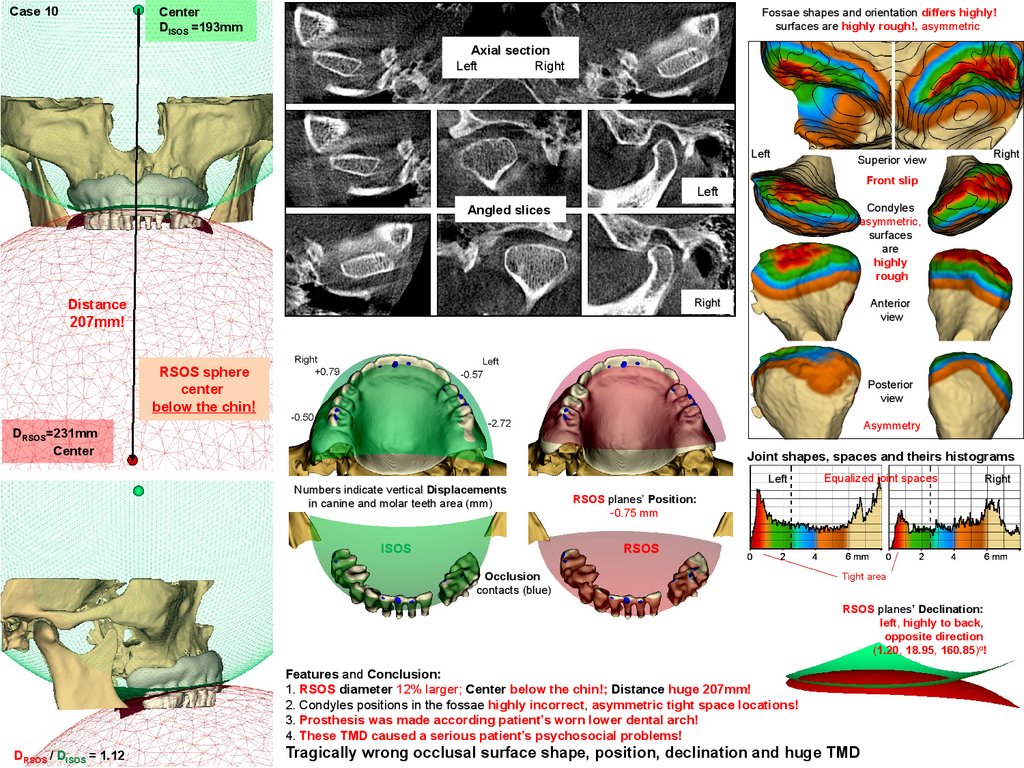
11.
Case 10Center
DISOS =193mm
Fossae shapes and orientation differs highly!
surfaces are highly rough!, asymmetric
Axial section
Left
Right
Left
Right
Superior view
Front slip
Left
Condyles
asymmetric,
surfaces
are
highly
rough
Angled slices
Right
Distance
207mm!
Anterior
view
RSOS sphere
center
below the chin!
Posterior
view
Asymmetry
DRSOS=231mm
Center
Joint shapes, spaces and theirs histograms
Numbers indicate vertical Displacements
in canine and molar teeth area (mm)
ISOS
Left
Equalized joint spaces
Right
RSOS planes’ Position:
-0.75 mm
RSOS
Occlusion
contacts (blue)
RSOS planes’ Declination:
left, highly to back,
opposite direction
(1.20, 18.95, 160.85)o!
Features and Conclusion:
1. RSOS diameter 12% larger; Center below the chin!; Distance huge 207mm!
2. Condyles positions in the fossae highly incorrect, asymmetric tight space locations!
3. Prosthesis was made according patient’s worn lower dental arch!
4. These TMD caused a serious patient’s psychosocial problems!
DRSOS / DISOS = 1.12
Tragically wrong occlusal surface shape, position, declination and huge TMD