


 Биология
Похожие презентации:
. Occurrence and classification")


Spirochaetales. Treponema Borrelia & Leptospira
1.
2.
Spirochaetales~~~~~~~~~~~~~~~~~~
Treponema
Borrelia &
Leptospira
3.
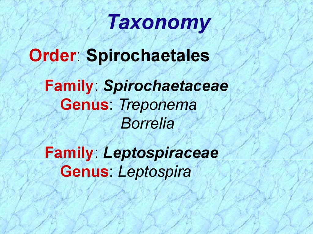
TaxonomyOrder: Spirochaetales
Family: Spirochaetaceae
Genus: Treponema
Borrelia
Family: Leptospiraceae
Genus: Leptospira
4.
General Overview of SpirochaetalesGram-negative spirochetes
• Spirochete from Greek for “coiled hair”
Extremely thin and can be very long
Tightly coiled helical cells with tapered ends
Motile by periplasmic flagella (a.k.a., axial fibrils
or endoflagella)
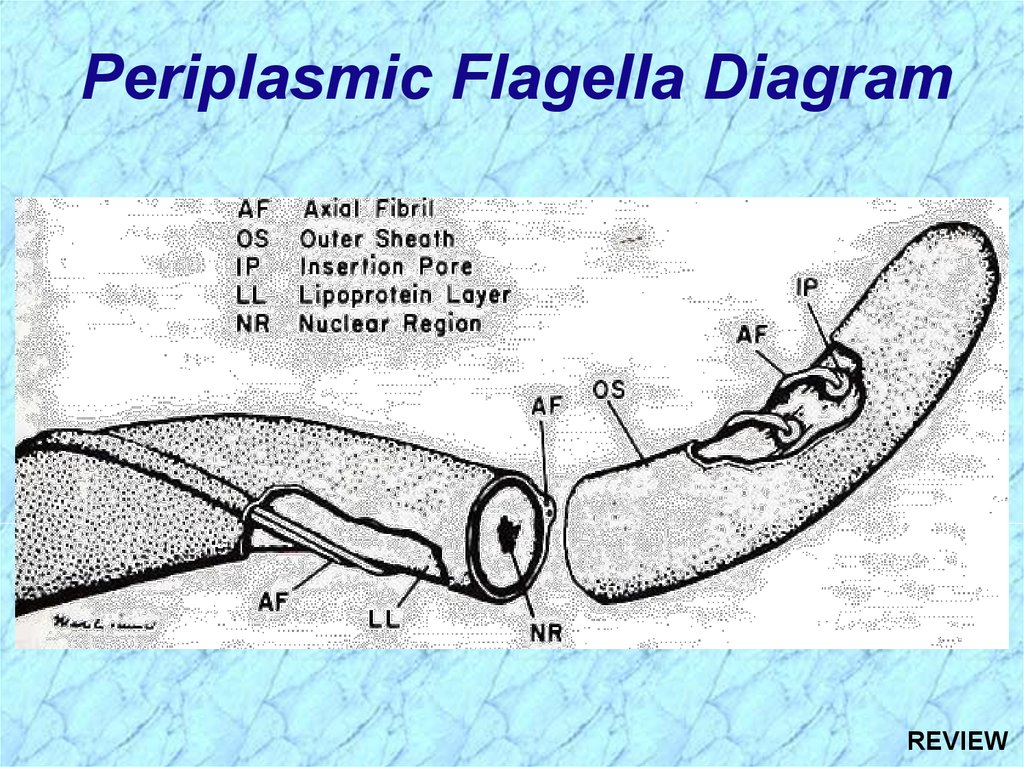
Outer sheath encloses axial fibrils wrapped around
protoplasmic cylinder
• Axial fibrils originate from insertion pores at both poles of cell
• May overlap at center of cell in Treponema and Borrelia, but
not in Leptospira
• Differering numbers of endoflagella according to genus &
species
5.
Periplasmic Flagella Diagram6.
Tightly Coiled SpirocheteOS = outer sheath
AF = axial fibrils
AF
Leptospira interrogans
7.
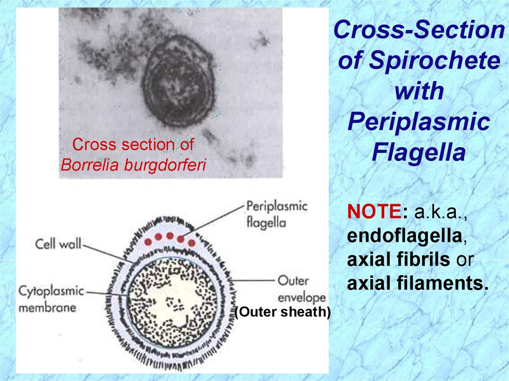
Cross-Sectionof Spirochete
with
Periplasmic
Flagella
Cross section of
Borrelia burgdorferi
NOTE: a.k.a.,
endoflagella,
axial fibrils or
axial filaments.
(Outer sheath)
8.
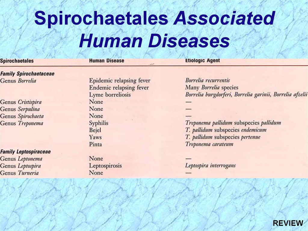
Spirochaetales AssociatedHuman Diseases
Genus
Species
Disease
Treponema pallidum ssp. pallidum
pallidum ssp. endemicum
pallidum ssp. pertenue
carateum
Syphilis
Bejel
Yaws
Pinta
Borrelia
burgdorferi
recurrentis
Many species
Lyme disease (borreliosis)
Epidemic relapsing fever
Endemic relapsing fever
Leptospira
interrogans
Leptospirosis
(Weil’s Disease)
9.
10.
Treponema spp.11.
NonvenerealTreponemal Diseases
Bejel, Yaws & Pinta
Primitive tropical and subtropical
regions
Primarily in impoverished children
12.
Treponema pallidum ssp. endemicumBejel (a.k.a. endemic syphilis)
• Initial lesions: nondescript oral lesions
• Secondary lesions: oral papules and mucosal patches
• Late: gummas (granulomas) of skin, bones &
nasopharynx
Transmitted person-to-person by contaminated
eating utensils
Primitive tropical/subtropical areas (Africa, Asia &
Australia)
13.
Treponema pallidum ssp. pertenue(May also see T. pertenue)
Yaws: granulomatous disease
• Early: skin lesions (see below)
• Late: destructive lesions of skin, lymph nodes & bones
Transmitted by direct contact with lesions
containing abundant spirochetes
Primitive tropical areas (S. America, Central Africa, SE Asia)
Papillomatous Lesions of
Yaws: painless nodules widely
distributed over body with
abundant contagious
spirochetes.
14.
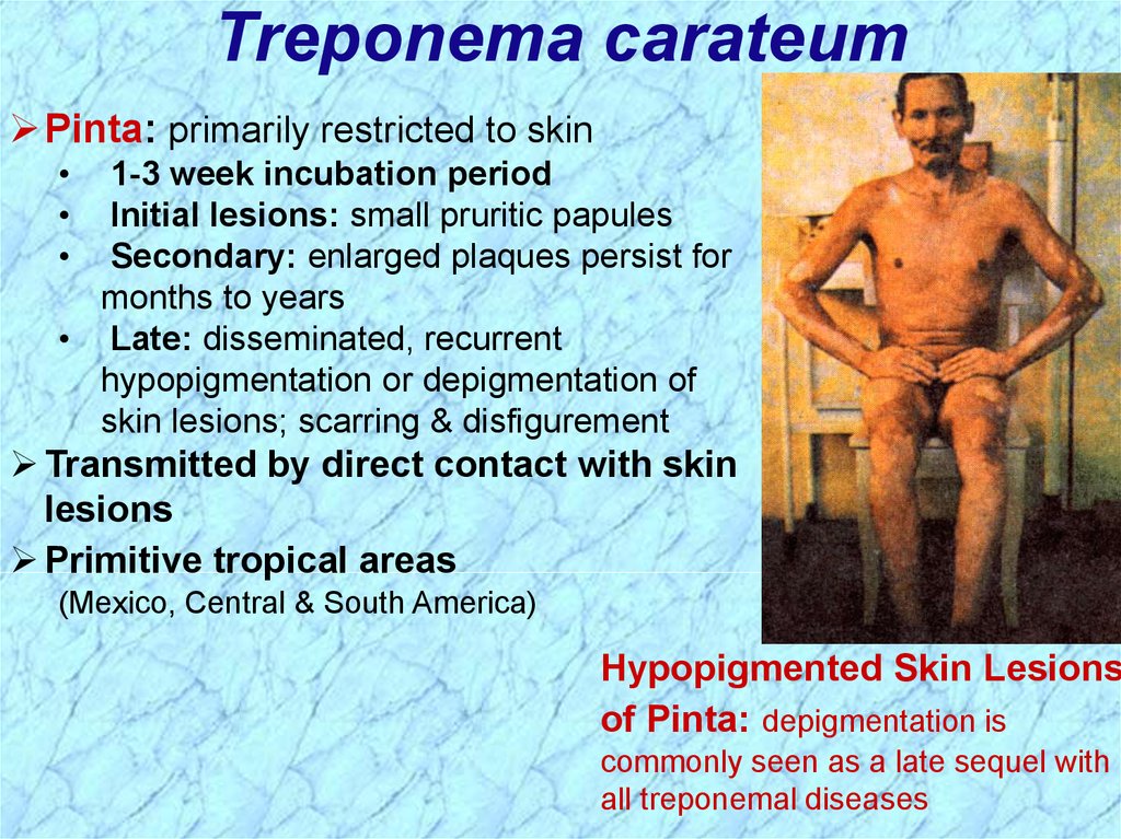
Treponema carateumPinta: primarily restricted to skin
1-3 week incubation period
Initial lesions: small pruritic papules
Secondary: enlarged plaques persist for
months to years
• Late: disseminated, recurrent
hypopigmentation or depigmentation of
skin lesions; scarring & disfigurement
Transmitted by direct contact with skin
lesions
Primitive tropical areas
(Mexico, Central & South America)
Hypopigmented Skin Lesions
of Pinta: depigmentation is
commonly seen as a late sequel with
all treponemal diseases
15.
16.
Treponema pallidumssp. pallidum
17.
Venereal TreponemalDisease
Syphilis
Primarily sexually transmitted disease
(STD)
May be transmitted congenitally
18.
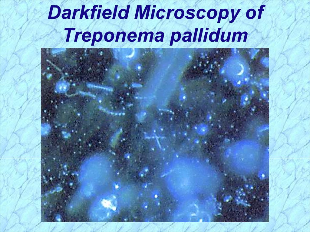
Darkfield Microscopy ofTreponema pallidum
19.
General Characteristics ofTreponema pallidum
Too thin to be seen with light microscopy in
specimens stained with Gram stain or Giemsa stain
• Motile spirochetes can be seen with darkfield
micoscopy
• Staining with anti-treponemal antibodies labeled with
fluorescent dyes
Intracellular pathogen
Cannot be grown in cell-free cultures in vitro
• Koch’s Postulates have not been met
Do not survive well outside of host
• Care must be taken with clinical specimens for
laboratory culture or testing
20.
Epidemiology of T. pallidumTransmitted from direct sexual contact or from
mother to fetus
Not highly contagious (~30% chance of acquiring
disease after single exposure to infected partner) but
transmission rate dependent upon stage of disease
Long incubation period during which time host is
non-infectious
• Useful epidemiologically for contact tracing and
administration of preventative therapy
Prostitution for drugs or for money to purchase drugs
remains central epidemiologic aspect of transmission
21.
Incidence of Syphilis in USA22.
Geographical Distribution ofSyphilis in USA
23.
Pathogenesis of T. pallidumTissue destruction and lesions are primarily a
consequence of patient’s immune response
Syphilis is a disease of blood vessels and of the
perivascular areas
In spite of a vigorous host immune response the
organisms are capable of persisting for decades
• Infection is neither fully controlled nor eradicated
• In early stages, there is an inhibition of cell-mediated
immunity
• Inhibition of CMI abates in late stages of disease, hence
late lesions tend to be localized
24.
Virulence Factors of T. pallidumOuter membrane proteins promote adherence
Hyaluronidase may facilitate perivascular
infiltration
Antiphagocytic coating of fibronectin
Tissue destruction and lesions are primarily
result of host’s immune response
(immunopathology)
25.
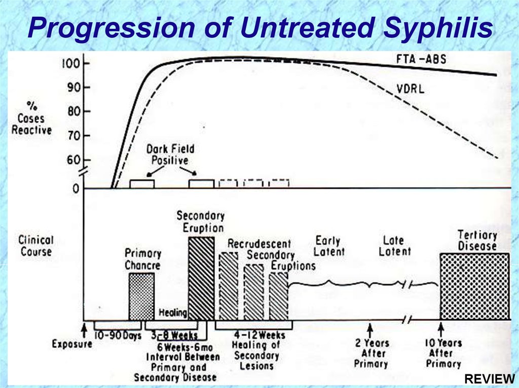
Pathogenesis of T. pallidum (cont.)Primary Syphilis
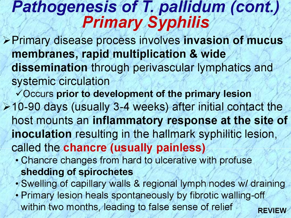
Primary disease process involves invasion of mucus
membranes, rapid multiplication & wide
dissemination through perivascular lymphatics and
systemic circulation
Occurs prior to development of the primary lesion
10-90 days (usually 3-4 weeks) after initial contact the
host mounts an inflammatory response at the site of
inoculation resulting in the hallmark syphilitic lesion,
called the chancre (usually painless)
• Chancre changes from hard to ulcerative with profuse
shedding of spirochetes
• Swelling of capillary walls & regional lymph nodes w/ draining
• Primary lesion heals spontaneously by fibrotic walling-off
within two months, leading to false sense of relief
26.
Pathogenesis of T. pallidum (cont.)Secondary Syphilis
Secondary disease 2-10 weeks after primary
lesion
Widely disseminated mucocutaneous rash
Secondary lesions of the skin and mucus
membranes are highly contagious
Generalized immunological response
27.
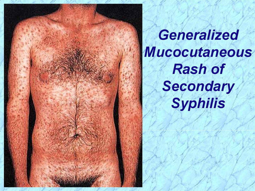
GeneralizedMucocutaneous
Rash of
Secondary
Syphilis
28.
Pathogenesis of T. pallidum (cont.)Latent Stage Syphilis
Following secondary disease, host enters latent
period
•First 4 years = early latent
•Subsequent period = late latent
About 40% of late latent patients progress to
late tertiary syphilitic disease
29.
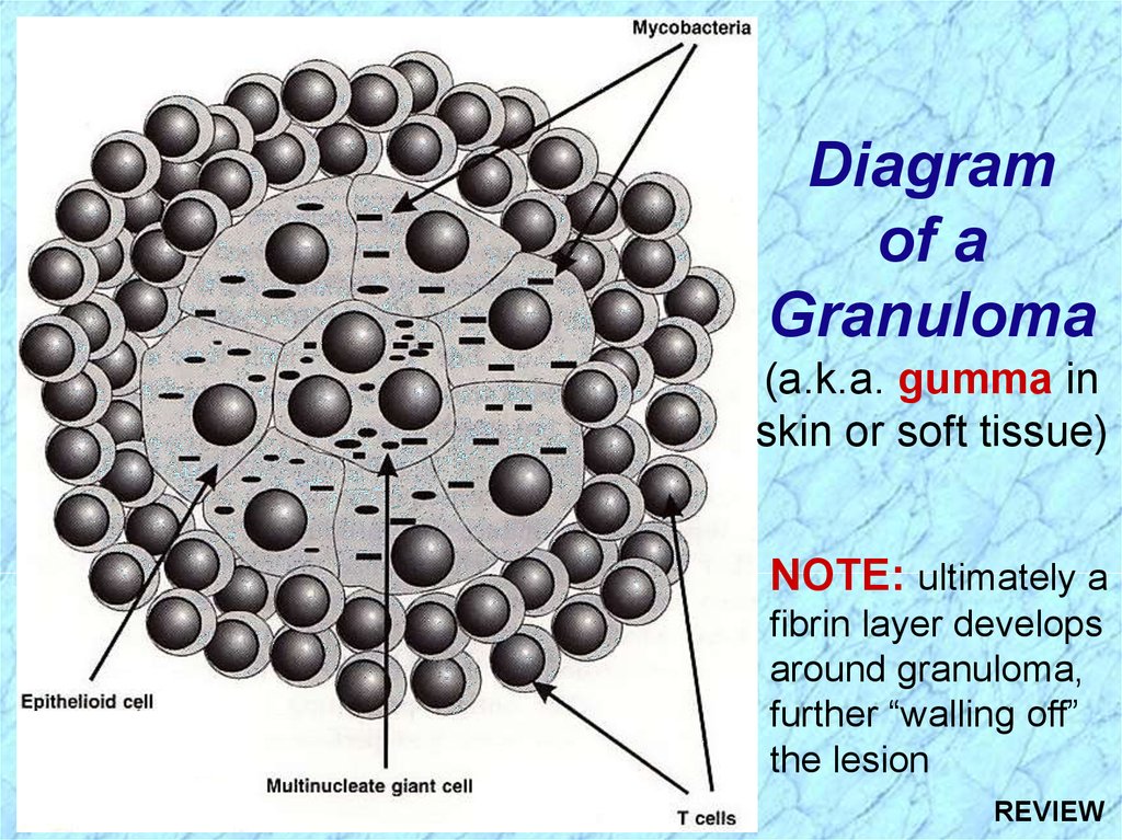
Pathogenesis of T. pallidum (cont.)Tertiary Syphilis
Tertiary syphilis characterized by localized
granulomatous dermal lesions (gummas) in which
few organisms are present
• Granulomas reflect containment by the immunologic
reaction of the host to chronic infection
Late neurosyphilis develops in about 1/6 untreated
cases, usually more than 5 years after initial infection
• Central nervous system and spinal cord involvement
• Dementia, seizures, wasting, etc.
Cardiovascular involvement appears 10-40 years
after initial infection with resulting myocardial
insufficiency and death
30.
Diagramof a
Granuloma
(a.k.a. gumma in
skin or soft tissue)
NOTE: ultimately a
fibrin layer develops
around granuloma,
further “walling off”
the lesion
31.
Progression of Untreated SyphilisLate benign Gummas in skin and soft tissues
Tertiary Stage
32.
Pathogenesis of T. pallidum (cont.)Congenital Syphilis
Congenital syphilis results from transplacental
infection
T. pallidum septicemia in the developing fetus and
widespread dissemination
Abortion, neonatal mortality, and late mental or
physical problems resulting from scars from the
active disease and progression of the active disease
state
33.
Comparison ofIncidence of 1o
& 2o Syphilis in
Women and
Congenital
Syphilis
34.
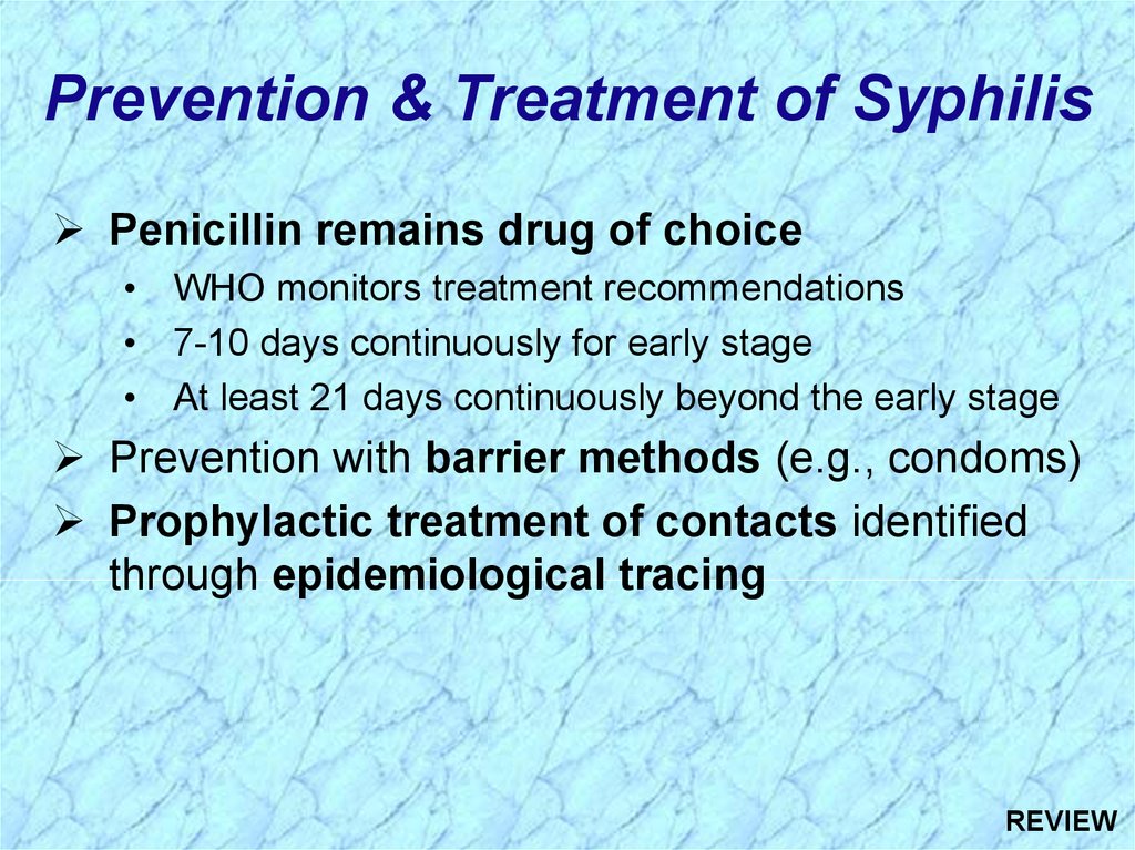
Prevention & Treatment of SyphilisPenicillin remains drug of choice
• WHO monitors treatment recommendations
• 7-10 days continuously for early stage
• At least 21 days continuously beyond the early stage
Prevention with barrier methods (e.g., condoms)
Prophylactic treatment of contacts identified
through epidemiological tracing
35.
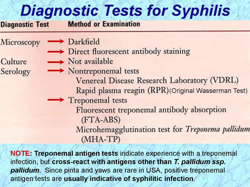
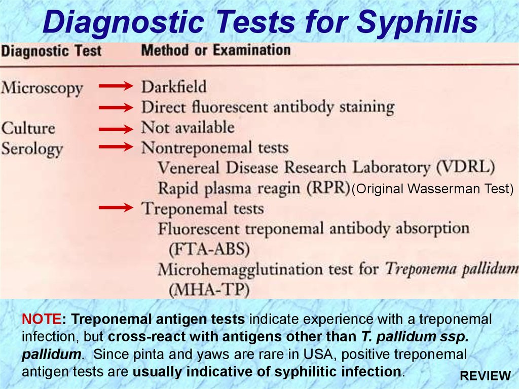
Diagnostic Tests for Syphilis(Original Wasserman Test)
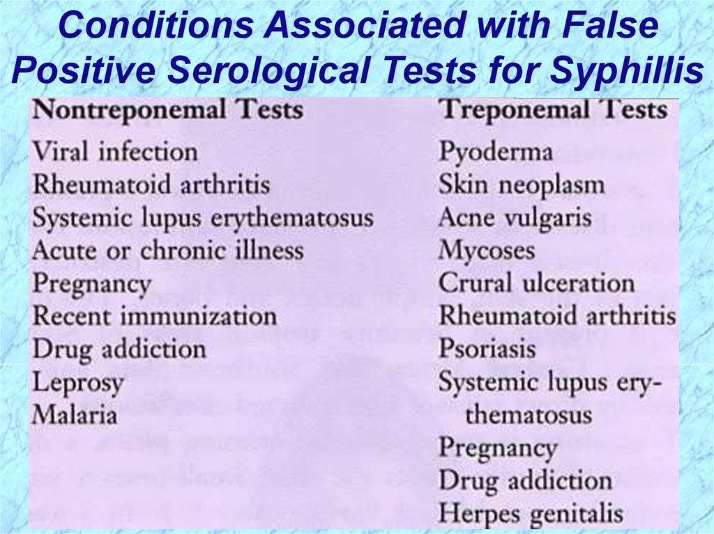
NOTE: Treponemal antigen tests indicate experience with a treponemal
infection, but cross-react with antigens other than T. pallidum ssp.
pallidum. Since pinta and yaws are rare in USA, positive treponemal
antigen tests are usually indicative of syphilitic infection.
36.
Sensitivity & Specificity ofSerologic Tests for Syphillis
37.
Review Handout onSensitivity & Specificity
of Diagnostic Tests
38.
Conditions Associated with FalsePositive Serological Tests for Syphillis
39.
Effect ofTreatment for
Syphillis on
Rapid Plasma
Reagin Test
Reactivity
40.
41.
Borrelia spp.42.
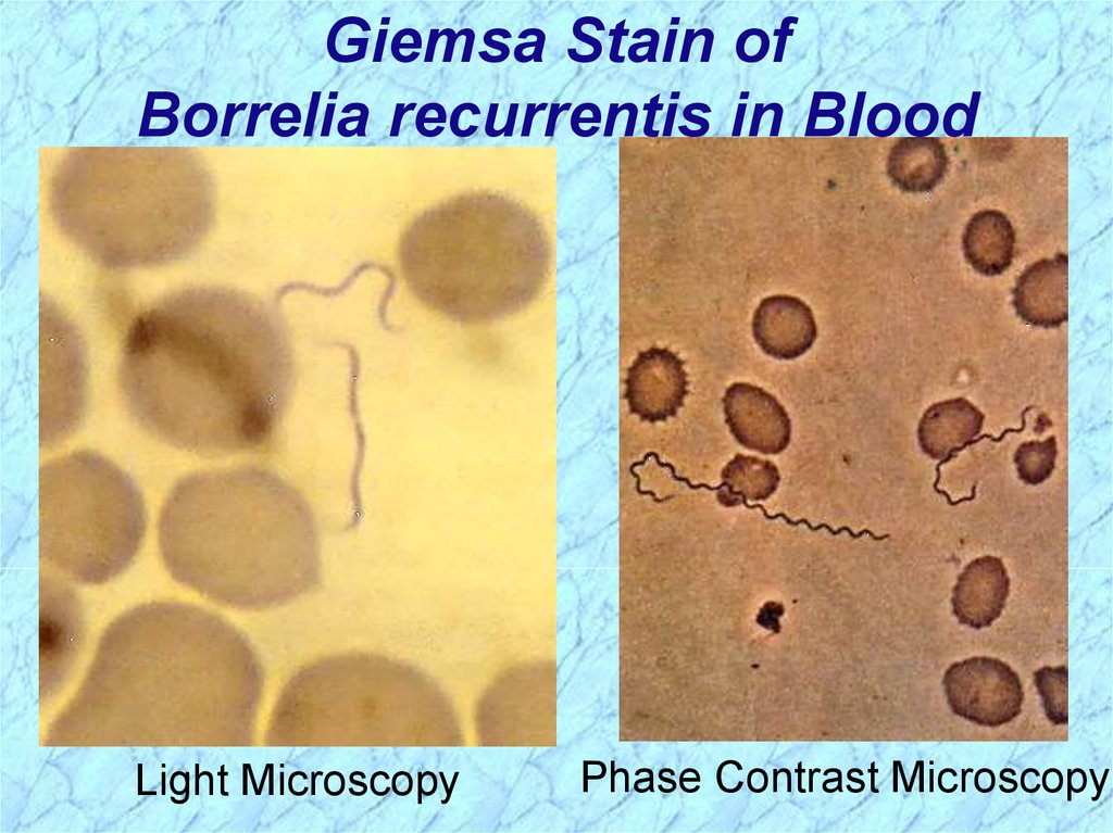
Giemsa Stain ofBorrelia recurrentis in Blood
Light Microscopy
Phase Contrast Microscopy
43.
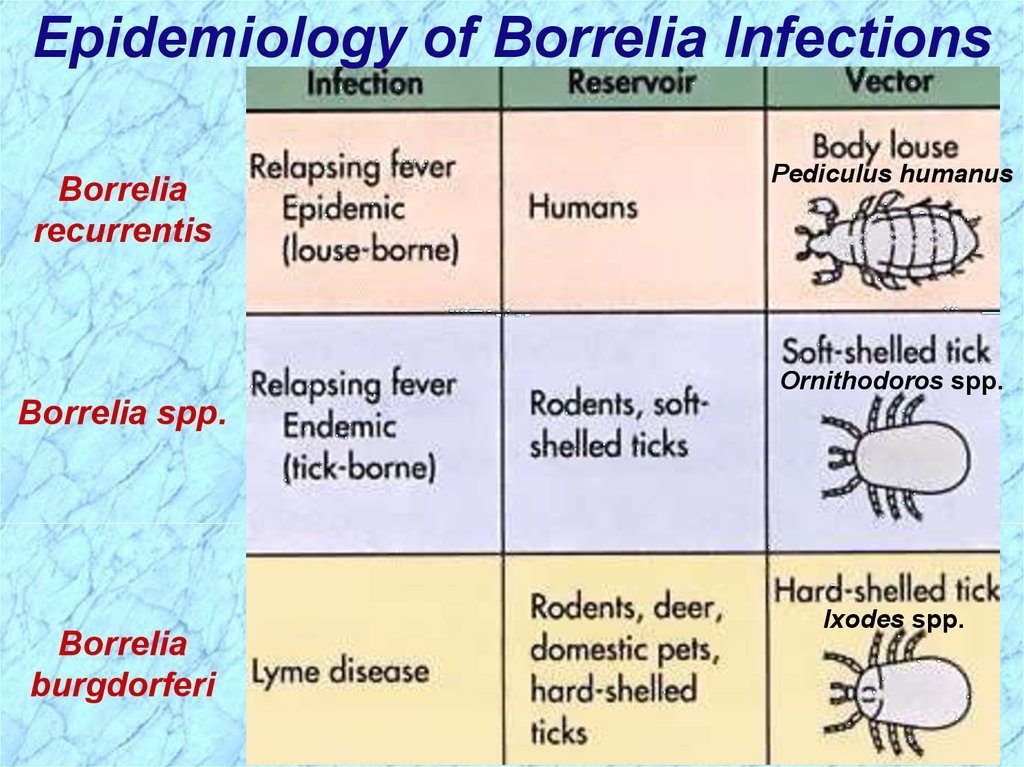
Epidemiology of Borrelia InfectionsBorrelia
recurrentis
Pediculus humanus
Ornithodoros spp.
Borrelia spp.
Borrelia
burgdorferi
Ixodes spp.
44.
Borrelia recurrentis& other Borrelia spp.
45.
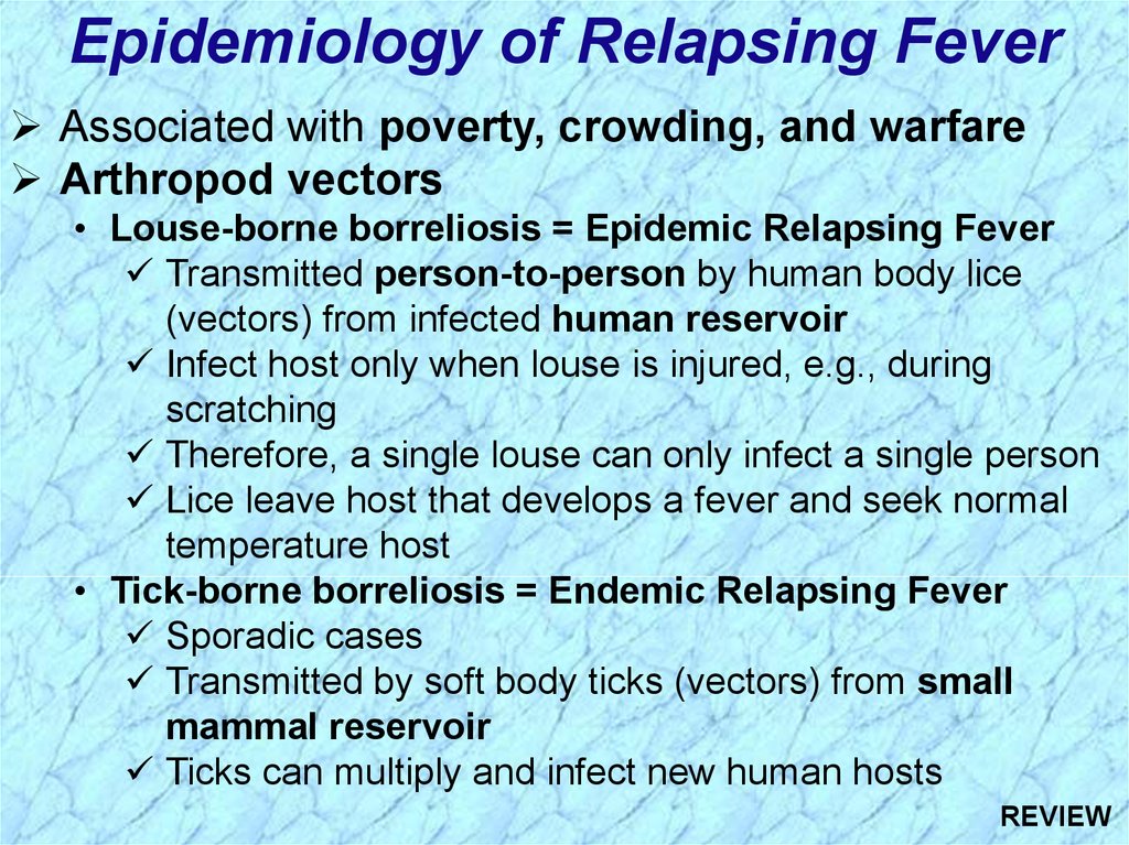
Epidemiology of Relapsing FeverAssociated with poverty, crowding, and warfare
Arthropod vectors
• Louse-borne borreliosis = Epidemic Relapsing Fever
Transmitted person-to-person by human body lice
(vectors) from infected human reservoir
Infect host only when louse is injured, e.g., during
scratching
Therefore, a single louse can only infect a single person
Lice leave host that develops a fever and seek normal
temperature host
• Tick-borne borreliosis = Endemic Relapsing Fever
Sporadic cases
Transmitted by soft body ticks (vectors) from small
mammal reservoir
Ticks can multiply and infect new human hosts
46.
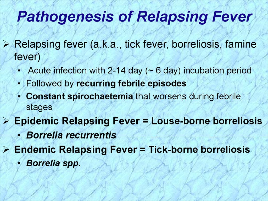
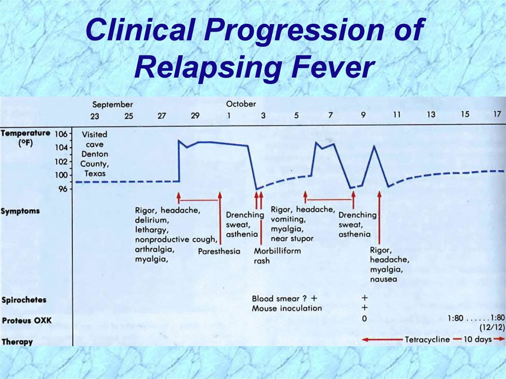
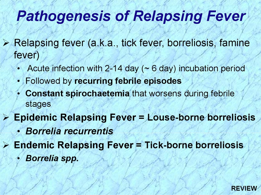
Pathogenesis of Relapsing FeverRelapsing fever (a.k.a., tick fever, borreliosis, famine
fever)
• Acute infection with 2-14 day (~ 6 day) incubation period
• Followed by recurring febrile episodes
• Constant spirochaetemia that worsens during febrile
stages
Epidemic Relapsing Fever = Louse-borne borreliosis
• Borrelia recurrentis
Endemic Relapsing Fever = Tick-borne borreliosis
• Borrelia spp.
47.
Clinical Progression ofRelapsing Fever
48.
Borrelia burgdorferi49.
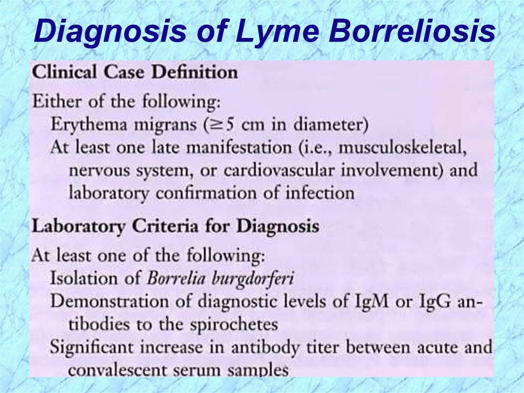
Pathogenesis of Lyme BorreliosisLyme disease characterized by three stages:
i.
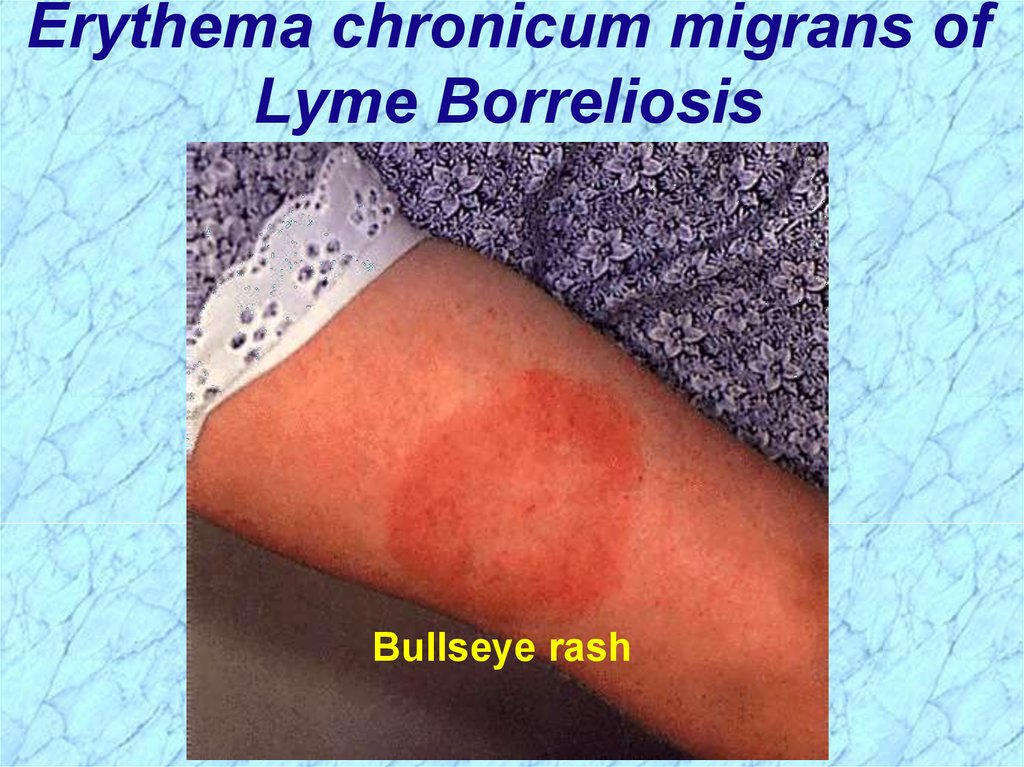
Initially a unique skin lesion (erythema chronicum
migrans (ECM)) with general malaise
ECM not seen in all infected hosts
ECM often described as bullseye rash
Lesions periodically reoccur
ii. Subsequent stage seen in 5-15% of patients with
neurological or cardiac involvement
iii. Third stage involves migrating episodes of nondestructive, but painful arthritis
Acute illness treated with phenoxymethylpenicillin
or tetracycline
50.
Erythema chronicum migrans ofLyme Borreliosis
Bullseye rash
51.
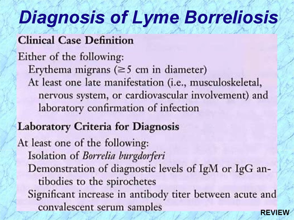
Diagnosis of Lyme Borreliosis52.
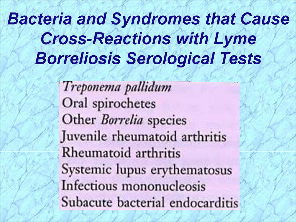
Bacteria and Syndromes that CauseCross-Reactions with Lyme
Borreliosis Serological Tests
53.
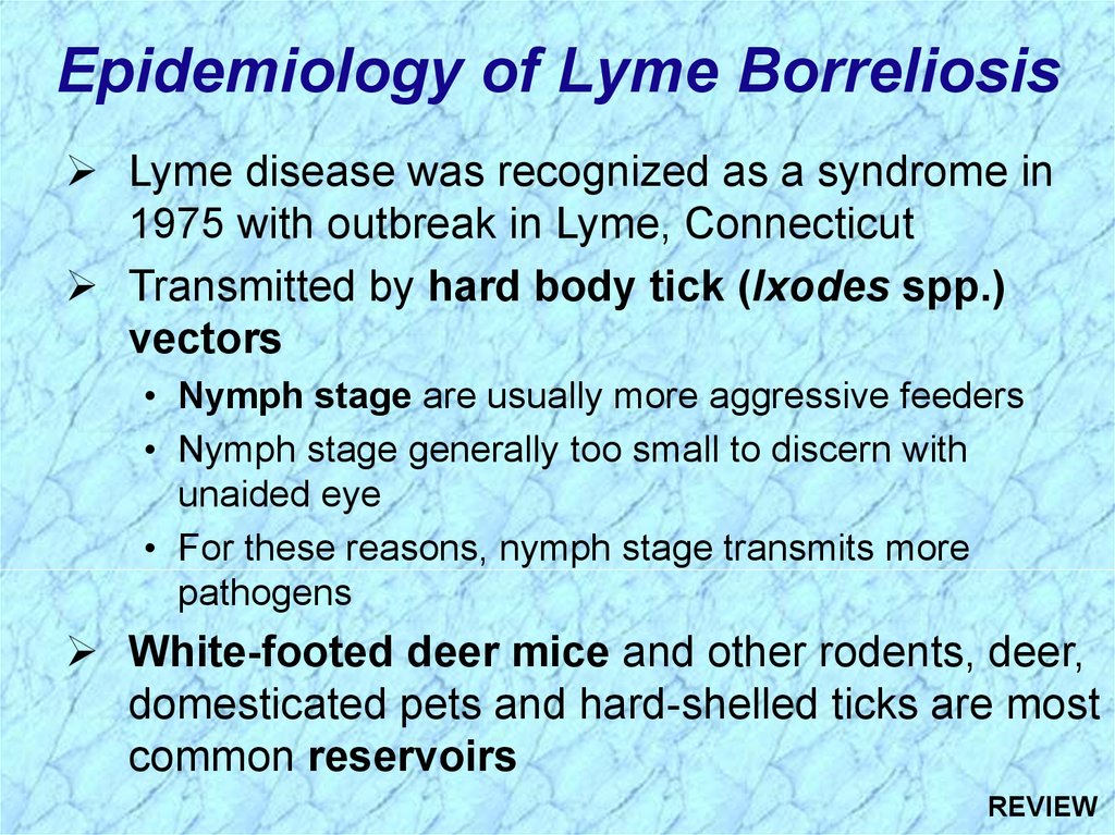
Epidemiology of Lyme BorreliosisLyme disease was recognized as a syndrome in
1975 with outbreak in Lyme, Connecticut
Transmitted by hard body tick (Ixodes spp.)
vectors
• Nymph stage are usually more aggressive feeders
• Nymph stage generally too small to discern with
unaided eye
• For these reasons, nymph stage transmits more
pathogens
White-footed deer mice and other rodents, deer,
domesticated pets and hard-shelled ticks are most
common reservoirs
54.
Incidence of Lyme Borreliosis in USA55.
56.
Leptospira interrogans57.
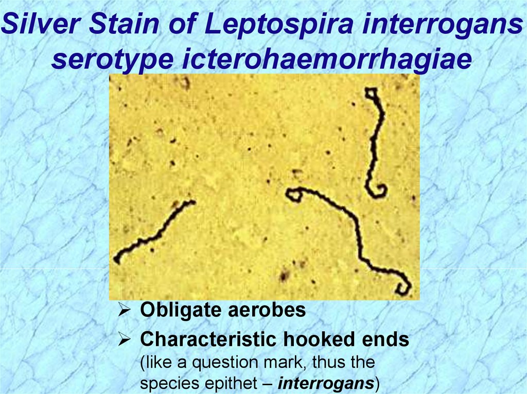
Silver Stain of Leptospira interrogansserotype icterohaemorrhagiae
Obligate aerobes
Characteristic hooked ends
(like a question mark, thus the
species epithet – interrogans)
58.
Leptospirosis Clinical SyndromesMild virus-like syndrome
(Anicteric leptospirosis) Systemic with aseptic
meningitis
(Icteric leptospirosis) Overwhelming disease
(Weil’s disease)
Vascular collapse
Thrombocytopenia
Hemorrhage
Hepatic and renal dysfunction
NOTE: Icteric refers to jaundice (yellowing of skin and mucus
membranes by deposition of bile) and liver involvement
59.
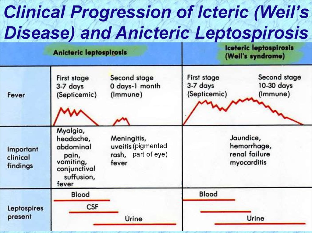
Pathogenesis of Icteric LeptospirosisLeptospirosis, also called Weil’s disease in humans
Direct invasion and replication in tissues
Characterized by an acute febrile jaundice &
immune complex glomerulonephritis
Incubation period usually 10-12 days with flu-like
illness usually progressing through two clinical
stages:
i. Leptospiremia develops rapidly after infection (usually
lasts about 7 days) without local lesion
ii. Infects the kidneys and organisms are shed in the urine
(leptospiruria) with renal failure and death not
uncommon
Hepatic injury & meningeal irritation is common
60.
Clinical Progression of Icteric (Weil’sDisease) and Anicteric Leptospirosis
(pigmented
part of eye)
61.
Epidemiology of LeptospirosisMainly a zoonotic disease
• Transmitted to humans from a variety of wild and
domesticated animal hosts
• In USA most common reservoirs rodents (rats), dogs,
farm animals and wild animals
Transmitted through breaks in the skin or intact
mucus membranes
Indirect contact (soil, water, feed) with infected
urine from an animal with leptospiruria
Occupational disease of animal handling
62.
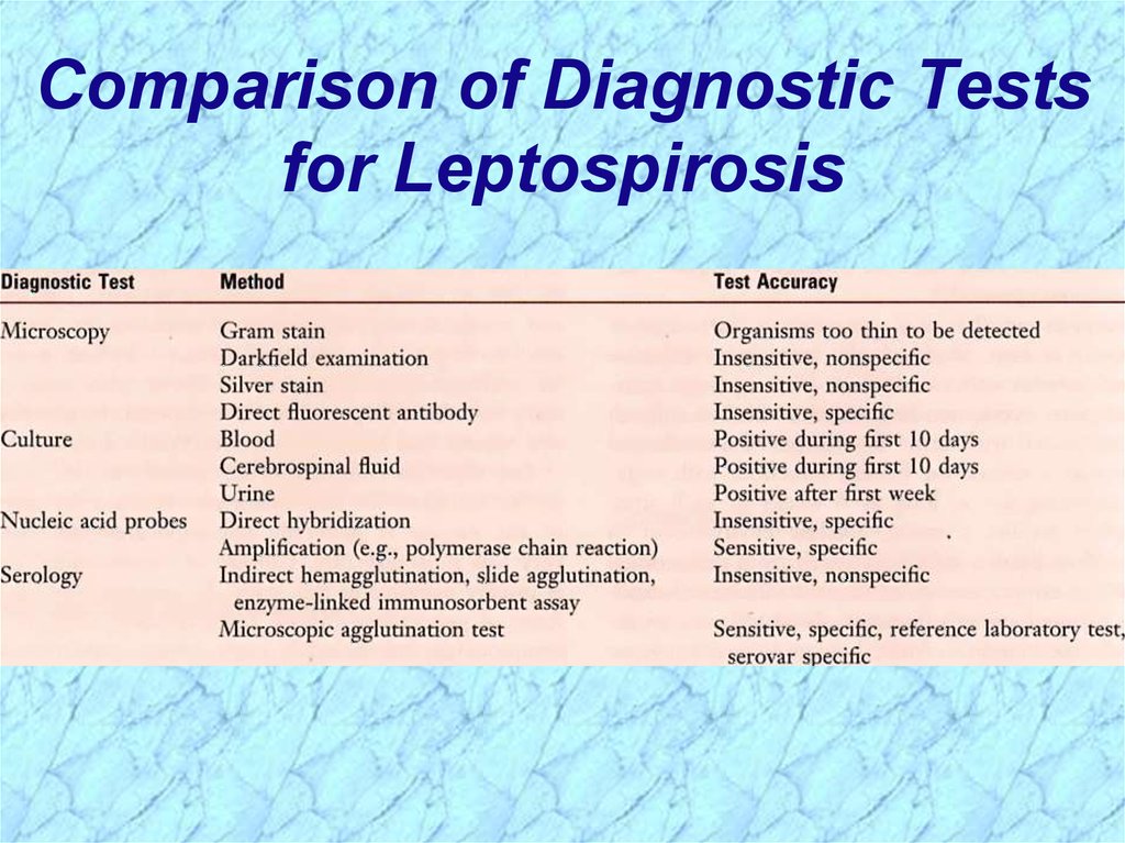
Comparison of Diagnostic Testsfor Leptospirosis
63.
64.
REVIEWof
Spirochaetales
65.
General Overview of SpirochaetalesGram-negative spirochetes
• Spirochete from Greek for “coiled hair”
Extremely thin and can be very long
Tightly coiled helical cells with tapered ends
Motile by periplasmic flagella (a.k.a., axial fibrils
or endoflagella)
Outer sheath encloses axial fibrils wrapped around
protoplasmic cylinder
• Axial fibrils originate from insertion pores at both poles of cell
• May overlap at center of cell in Treponema and Borrelia, but
not in Leptospira
• Differering numbers of endoflagella according to genus &
species
REVIEW
66.
Periplasmic Flagella DiagramREVIEW
67.
Spirochaetales AssociatedHuman Diseases
REVIEW
68.
Review ofTreponema
69.
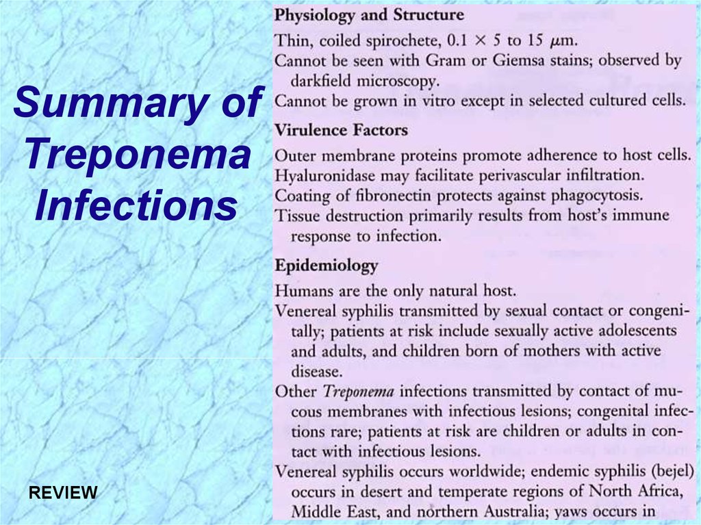
Summary ofTreponema
Infections
REVIEW
70.
Summary ofTreponema
Infections
(cont.)
REVIEW
71.
NonvenerealTreponemal Diseases
Bejel, Yaws & Pinta
Primitive tropical and subtropical
regions
Primarily in impoverished children
REVIEW
72.
Review ofTreponema pallidum
ssp. pallidum
73.
General Characteristics ofTreponema pallidum
Too thin to be seen with light microscopy in
specimens stained with Gram stain or Giemsa stain
• Motile spirochetes can be seen with darkfield
micoscopy
• Staining with anti-treponemal antibodies labeled with
fluorescent dyes
Intracellular pathogen
Cannot be grown in cell-free cultures in vitro
• Koch’s Postulates have not been met
Do not survive well outside of host
• Care must be taken with clinical specimens for
laboratory culture or testing
REVIEW
74.
Epidemiology of T. pallidumTransmitted from direct sexual contact or from
mother to fetus
Not highly contagious (~30% chance of acquiring
disease after single exposure to infected partner) but
transmission rate dependent upon stage of disease
Long incubation period during which time host is
non-infectious
• Useful epidemiologically for contact tracing and
administration of preventative therapy
Prostitution for drugs or for money to purchase drugs
remains central epidemiologic aspect of transmission
REVIEW
75.
Pathogenesis of T. pallidumTissue destruction and lesions are primarily a
consequence of patient’s immune response
Syphilis is a disease of blood vessels and of the
perivascular areas
In spite of a vigorous host immune response the
organisms are capable of persisting for decades
• Infection is neither fully controlled nor eradicated
• In early stages, there is an inhibition of cell-mediated
immunity
• Inhibition of CMI abates in late stages of disease, hence
late lesions tend to be localized
REVIEW
76.
Virulence Factors of T. pallidumOuter membrane proteins promote adherence
Hyaluronidase may facilitate perivascular
infiltration
Antiphagocytic coating of fibronectin
Tissue destruction and lesions are primarily
result of host’s immune response
(immunopathology)
REVIEW
77.
Pathogenesis of T. pallidum (cont.)Primary Syphilis
Primary disease process involves invasion of mucus
membranes, rapid multiplication & wide
dissemination through perivascular lymphatics and
systemic circulation
Occurs prior to development of the primary lesion
10-90 days (usually 3-4 weeks) after initial contact the
host mounts an inflammatory response at the site of
inoculation resulting in the hallmark syphilitic lesion,
called the chancre (usually painless)
• Chancre changes from hard to ulcerative with profuse
shedding of spirochetes
• Swelling of capillary walls & regional lymph nodes w/ draining
• Primary lesion heals spontaneously by fibrotic walling-off
within two months, leading to false sense of relief
REVIEW
78.
Pathogenesis of T. pallidum (cont.)Secondary Syphilis
Secondary disease 2-10 weeks after primary
lesion
Widely disseminated mucocutaneous rash
Secondary lesions of the skin and mucus
membranes are highly contagious
Generalized immunological response
REVIEW
79.
Pathogenesis of T. pallidum (cont.)Latent Stage Syphilis
Following secondary disease, host enters latent
period
•First 4 years = early latent
•Subsequent period = late latent
About 40% of late latent patients progress to
late tertiary syphilitic disease
REVIEW
80.
Pathogenesis of T. pallidum (cont.)Tertiary Syphilis
Tertiary syphilis characterized by localized
granulomatous dermal lesions (gummas) in which
few organisms are present
• Granulomas reflect containment by the immunologic
reaction of the host to chronic infection
Late neurosyphilis develops in about 1/6 untreated
cases, usually more than 5 years after initial infection
• Central nervous system and spinal cord involvement
• Dementia, seizures, wasting, etc.
Cardiovascular involvement appears 10-40 years
after initial infection with resulting myocardial
insufficiency and death
REVIEW
81.
Diagramof a
Granuloma
(a.k.a. gumma in
skin or soft tissue)
NOTE: ultimately a
fibrin layer develops
around granuloma,
further “walling off”
the lesion
REVIEW
82.
Progression of Untreated SyphilisLate benign Gummas in skin and soft tissues
Tertiary Stage
REVIEW
83.
Progression of Untreated SyphilisREVIEW
84.
Pathogenesis of T. pallidum (cont.)Congenital Syphilis
Congenital syphilis results from transplacental
infection
T. pallidum septicemia in the developing fetus and
widespread dissemination
Abortion, neonatal mortality, and late mental or
physical problems resulting from scars from the
active disease and progression of the active disease
state
REVIEW
85.
Prevention & Treatment of SyphilisPenicillin remains drug of choice
• WHO monitors treatment recommendations
• 7-10 days continuously for early stage
• At least 21 days continuously beyond the early stage
Prevention with barrier methods (e.g., condoms)
Prophylactic treatment of contacts identified
through epidemiological tracing
REVIEW
86.
Diagnostic Tests for Syphilis(Original Wasserman Test)
NOTE: Treponemal antigen tests indicate experience with a treponemal
infection, but cross-react with antigens other than T. pallidum ssp.
pallidum. Since pinta and yaws are rare in USA, positive treponemal
antigen tests are usually indicative of syphilitic infection.
REVIEW
87.
Review Handout onSensitivity & Specificity
of Diagnostic Tests
88. Analytic Performance of a Diagnostic Test
ACTUALACTUAL
POSITIVE NEGATIVE
TEST
POSITIVE
80
True
Positives
TEST
20
NEGATIVE
False
Negatives
100
TOTALS
Actual
Positives
25
False
Positives
75
True
Negatives
100
Actual
Negatives
TOTALS
105
Test
Positives
95
Test
Negatives
200
REVIEW
89.
Analytic Performanceof a Diagnostic Test (cont.)
Sensitivity = Measure of True Positive Rate (TPR)
= No. of True Pos. =
No. of True Pos.
=
80 = 80%
No. of Actual Pos.
No. of (True Pos. + False Neg.) 80+20 Sensitivity
In conditional probability terms, the probability of a positive
test given an actual positive sample/patient.
Specificity = Measure of True Negative Rate (TNR)
= No. of True Neg. =
No. of True Neg.
= 75 = 75%
No. of Actual Neg. No. of (True Neg. + False Pos.) 75+25 Specificity
In conditional probability terms, the probability of a negative
test given an actual negative sample/patient.
REVIEW
90.
Review of Borrelia91.
Summary ofBorellia
Infections
REVIEW
92.
Summary ofBorellia
Infections
(cont.)
REVIEW
93.
Epidemiology of Borrelia InfectionsBorrelia
recurrentis
Pediculus humanus
Ornithodoros spp.
Borrelia spp.
Borrelia
burgdorferi
REVIEW
Ixodes spp.
94.
Review ofBorrelia recurrentis
& other Borrelia spp.
95.
Epidemiology of Relapsing FeverAssociated with poverty, crowding, and warfare
Arthropod vectors
• Louse-borne borreliosis = Epidemic Relapsing Fever
Transmitted person-to-person by human body lice
(vectors) from infected human reservoir
Infect host only when louse is injured, e.g., during
scratching
Therefore, a single louse can only infect a single person
Lice leave host that develops a fever and seek normal
temperature host
• Tick-borne borreliosis = Endemic Relapsing Fever
Sporadic cases
Transmitted by soft body ticks (vectors) from small
mammal reservoir
Ticks can multiply and infect new human hosts
REVIEW
96.
Pathogenesis of Relapsing FeverRelapsing fever (a.k.a., tick fever, borreliosis, famine
fever)
• Acute infection with 2-14 day (~ 6 day) incubation period
• Followed by recurring febrile episodes
• Constant spirochaetemia that worsens during febrile
stages
Epidemic Relapsing Fever = Louse-borne borreliosis
• Borrelia recurrentis
Endemic Relapsing Fever = Tick-borne borreliosis
• Borrelia spp.
REVIEW
97.
Review ofBorrelia burgdorferi
98.
Pathogenesis of Lyme BorreliosisLyme disease characterized by three stages:
i.
Initially a unique skin lesion (erythema chronicum
migrans (ECM)) with general malaise
ECM not seen in all infected hosts
ECM often described as bullseye rash
Lesions periodically reoccur
ii. Subsequent stage seen in 5-15% of patients with
neurological or cardiac involvement
iii. Third stage involves migrating episodes of nondestructive, but painful arthritis
Acute illness treated with phenoxymethylpenicillin
or tetracycline
REVIEW
99.
Diagnosis of Lyme BorreliosisREVIEW
100.
Epidemiology of Lyme BorreliosisLyme disease was recognized as a syndrome in
1975 with outbreak in Lyme, Connecticut
Transmitted by hard body tick (Ixodes spp.)
vectors
• Nymph stage are usually more aggressive feeders
• Nymph stage generally too small to discern with
unaided eye
• For these reasons, nymph stage transmits more
pathogens
White-footed deer mice and other rodents, deer,
domesticated pets and hard-shelled ticks are most
common reservoirs
REVIEW
101.
Review of Leptospira102.
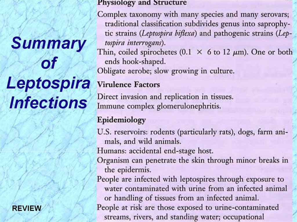
Summaryof
Leptospira
Infections
REVIEW
103.
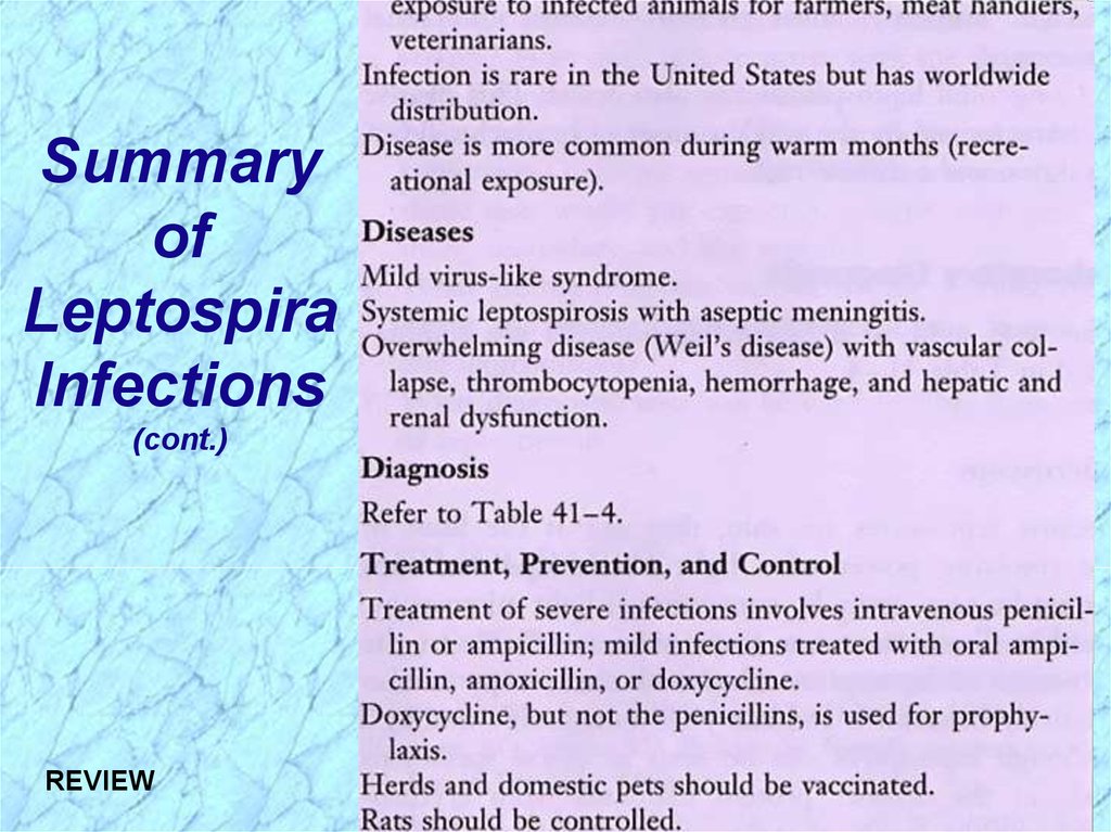
Summaryof
Leptospira
Infections
(cont.)
REVIEW
104.
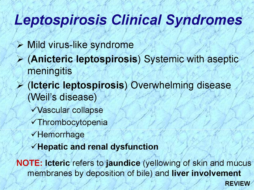
Leptospirosis Clinical SyndromesMild virus-like syndrome
(Anicteric leptospirosis) Systemic with aseptic
meningitis
(Icteric leptospirosis) Overwhelming disease
(Weil’s disease)
Vascular collapse
Thrombocytopenia
Hemorrhage
Hepatic and renal dysfunction
NOTE: Icteric refers to jaundice (yellowing of skin and mucus
membranes by deposition of bile) and liver involvement
REVIEW
105.
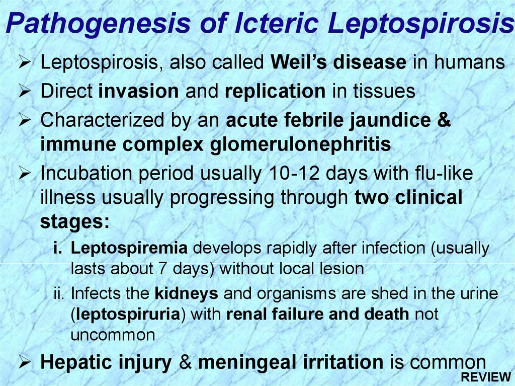
Pathogenesis of Icteric LeptospirosisLeptospirosis, also called Weil’s disease in humans
Direct invasion and replication in tissues
Characterized by an acute febrile jaundice &
immune complex glomerulonephritis
Incubation period usually 10-12 days with flu-like
illness usually progressing through two clinical
stages:
i. Leptospiremia develops rapidly after infection (usually
lasts about 7 days) without local lesion
ii. Infects the kidneys and organisms are shed in the urine
(leptospiruria) with renal failure and death not
uncommon
Hepatic injury & meningeal irritation is common
REVIEW
106.
Epidemiology of LeptospirosisMainly a zoonotic disease
• Transmitted to humans from a variety of wild and
domesticated animal hosts
• In USA most common reservoirs rodents (rats), dogs,
farm animals and wild animals
Transmitted through breaks in the skin or intact
mucus membranes
Indirect contact (soil, water, feed) with infected
urine from an animal with leptospiruria
Occupational disease of animal handling
REVIEW