


 Английский язык
Похожие презентации:


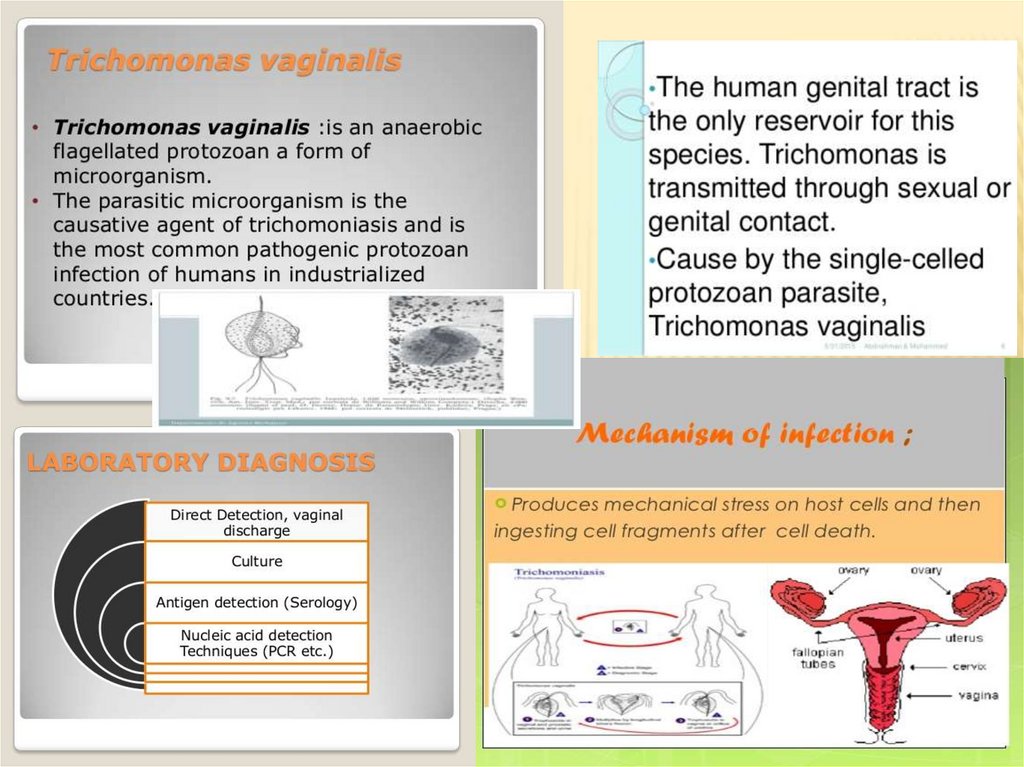
Tissue parasites
1. Tissue parasites
TISSUE PARASITES2.
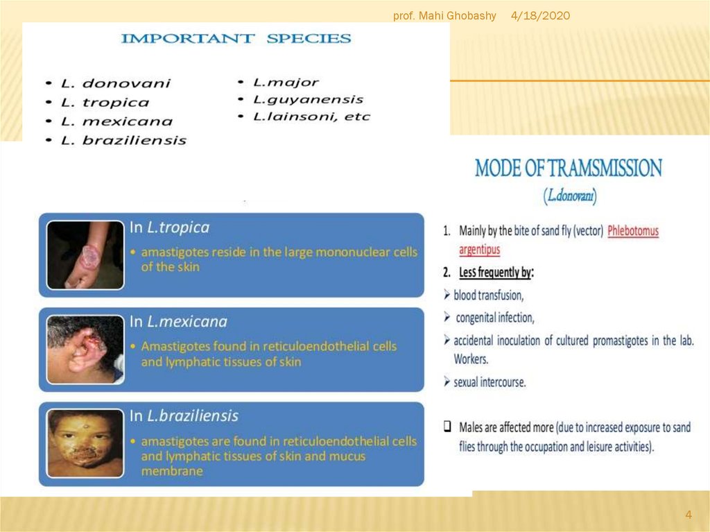
3. Leishmania
prof. Mahi Ghobashy4/18/2020
LEISHMANIA
3
4.
prof. Mahi Ghobashy4/18/2020
4
5.
6. دورة حياة الليشمانيا
4/18/20206
prof. Mahi Ghobashy
دورة حياة الليشمانيا
7.
8.
prof. Mahi Ghobashy4/18/2020
Clinical Features:
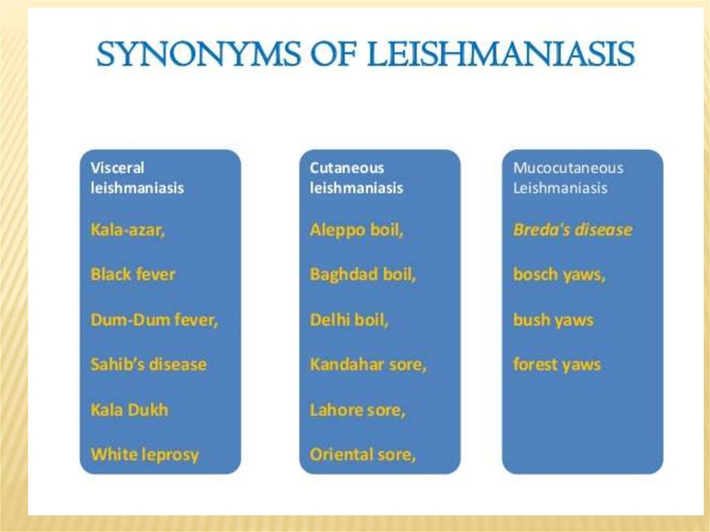
Human leishmanial infections can result in 2 main forms of
disease, cutaneous leishmaniasis and visceral
leishmaniasis (kala-azar). The factors determining the form
of disease include leishmanial species, geographic location,
and immune response of the host. Cutaneous
leishmaniasis is characterized by one or more cutaneous
lesions on areas where sandflies have fed. Persons who
have cutaneous leishmaniasis have one or more sores on
their skin. The sores can change in size and appearance
over time. They often end up looking somewhat like a
volcano, with a raised edge and central crater. A scab
covers some sores. The sores can be painless or painful.
Some people have swollen glands near the sores (for
example, in the armpit if the sores are on the arm or hand).
Laboratory Diagnosis:
Examination of Giemsa stained slides of the relevant tissue
is still the technique most commonly used to detect the
parasite.
Diagnostic findings
Microscopy
Isolation of the organism in culture (using for example the
diphasic NNN medium) or in experimental animals
(hamsters) constitutes another method of parasitilogic
confirmation of the diagnosis, and in addition can provide
material for further investigations (e.g., isoenzyme analysis).
Antibody detection can prove useful in visceral
leishmaniasis but is of limited value in cutaneous disease,
where most patients do not develop a significant antibody
response
molecular (PCR) approaches. Such techniques, however, are
not readily available in general diagnostic laboratories.
8
9. Toxoplasma gondii
prof. Mahi Ghobashy4/18/2020
TOXOPLASMA GONDII
Causal Agent:
Toxoplasma gondii that infects most species of warm blooded animals, including
humans, . A single-celled parasite (a protozoan parasite ) causes a disease known
as toxoplasmosis. While the parasite is found throughout the world, more than 60
million people in the United States may be infected with the Toxoplasma parasite.
Of those who are infected, very few have symptoms because a healthy person's
immune system usually keeps the parasite from causing illness. However, pregnant
women and individuals who have compromised immune systems should be
cautious; for them, aToxoplasma infection could cause serious health problems.
Geographic Distribution:
Serologic prevalence data indicate that toxoplasmosis is one of the most common
of humans infections throughout the world. A high prevalence of infection in France
has been related to a preference for eating raw or undercooked meat, while a high
prevalence in Central America has been related to the frequency of stray cats in a
climate favoring survival of oocysts and soil exposure. The overall seroprevalence in
the United States among adolescents and adults, as determined with specimens
collected by the third National Health and Nutrition Examination Survey (NHANES
III) between 1988 and 1994, was found to be 22.5%, with a seroprevalence among
women of childbearing age (15 to 44 years) of 15%.
9
10.
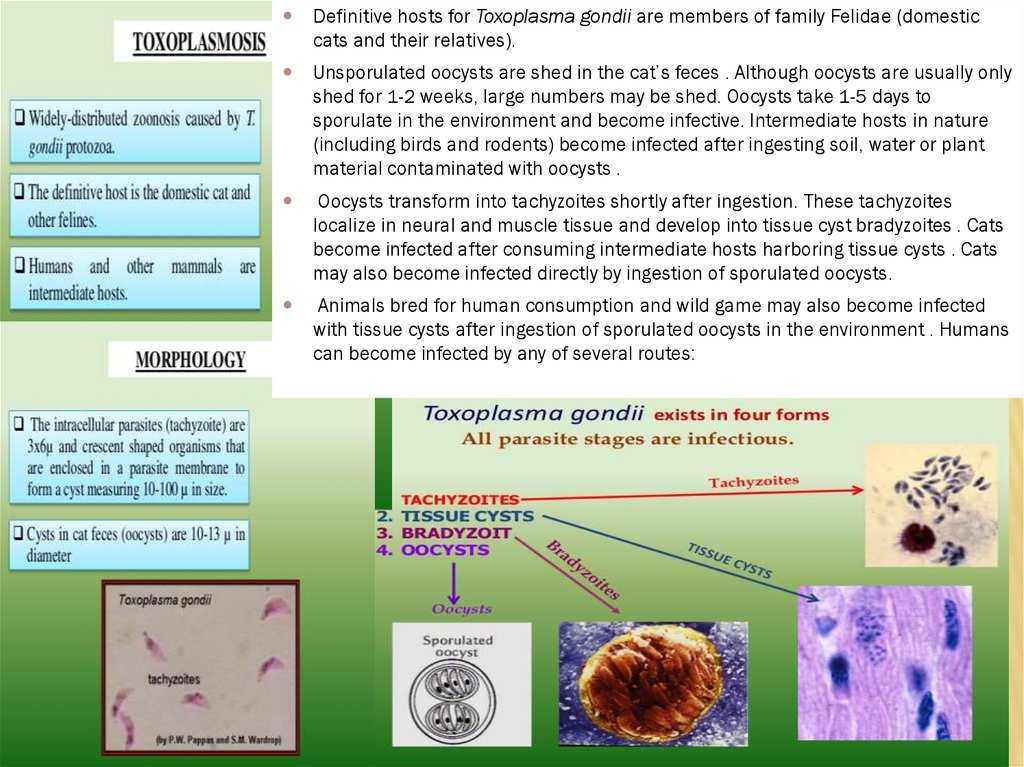
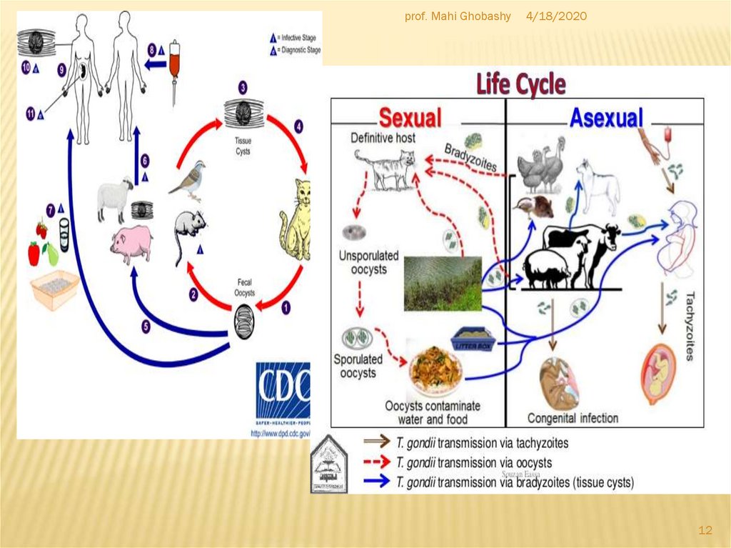
Definitive hosts for Toxoplasma gondii are members of family Felidae (domesticcats and their relatives).
Unsporulated oocysts are shed in the cat’s feces . Although oocysts are usually only
shed for 1-2 weeks, large numbers may be shed. Oocysts take 1-5 days to
sporulate in the environment and become infective. Intermediate hosts in nature
(including birds and rodents) become infected after ingesting soil, water or plant
material contaminated with oocysts .
Oocysts transform into tachyzoites shortly after ingestion. These tachyzoites
localize in neural and muscle tissue and develop into tissue cyst bradyzoites . Cats
become infected after consuming intermediate hosts harboring tissue cysts . Cats
may also become infected directly by ingestion of sporulated oocysts.
Animals bred for human consumption and wild game may also become infected
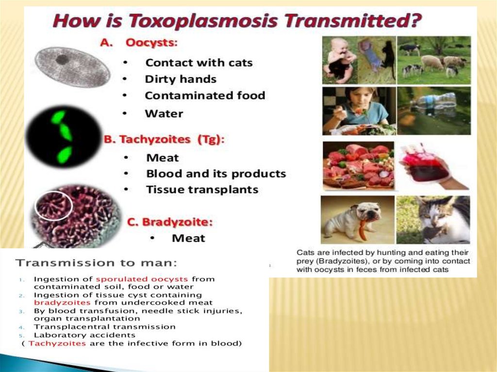
with tissue cysts after ingestion of sporulated oocysts in the environment . Humans
can become infected by any of several routes:
11.
12.
prof. Mahi Ghobashy4/18/2020
12
13.
prof. Mahi Ghobashy4/18/2020
In the human host, the parasites form tissue cysts, most
commonly in skeletal muscle, myocardium, brain, and eyes; these
cysts may remain throughout the life of the host
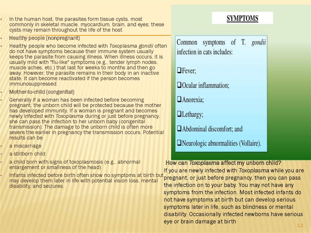
Healthy people (nonpregnant)
Healthy people who become infected with Toxoplasma gondii often
do not have symptoms because their immune system usually
keeps the parasite from causing illness. When illness occurs, it is
usually mild with "flu-like" symptoms (e.g., tender lymph nodes,
muscle aches, etc.) that last for weeks to months and then go
away. However, the parasite remains in their body in an inactive
state. It can become reactivated if the person becomes
immunosuppressed.
Mother-to-child (congenital)
Generally if a woman has been infected before becoming
pregnant, the unborn child will be protected because the mother
has developed immunity. If a woman is pregnant and becomes
newly infected with Toxoplasma during or just before pregnancy,
she can pass the infection to her unborn baby (congenital
transmission). The damage to the unborn child is often more
severe the earlier in pregnancy the transmission occurs. Potential
results can be
a miscarriage
a stillborn child
a child born with signs of toxoplasmosis (e.g., abnormal
How can Toxoplasma affect my unborn child?
enlargement or smallness of the head)
If you are newly infected with Toxoplasma while you are
Infants infected before birth often show no symptoms at birth but pregnant, or just before pregnancy, then you can pass
may develop them later in life with potential vision loss, mental
the infection on to your baby. You may not have any
disability, and seizures.
symptoms from the infection. Most infected infants do
not have symptoms at birth but can develop serious
symptoms later in life, such as blindness or mental
disability. Occasionally infected newborns have serious
eye or brain damage at birth
13
14.
prof. Mahi Ghobashy4/18/2020
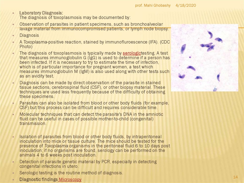
Laboratory Diagnosis:
The diagnosis of toxoplasmosis may be documented by:
Observation of parasites in patient specimens, such as bronchoalveolar
lavage material from immunocompromised patients, or lymph node biopsy.
Diagnosis
A Toxoplasma-positive reaction, stained by immunofluroescence (IFA). (CDC
Photo)
The diagnosis of toxoplasmosis is typically made by serologictesting. A test
that measures immunoglobulin G (IgG) is used to determine if a person has
been infected. If it is necessary to try to estimate the time of infection,
which is of particular importance for pregnant women, a test which
measures immunoglobulin M (IgM) is also used along with other tests such
as an avidity test.
Diagnosis can be made by direct observation of the parasite in stained
tissue sections, cerebrospinal fluid (CSF), or other biopsy material. These
techniques are used less frequently because of the difficulty of obtaining
these specimens.
Parasites can also be isolated from blood or other body fluids (for example,
CSF) but this process can be difficult and requires considerable time.
Molecular techniques that can detect the parasite's DNA in the amniotic
fluid can be useful in cases of possible mother-to-child (congenital)
transmission.
Isolation of parasites from blood or other body fluids, by intraperitoneal
inoculation into mice or tissue culture. The mice should be tested for the
presence of Toxoplasma organisms in the peritoneal fluid 6 to 10 days post
inoculation; if no organisms are found, serology can be performed on the
animals 4 to 6 weeks post inoculation.
Detection of parasite genetic material by PCR, especially in detecting
congenital infections in utero.
Serologic testing is the routine method of diagnosis.
Diagnostic findings Microscopy
14
15.
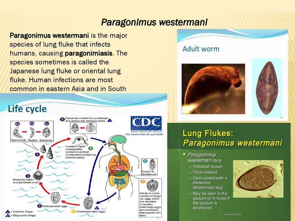
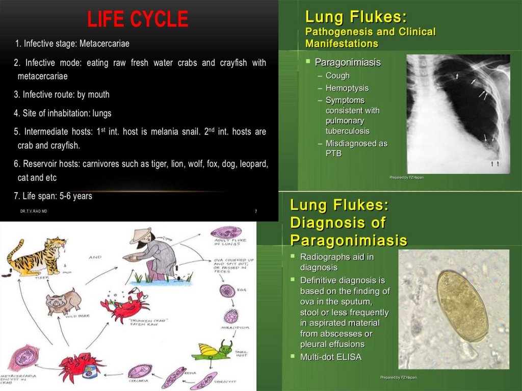
Paragonimus westermaniParagonimus westermani is the major
species of lung fluke that infects
humans, causing paragonimiasis. The
species sometimes is called the
Japanese lung fluke or oriental lung
fluke. Human infections are most
common in eastern Asia and in South
America.
16.
17.
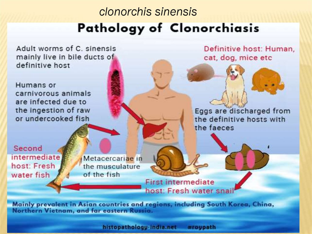
clonorchis sinensis18.
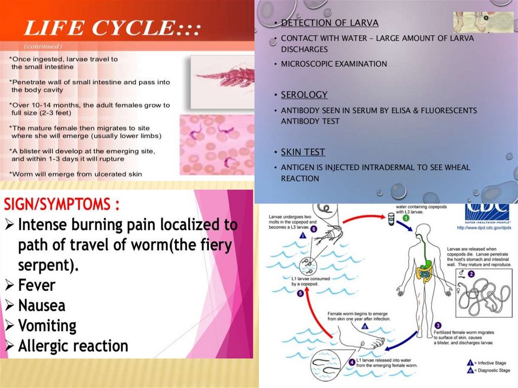
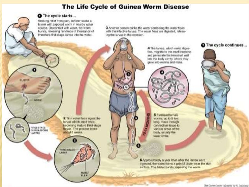
19. Dracunculus medinensis
prof. Mahi Ghobashy4/18/2020
DRACUNCULUS MEDINENSIS
19
20.
21.
22.
Thankswish you all the best