


 Физика
Похожие презентации:


Radiation
1. Radiation
Dr. Rasha SalamaPhD Community Medicine
Suez Canal University
Egypt
2. Definition of Radiation
“Radiation is an energy in the form ofelectro-magnetic waves or particulate
matter, traveling in the air.”
3.
Forces: There are many interactionsamong nuclei. It turns out that there are
forces other than the electromagnetic
force and the gravitational force which
govern the interactions among nuclei.
Einstein in 1905m showed 2 more laws:
energy/mass, and binding energy
4.
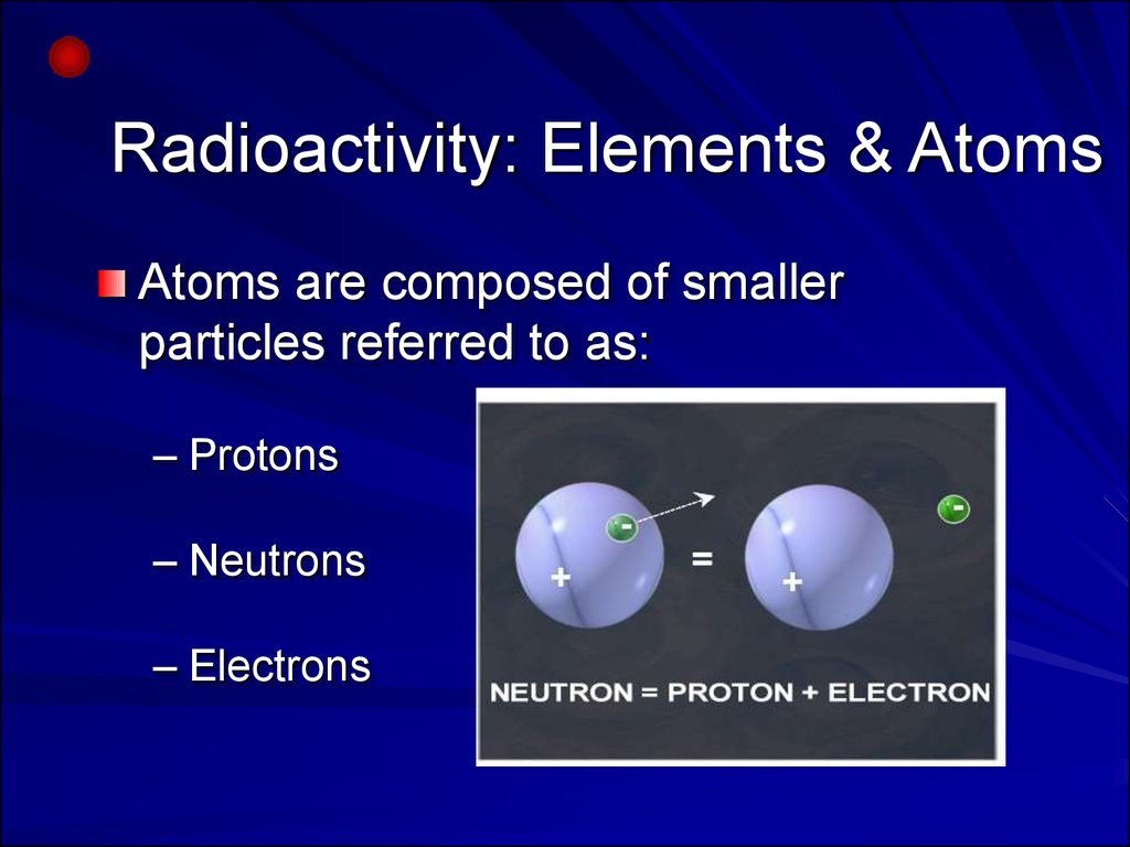
Radioactivity: Elements & AtomsAtoms are composed of smaller
particles referred to as:
– Protons
– Neutrons
– Electrons
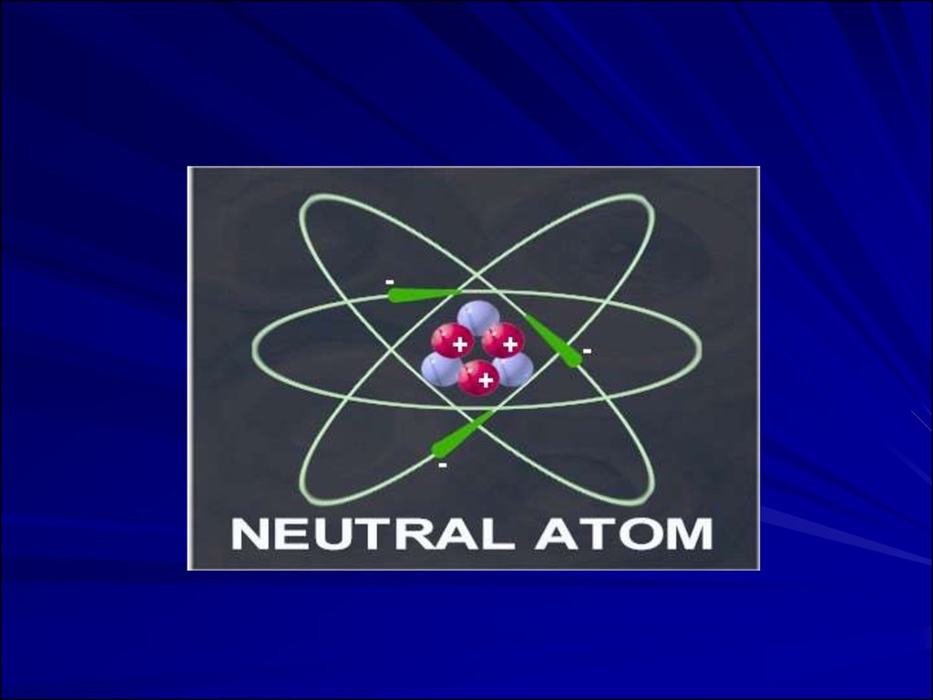
5. Basic Model of a Neutral Atom.
Electrons (-) orbiting nucleus of protons (+)and neutrons. Same number of electrons
as protons; net charge = 0.
Atomic number (number of protons)
determines element.
Mass number (protons + neutrons)
6.
7. Radioactivity
If a nucleus is unstable for any reason, itwill emit and absorb particles. There are
many types of radiation and they are all
pertinent to everyday life and health as
well as nuclear physical applications.
8.
IonizationIonizing radiation is produced by unstable
atoms. Unstable atoms differ from stable
atoms because they have an excess of
energy or mass or both.
Unstable atoms are said to be radioactive. In
order to reach stability, these atoms give off,
or emit, the excess energy or mass. These
emissions are called radiation.
9.
10.
11.
12.
13.
Types or Products of IonizingRadiation
neutron
or X-ray
14.
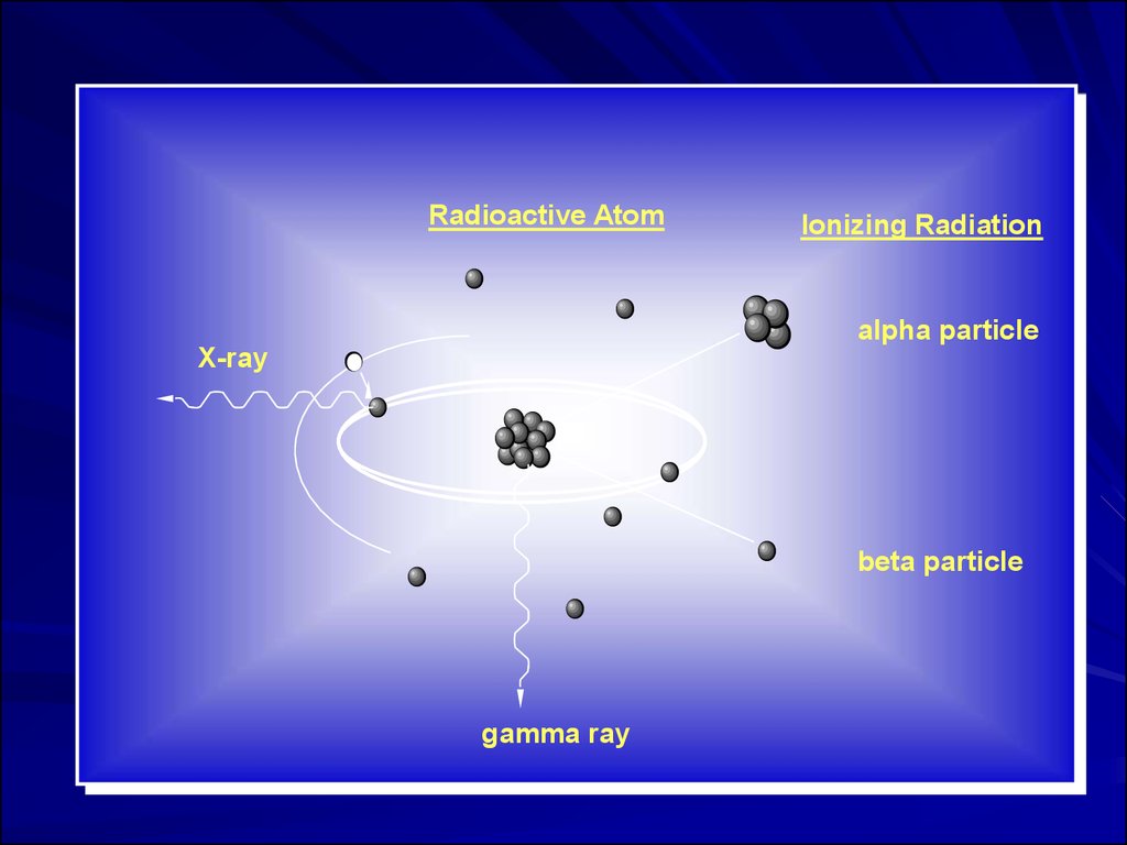
Radioactive AtomIonizing Radiation
alpha particle
X-ray
beta particle
gamma ray
15.
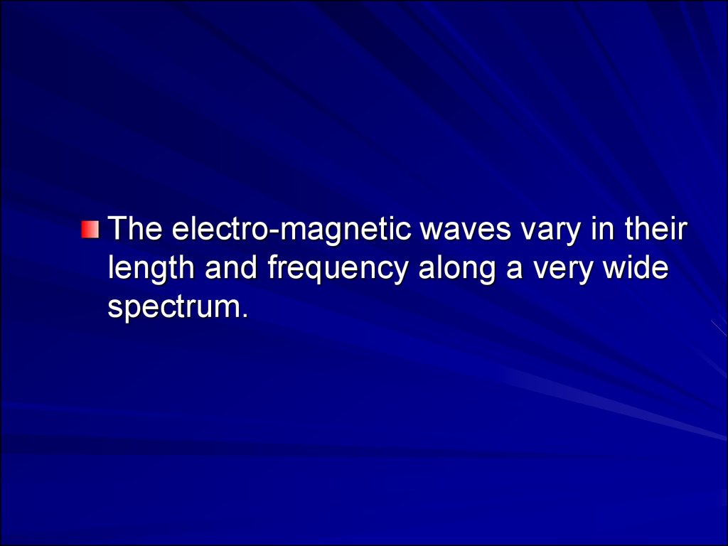
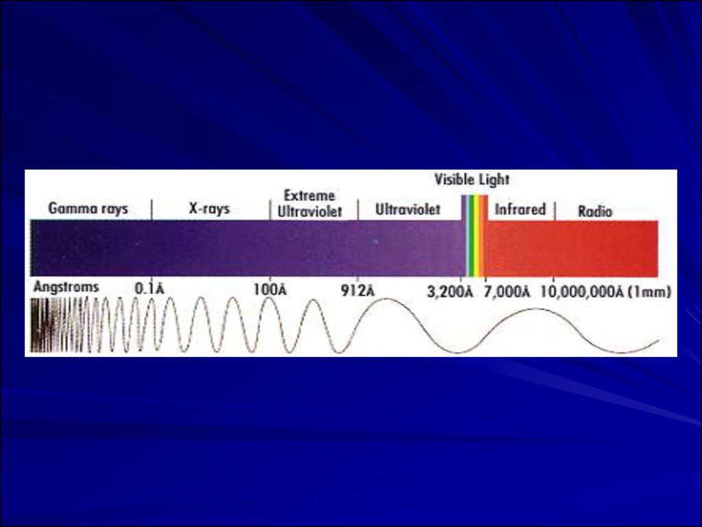
The electro-magnetic waves vary in theirlength and frequency along a very wide
spectrum.
16.
17.
18.
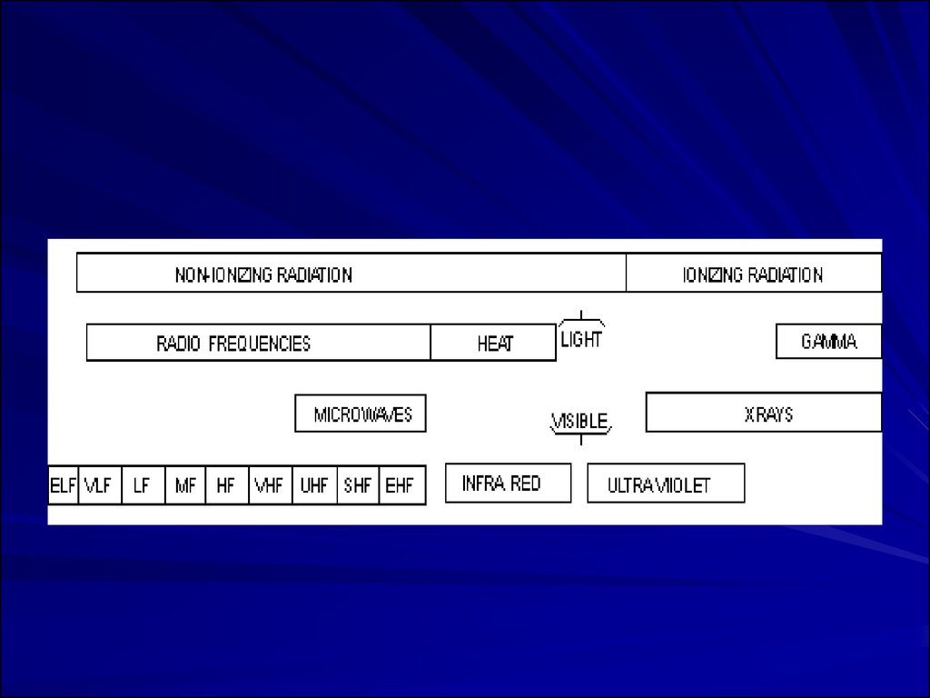
19. Types of Radiation
Radiation is classified into:–Ionizing radiation
–Non-ionizing radiation
20.
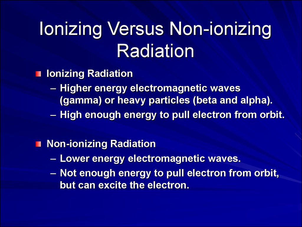
Ionizing Versus Non-ionizingRadiation
Ionizing Radiation
– Higher energy electromagnetic waves
(gamma) or heavy particles (beta and alpha).
– High enough energy to pull electron from orbit.
Non-ionizing Radiation
– Lower energy electromagnetic waves.
– Not enough energy to pull electron from orbit,
but can excite the electron.
21. Ionizing Radiation
Definition:“ It is a type of radiation that is able to
disrupt atoms and molecules on which
they pass through, giving rise to ions and
free radicals”.
22. Another Definition
Ionizing radiationA radiation is said to be ionizing when it has enough
energy to eject one or more electrons from the atoms
or molecules in the irradiated medium. This is the
case of a and b radiations, as well as of
electromagnetic radiations such as gamma
radiations, X-rays and some ultra-violet rays. Visible
or infrared light are not, nor are microwaves or radio
waves.
23.
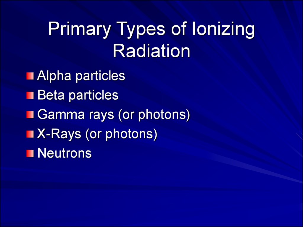
Primary Types of IonizingRadiation
Alpha particles
Beta particles
Gamma rays (or photons)
X-Rays (or photons)
Neutrons
24.
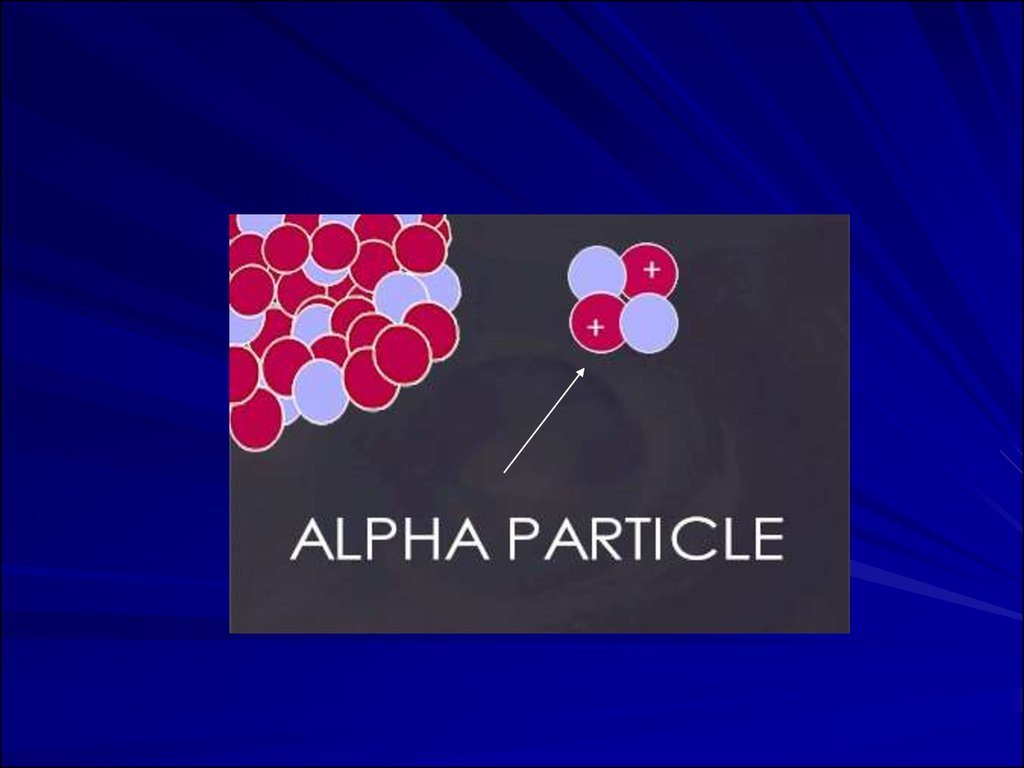
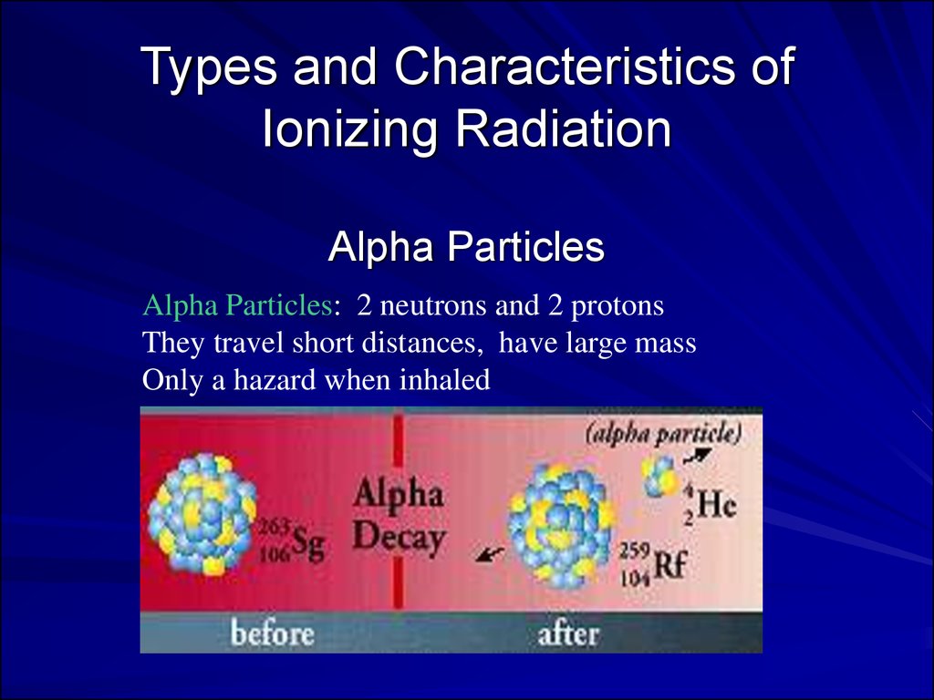
Types and Characteristics ofIonizing Radiation
Alpha Particles
Alpha Particles: 2 neutrons and 2 protons
They travel short distances, have large mass
Only a hazard when inhaled
25.
Alpha Particles (or Alpha Radiation):Helium nucleus (2 neutrons and 2
protons); +2 charge; heavy (4
AMU). Typical Energy = 4-8 MeV;
Limited range (<10cm in air; 60µm in
tissue); High LET (QF=20) causing heavy
damage (4K-9K ion pairs/µm in tissue).
Easily shielded (e.g., paper, skin) so an
internal radiation hazard. Eventually lose
too much energy to ionize; become He.
26.
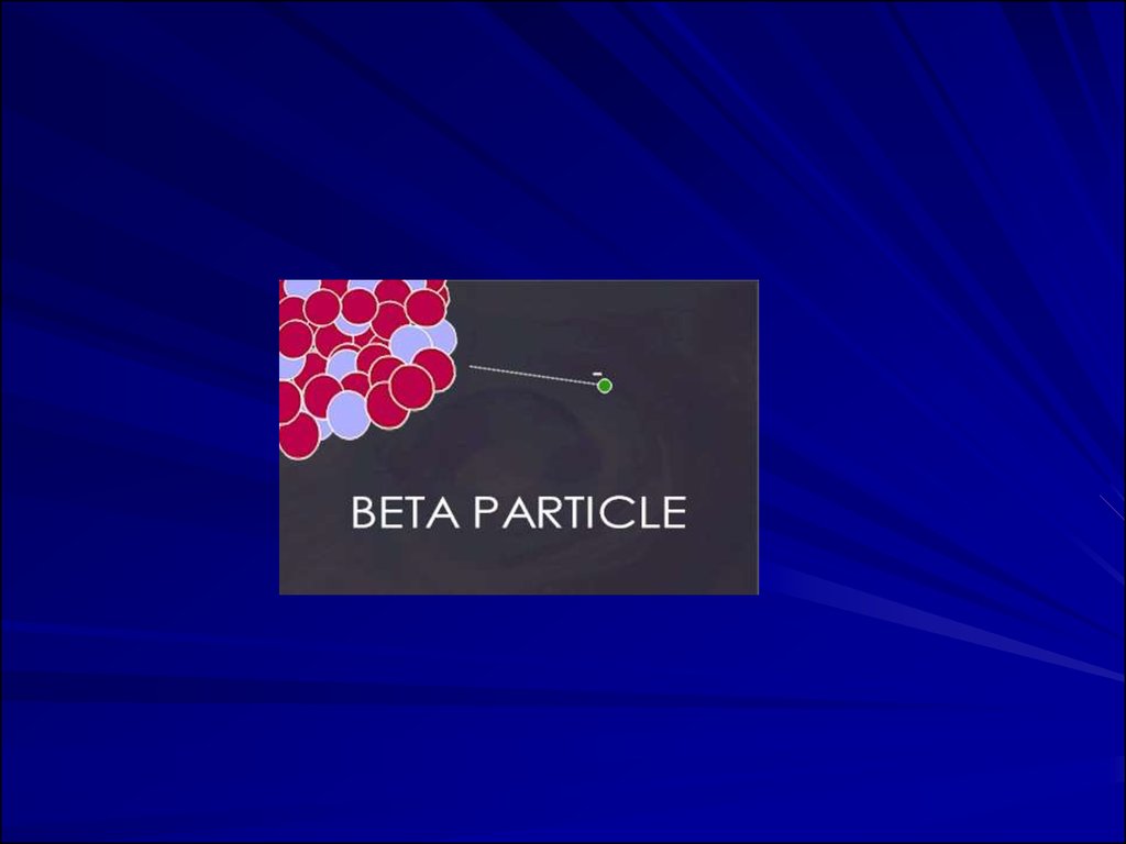
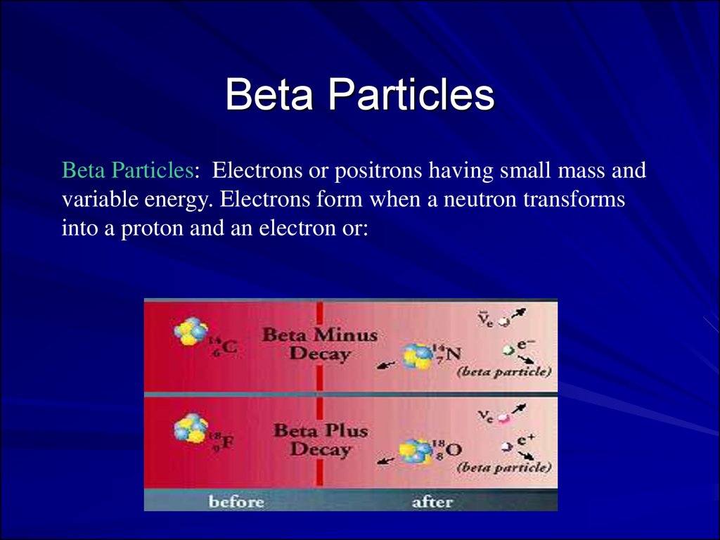
Beta ParticlesBeta Particles: Electrons or positrons having small mass and
variable energy. Electrons form when a neutron transforms
into a proton and an electron or:
27.
Beta Particles: High speed electron ejected fromnucleus; -1 charge, light 0.00055 AMU; Typical
Energy = several KeV to 5 MeV; Range approx.
12'/MeV in air, a few mm in tissue; Low LET (QF=1)
causing light damage (6-8 ion pairs/µm in tissue).
Primarily an internal hazard, but high beta can be an
external hazard to skin. In addition, the high speed
electrons may lose energy in the form of X-rays when
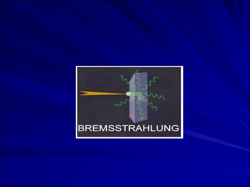
they quickly decelerate upon striking a heavy
material. This is called Bremsstralung (or Breaking)
Radiation. Aluminum and other light (<14)
materials are used for shielding.
28.
29.
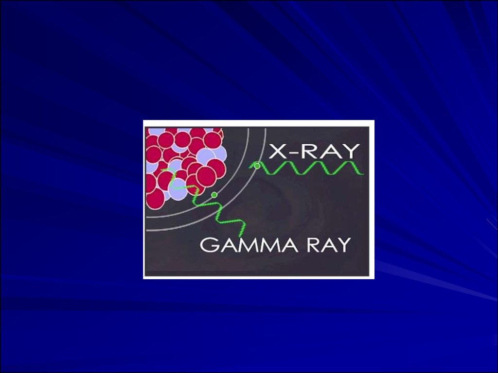
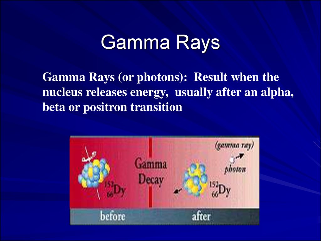
Gamma RaysGamma Rays (or photons): Result when the
nucleus releases energy, usually after an alpha,
beta or positron transition
30.
X-RaysX-Rays: Occur whenever an inner shell
orbital electron is removed and
rearrangement of the atomic electrons
results with the release of the elements
characteristic X-Ray energy
31.
X- and Gamma Rays: X-rays are photons(Electromagnetic radiations) emitted from
electron orbits. Gamma rays are
photons emitted from the nucleus, often
as part of radioactive decay. Gamma rays
typically have higher energy (Mev's) than
X-rays (KeV's), but both are unlimited.
32.
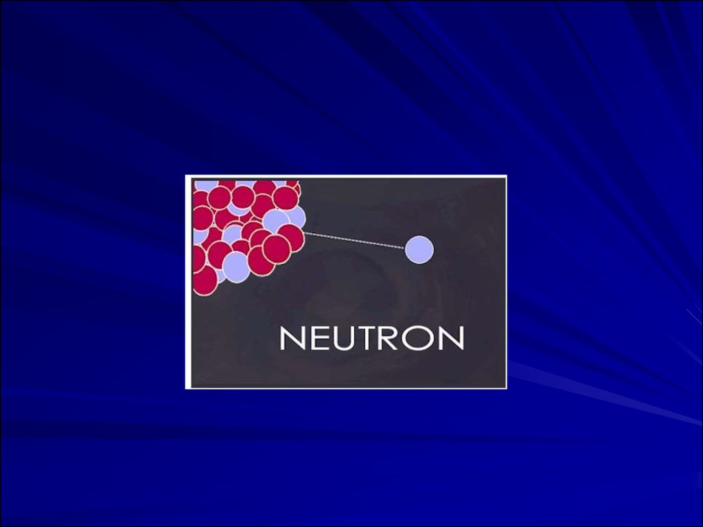
NeutronsNeutrons: Have the same mass as
protons but are uncharged
33.
34.
35. QUANTIFICATION OF RADIATION
A. Quantifying Radioactive DecayB. Quantifying Exposure and Dose
36. A. Quantifying Radioactive Decay
Measurement of Activity in disintegrationsper second (dps);
1 Becquerel (Bq) = 1 dps;
1 Curie (Ci) = 3.7 x 10¹⁰ dps;
Activity of substances are expressed as
activity per weight or volume (e.g., Bq/gm
or Ci/l).
37. B. Quantifying Exposure and Dose
Exposure: Roentgen 1 Roentgen (R) = amount of X orgamma radiation that produces ionization resulting in 1
electrostatic unit of charge in 1 cm3 of dry
air. Instruments often measure exposure rate in mR/hr.
Absorbed Dose: rad (Roentgen absorbed dose) =
absorption of 100 ergs of energy from any radiation in 1
gram of any material; 1 Gray (Gy) = 100 rads = 1
Joule/kg; Exposure to 1 Roentgen approximates 0.9 rad
in air.
Biologically Equivalent Dose: Rem (Roentgen
equivalent man) = dose in rads x QF, where QF =
quality factor. 1 Sievert (Sv) = 100 rems.
38.
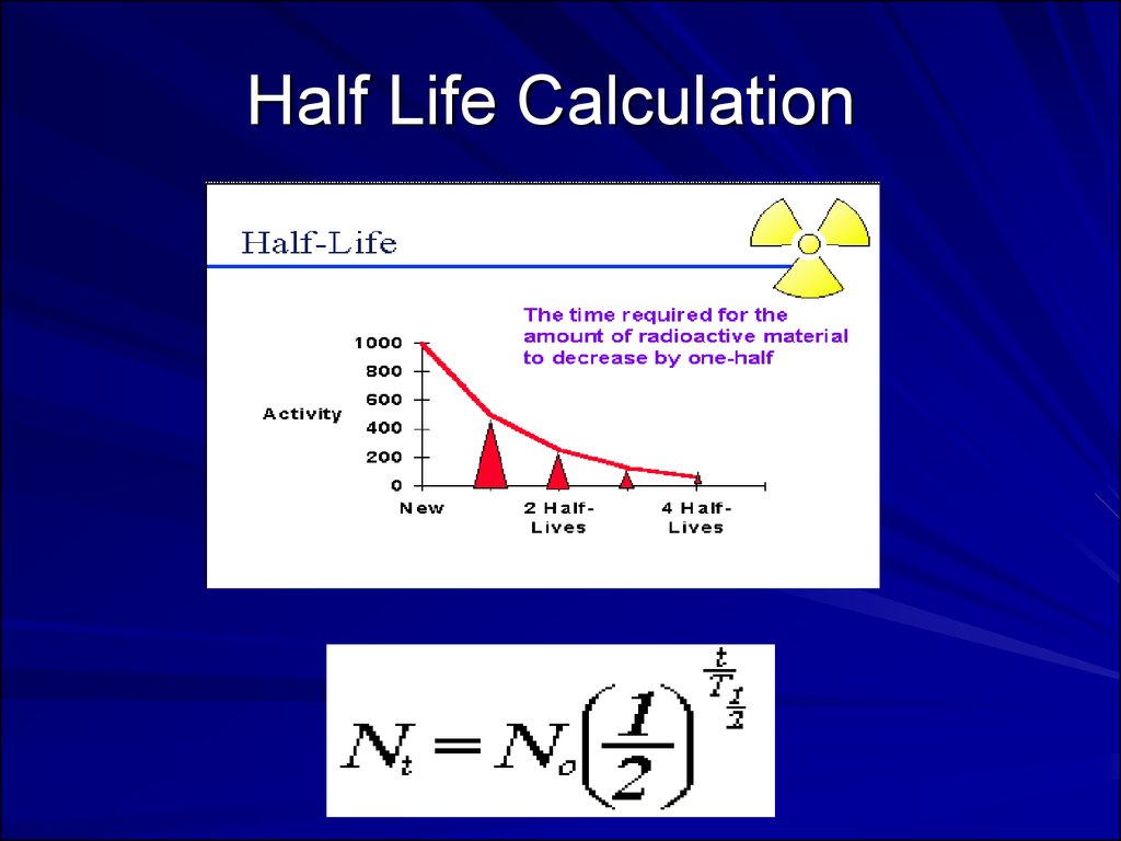
Half Life Calculation39.
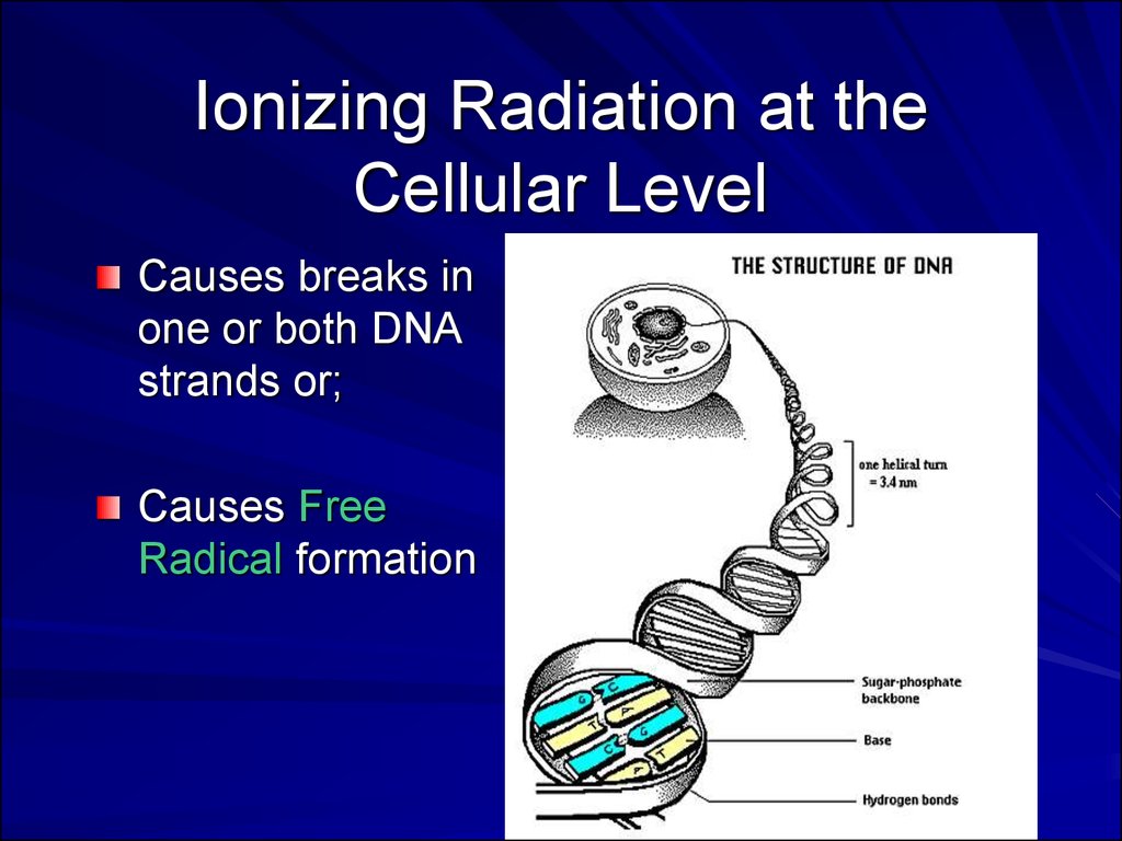
Ionizing Radiation at theCellular Level
Causes breaks in
one or both DNA
strands or;
Causes Free
Radical formation
40. Exposure Limits
OSHA Limits: Whole body limit = 1.25rem/qtr or 5 rem (50 mSv) per year.
Hands and feet limit = 18.75 rem/qtr.
Skin of whole body limit = 7.5 rem/qtr.
Total life accumulation = 5 x (N-18) rem
where N = age. Can have 3 rem/qtr if total
life accumulation not exceeded.
Note: New recommendations reduce the 5
rem to 2 rem.
41.
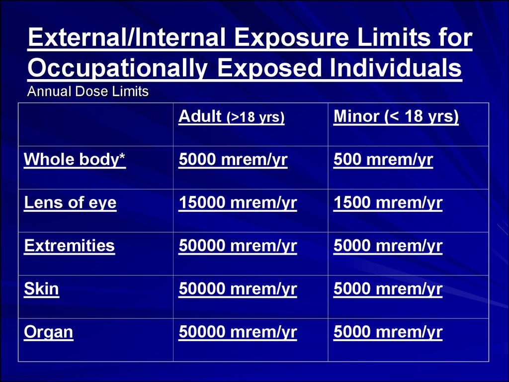
External/Internal Exposure Limits forOccupationally Exposed Individuals
Annual Dose Limits
Adult (>18 yrs)
Minor (< 18 yrs)
Whole body*
5000 mrem/yr
500 mrem/yr
Lens of eye
15000 mrem/yr
1500 mrem/yr
Extremities
50000 mrem/yr
5000 mrem/yr
Skin
50000 mrem/yr
5000 mrem/yr
Organ
50000 mrem/yr
5000 mrem/yr
42.
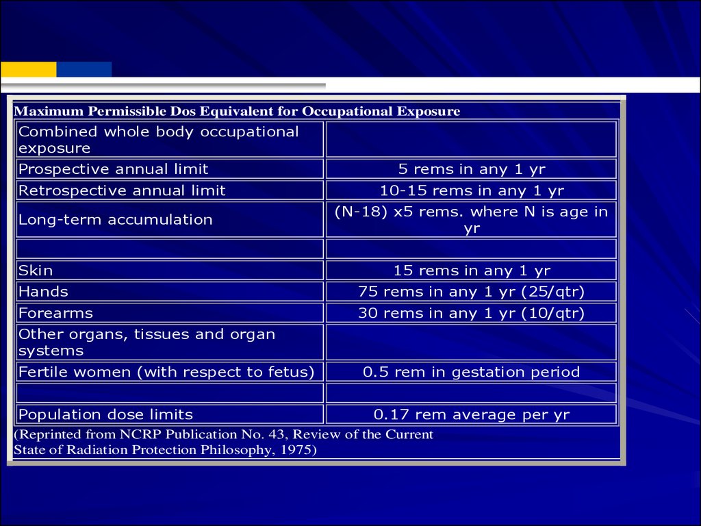
Maximum Permissible Dos Equivalent for Occupational ExposureCombined whole body occupational
exposure
Prospective annual limit
Retrospective annual limit
Long-term accumulation
Skin
5 rems in any 1 yr
10-15 rems in any 1 yr
(N-18) x5 rems. where N is age in
yr
15 rems in any 1 yr
Hands
75 rems in any 1 yr (25/qtr)
Forearms
30 rems in any 1 yr (10/qtr)
Other organs, tissues and organ
systems
Fertile women (with respect to fetus)
0.5 rem in gestation period
Population dose limits
0.17 rem average per yr
(Reprinted from NCRP Publication No. 43, Review of the Current
State of Radiation Protection Philosophy, 1975)
43. Community Emergency Radiation
Hazardous Waste Sites:Radiation above background (0.01-0.02 m
rem/hr) signifies possible presence which
must be monitored. Radiation above 2 m
rem/hr indicates potential hazard.
Evacuate site until controlled.
44.
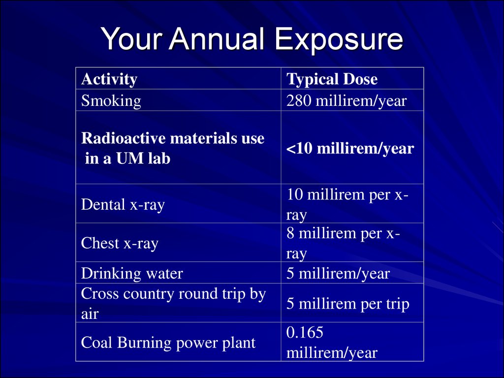
Your Annual ExposureActivity
Smoking
Typical Dose
280 millirem/year
Radioactive materials use
in a UM lab
<10 millirem/year
Dental x-ray
Chest x-ray
Drinking water
Cross country round trip by
air
Coal Burning power plant
10 millirem per xray
8 millirem per xray
5 millirem/year
5 millirem per trip
0.165
millirem/year
45.
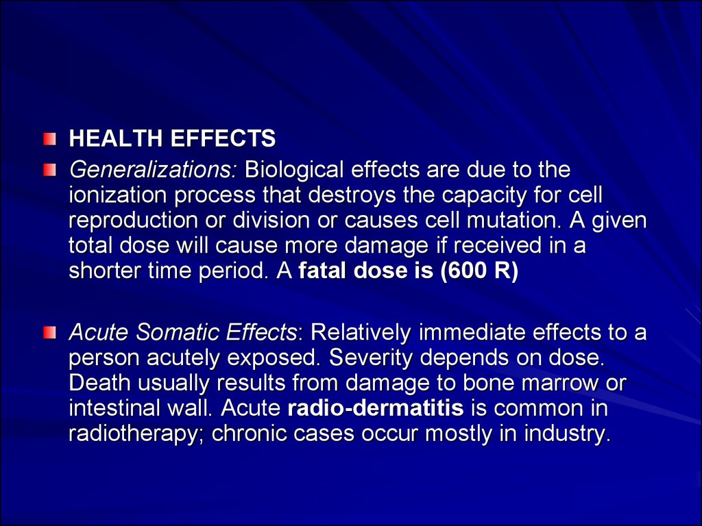
HEALTH EFFECTSGeneralizations: Biological effects are due to the
ionization process that destroys the capacity for cell
reproduction or division or causes cell mutation. A given
total dose will cause more damage if received in a
shorter time period. A fatal dose is (600 R)
Acute Somatic Effects: Relatively immediate effects to a
person acutely exposed. Severity depends on dose.
Death usually results from damage to bone marrow or
intestinal wall. Acute radio-dermatitis is common in
radiotherapy; chronic cases occur mostly in industry.
46.
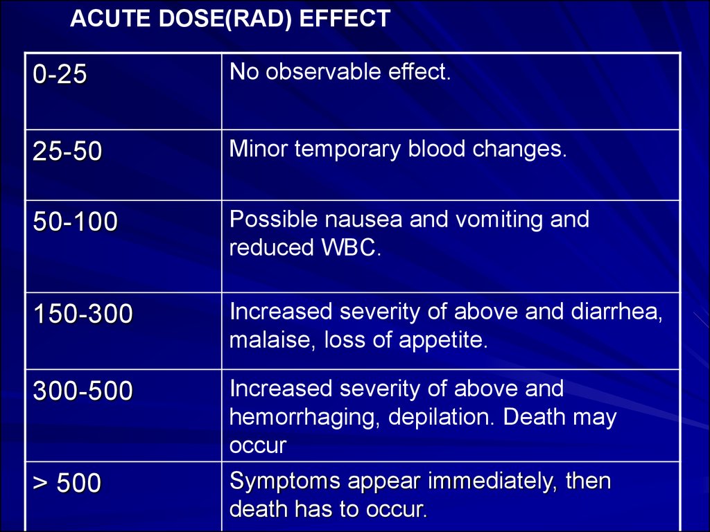
ACUTE DOSE(RAD) EFFECT0-25
No observable effect.
25-50
Minor temporary blood changes.
50-100
Possible nausea and vomiting and
reduced WBC.
150-300
Increased severity of above and diarrhea,
malaise, loss of appetite.
300-500
Increased severity of above and
hemorrhaging, depilation. Death may
occur
Symptoms appear immediately, then
death has to occur.
> 500
47.
Delayed Somatic Effects: Delayed effects to exposedperson include: Cancer, leukemia, cataracts, life
shortening from organ failure, and abortion.
Probability of an effect is proportional to dose (no
threshold). Severity is independent of dose. Doubling
dose for cancer is approximately 10-100 rems.
Genetic Effects: Genetic effects to off-spring of
exposed persons are irreversible and nearly always
harmful. Doubling dose for mutation rate is
approximately 50-80 rems. (Spontaneous mutation
rate is approx. 10-100 mutations per million
population per generation.)
48.
Critical Organs: Organs generally mostsusceptible to radiation damage include:
Lymphocytes, bone marrow, gastro-intestinal,
gonads, and other fast-growing cells. The
central nervous system is relatively resistant.
Many nuclides concentrate in certain organs
rather than being uniformly distributed over the
body, and the organs may be particularly
sensitive to radiation damage, e.g., isotopes of
iodine concentrate in the thyroid gland. These
organs are considered "critical" for the specific
nuclide.
49. Non-ionizing Radiation
Definition:“ They are electromagnetic waves incapable
of producing ions while passing through
matter, due to their lower energy.”
50.
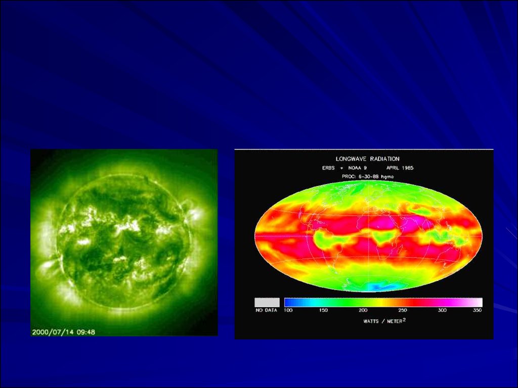
– All earth surface system components emit radiation--the sun and the earth are the components we aremost interested in
– The sun emits radiation composed of high energy
infrared radiation, visible light, and ultraviolet radiation
collectively known as shortwave radiation (SW)
– The earth emits radiation composed of lower energy
infrared radiation collectively known as long-wave
radiation (LW)
51.
52.
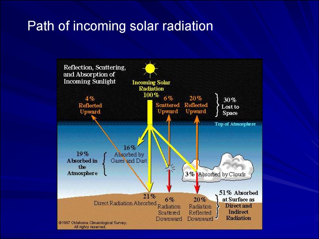
Path of incoming solar radiation53.
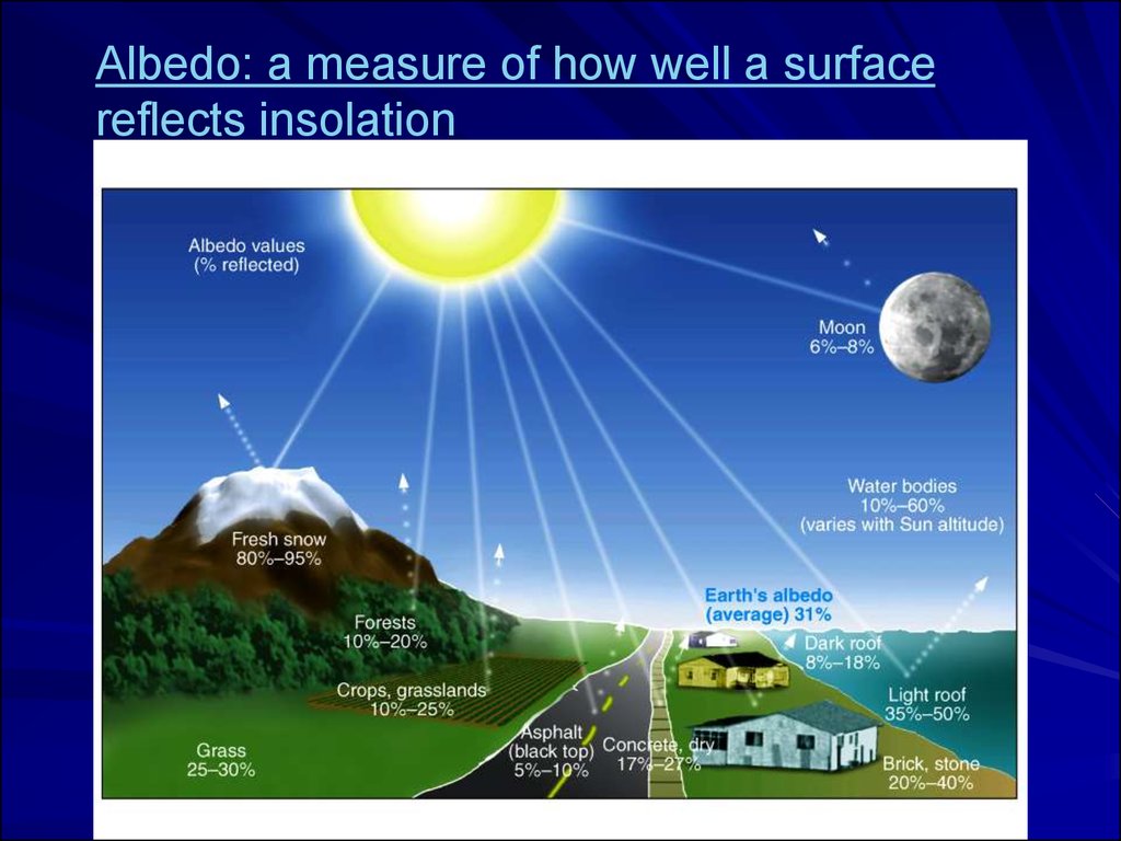
Albedo: a measure of how well a surfacereflects insolation
54.
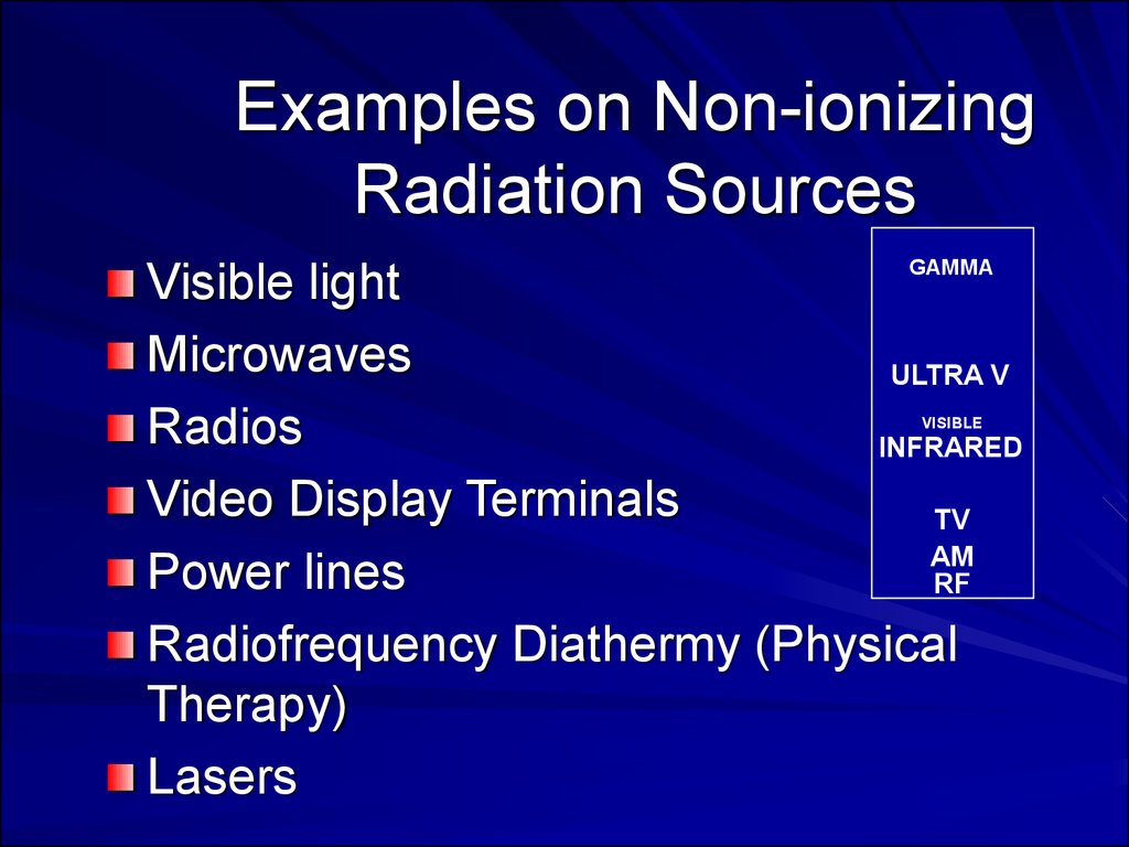
Examples on Non-ionizingRadiation Sources
GAMMA
Visible light
Microwaves
ULTRA V
Radios
INFRARED
Video Display Terminals
TV
AM
Power lines
RF
Radiofrequency Diathermy (Physical
Therapy)
Lasers
VISIBLE
MICROWAVE
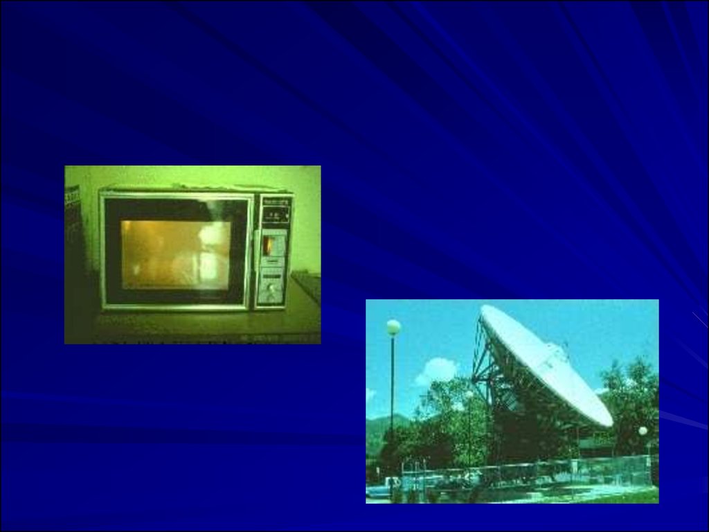
55. Other Manmade Sources of Non-Ionizing Radiation
Other Manmade Sources of NonIonizing Radiation56.
57.
58. Effects
Radiofrequency Ranges (10 kHz to 300 GHz)–
–
–
–
Effects only possible at ten times the permissible
exposure limit
Heating of the body (thermal effect)
Cataracts
Some studies show effects of teratoginicity and
carcinogenicity.
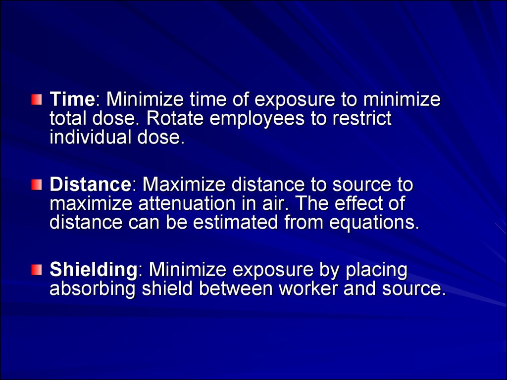
59. RADIATION CONTROLS
A. Basic Control Methods for ExternalRadiation
Decrease Time
Increase Distance
Increase Shielding
60.
Time: Minimize time of exposure to minimizetotal dose. Rotate employees to restrict
individual dose.
Distance: Maximize distance to source to
maximize attenuation in air. The effect of
distance can be estimated from equations.
Shielding: Minimize exposure by placing
absorbing shield between worker and source.
61.
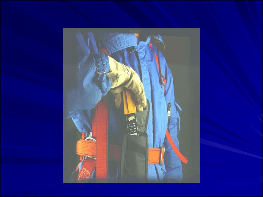
62. B. Monitoring
Personal Dosimeters: Normally they donot prevent exposures (no alarm), just
record it. They can provide a record of
accumulated exposure for an individual
worker over extended periods of time
(hours, days or weeks), and are small
enough for measuring localized exposures
Common types: Film badges;
Thermoluminescence detectors (TLD);
and pocket dosimeters.
63.
64.
65.
66.
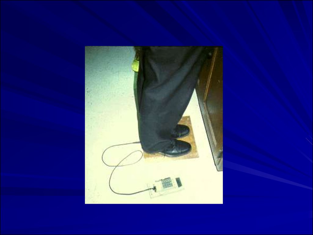
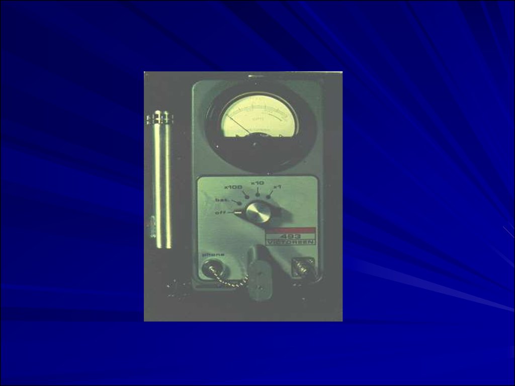
Direct Reading Survey Meters and Counters: Useful inidentifying source of exposures recorded by personal
dosimeters, and in evaluating potential sources, such as
surface or sample contamination, source leakage,
inadequate decontamination procedures, background
radiation.
Common types:
Alpha Proportional or Scintillation counters
Beta, gamma Geiger-Mueller or Proportional
counters
X-ray, Gamma Ionization chambers
Neutrons Proportional counters
67.
68.
Continuous Monitors: Continuous direct readingionization detectors (same detectors as above)
can provide read-out and/or alarm to monitor
hazardous locations and alert workers to
leakage, thereby preventing exposures.
Long-Term Samplers: Used to measure average
exposures over a longer time period. For
example, charcoal canisters or electrets are set
out for days to months to measure radon in
basements (should be <4 pCi/L).
69. Elements of Radiation Protection Program
Monitoring of exposures: Personal, area, and screeningmeasurements; Medical/biologic monitoring.
Task-Specific Procedures and Controls: Initial, periodic,
and post-maintenance or other non-scheduled events.
Engineering (shielding) vs. PPE vs. administrative
controls. Including management and employee
commitment and authority to enforce procedures and
controls.
Emergency procedures: Response, "clean-up", post
clean-up testing and spill control.
Training and Hazard Communications including signs,
warning lights, lockout/tagout, etc. Criteria for need,
design, and information given.
Material Handling: Receiving, inventory control, storage,
and disposal.