


 Биология
Похожие презентации:


History of Microbiology. Classification of Microorganisms. Morphology and Structure of Bacteria, Fungi, Spirochetes, Chlamydia
1. History of Microbiology. Classification of Microorganisms. Morphology and Structure of Bacteria, Fungi, Spirochetes, Chlamydia,
Rickettsia.Department of Microbiology, Immunology and Virology.
2. Historical Introduction
Antony vanLeeuwenhoek
John Tyndall
Louis Pasteur
First to observe live microorganisms, using a simple
microscope (1685)
Developed tyndallization to destroy spores (1660)
Disproved the theory of spontaneous generation (1861)
Contributed to the understanding of fermentation (1858)
Developed technique for selective destruction of the microorganisms
(pasteurization) (1866). Study of bacterial contamination of wine (1866)
and diseases of silkworms (1868). Attenuated vaccines for anthrax (1881)
and chicken cholera. Immunization against rabies (1885)
Joseph Lister
Contributed to concept of aseptic technique (1865-1870)
Robert Koch
Developed postulates for proving the cause of infectious diseases (1884)
and pure culture concept. Observed anthrax bacilli (1876). Developed solid
culture media (1882). Discovered organisms causing tuberculosis (1882)
Paul Ehrlich
Formulated humoral theory of resistance. Developed new staining
techniques. Developed first chemotherapeutic agent (1890s to 1900)
Elie Metchnikoff Formulated cellular theory of resistance (1890s)
3. Definition of Microbiology
Medical microbiology is the study of microbes that infect humans,the diseases they cause, their diagnosis, prevention measures, aseptic techniques,
treatment of infectious diseases, immunology, and production of vaccines to protect against
infectious diseases.
4. Classification of Microorganisms
Scientific nomenclature includes a hierarchial scheme. The lower down in thesystem the more specific or narrowly defined is the group.
Kingdom
Phylum
Class
Order
Family
Genus
Species
Species- is fundamental unit as outlined above the concept is that all bacteria,
which share a specific set of defined properties, belong to a particular species.
Classification of Bacteria is based on Gram staining characteristic, morphology,
and metabolism type. Bergey’s Manual of Systematic Bacteriology is the bible of
bacterial taxonomy.
Classification of Viruses is based on nucleic acid type, host organism, and
morphology.
5. Family Tree of Microorganisms
ProtozoaFungi
Bacteria
Algae
Cyanobacteria
Archaebacteria
Eukaryotes
Prokaryotes
Primitive Cells
6. Differences between Prokaryotic and Eukaryotic Cells
StructureNucleus
Nuclear membrane
Nucleus
Chromosome
Deoxyribonucleprotein
Division
Prokaryotes
Eukaryotes
Absent
Absent
One
Absent
By binary fission
Present
Present
More than one
Present
By mitosis
All are absent
All are present
Absent
Present
Present
Absent
Cytoplasm
Mitochondria, Golgi apparatus,
Lysosomes, Pinocytosis,
Endoplasmic reticulum
Chemical composition
Sterols
Muramic acid
7. Prokaryotic Cell Structure
Prokaryotes are unicellular organisms of relatively simple construction.A prokaryotic cell has five essential structural components: a genome (DNA),
ribosomes, cell membrane, cell wall, and some sort of surface layer.
Structurally a prokaryotic cell has three architectural regions: appendages
(attachment to the cell surface) in the form of flagella and pili (or fimbriae);
a cell envelope consisting of a capsule, cell wall and plasma membrane;
and a cytoplasmic region that contains the cell genome (DNA) and
ribosomes and various sort of inclusions.
8. Cell structure
9. Characteristic of typical bacterial cell structures
SturctureFunction(s)
Flagella
Pili
Sex pilus
Common pili or
Fimbriae
Swimming movement
Mediates DNA transfer during conjugation
Attachment to surfaces; protection against
Phagotrophic engulfment
Predominant chemical composition
Protein
Protein
Protein
Capsules (includes Attachment to surfaces; protection against
Usually polysaccharide; occasionally
“slime layers” and phagocytic engulfment, occasionally killing
polypeptide
glycocalyx)
or digestion; reserve of nutrients or protection
against desiccation
Cell wall
Gram-positive
Prevents osmotic lysis of cell protoplast and
Peptidoglycan (murein) complexed
bacteria
confers rigidity and shape on cells
with teichoic acids
Gram-negative
bacteria
Peptidoglycan prevents osmotic lysis and
Peptidoglycan (murein) surrounded
confers rigidity and shape; outer membrane is
by phospholipid proteinpermiability barrier; associated LPS and proteins lipopolysacharide “outer membrane”
have various functions
10.
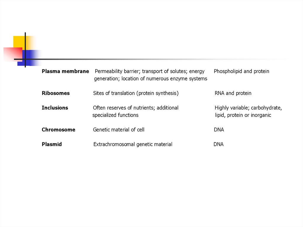
Plasma membranePermeability barrier; transport of solutes; energy
generation; location of numerous enzyme systems
Phospholipid and protein
Ribosomes
Sites of translation (protein synthesis)
RNA and protein
Inclusions
Often reserves of nutrients; additional
specialized functions
Highly variable; carbohydrate,
lipid, protein or inorganic
Chromosome
Genetic material of cell
DNA
Plasmid
Extrachromosomal genetic material
DNA
11. Appendages
Flagella-are filamentous protein structures attachedto the cell surface that provide the swimming
movement for most motile prokaryotes.
The diameter is about 20 nanometers.
The flagellar apparatus consists of several
distinct proteins: a system of rings
embedded in the cell envelope (the basal
body), a hook-like structure near the cell
surface, and the flagellar filament.
The innermost rings, the M and S rings located
in the plasma membrane, comprise the motor
apparatus.
The outermost rings, the P and L rings, located
in the periplasm, function as bushings to
support the rod where it is joined to the hook of
the filament on the cell surface.
12.
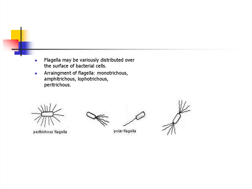
Flagella may be variously distributed overthe surface of bacterial cells.
Arraingment of flagella: monotrichous,
amphitrichous, lophotrichous,
peritrichous.
13. Detecting Bacterial Motility
Flagellar stains (show their pattern ofdistribution)
Motility test medium demonstrates if cells
can swim in a semisolid medium (Proteus)
Direct microscopic observation of living
bacteria in a wet mount
Dark ground Illumination
Electron microscopy
14. Fimbriae
Fimbriae and pili are short, hair-like structuresthey are composed of protein
shorter, stiffer, smaller in diameter
very common in Gram-negative bacteria, but occur in
some archaea and Gram-positive bacterias
involved in adherence of bacteria to surfaces, substrates
and other cells or tissues in nature
F or sex pilus, specialized type of pilus in E.coli mediates the
transfer of DNA between mating bacteria during the
process of conjugation,
Common pili (almost always called fimbriae)
usually involved in specific attachment of prokaryotes to
surface in nature
they are major determinants of bacterial virulence: they
allow pathogens to attach to (colonize) tissues, resist
attack by phagocytic white blood cells
Col I (colicin) pili
15. The Cell Envelope
The cell envelope consists:plasma membrane
a cell wall
a capsule
16. Capsules
Polysaccharide layer outside of the cell wall polymer17.
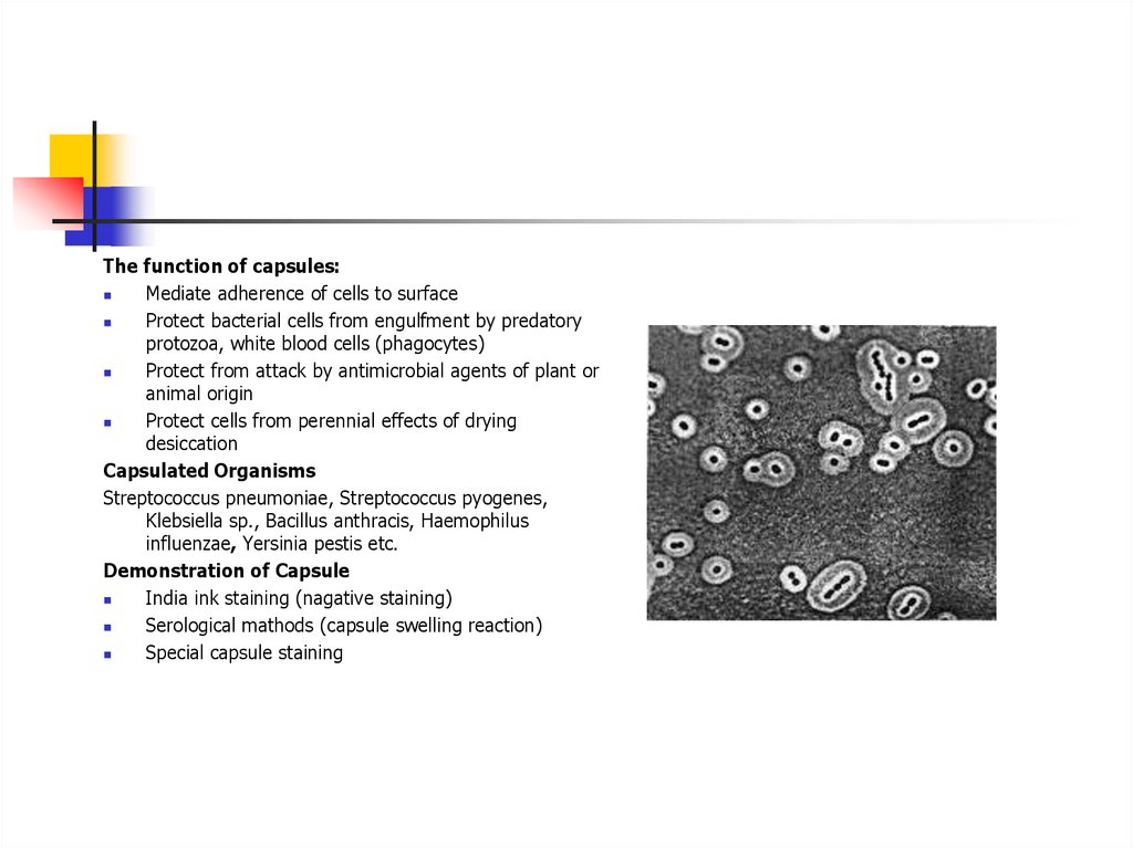
The function of capsules:Mediate adherence of cells to surface
Protect bacterial cells from engulfment by predatory
protozoa, white blood cells (phagocytes)
Protect from attack by antimicrobial agents of plant or
animal origin
Protect cells from perennial effects of drying
desiccation
Capsulated Organisms
Streptococcus pneumoniae, Streptococcus pyogenes,
Klebsiella sp., Bacillus anthracis, Haemophilus
influenzae, Yersinia pestis etc.
Demonstration of Capsule
India ink staining (nagative staining)
Serological mathods (capsule swelling reaction)
Special capsule staining
18. Cell Wall
is essential rigid structure for viability (protection cell protoplasm from mechanicaldamage and osmotic rupture or lysis)
composed of unique components found nowhere else in nature
one of the most important sites for attack by antibiotics
provide ligands for adherence and receptor sites for drugs or viruses
cause symptoms of disease in animals
provide for immunological distinction and immunological variation among strains of
bacteria
It is 10-25 nm in thickness and weighs about 20-25% of the dry weight cell wall.
19.
Cell wall structurecontains a unique type of peptidoglycan called murein- (N-acetylmuramic acid)
By cell wall structure there are two groups of bacterias
Gram-Positive Cell Envelope(15-80nm)
consists in two or three layers:
cytoplasmic membrane,
a thick peptidoglycan layer,
and outer layer or, capsule or,
glycoprotein (S-layer)
Gram-Negative Cell Envelope (10nm)
cytoplasmic membrane (inner membrane),
single planar sheet of peptidoglycan,
outer membrane contains a unique component
lipopolysaccharide (LPS or endotoxin),
the space between inner and outer membrane is
the periplasmic space
20. Bacteria with Defective Cell Wall
The synthesis of cell wall may be inhibited or interfered by many factors such as,antibiotics, bacteriphages, and lysozyme.
Mycoplasma: This is a naturally occuring bacteria without cell walls. They don’t require
hypertonic environment for maintenance and are stable in culture medium
L-forms: L-forms develop either spontaneously or in the presence of penicillin or other
agents that interfere with synthesis of cell wall. These are difficult to cultivate and require
agar containing solid medium having right osmotic strength. L-forms are more stable than
protoplasts and spheroplasts
Protoplasts: These are derived from Gram positive bacteria. They contain cytoplasmic
membrane and cell wall is totally lacking. These are produced artificially by lysozyme in a
hypertonic medium. These are unstable.
Spheroplasts: These are derived from Gram positive bacteria. They are produced in
presence of penicillin. They are osmotically fragile and must be maintained in hypertonic
culture medium. They differ from protoplast in that some cell wall material retained.
21. The Plasma Membrane
Functions of the prokaryotic plasma membrane.1. Osmotic or permeability barrier
2. Location of transport systems for specific solutes (nutrients and ions)
3. Energy generating functions, involving respiratory and photosynthetic electron transport
systems, establishment of proton motive force, and transmembranous, ATP-synthesizing
ATPase
4. Synthesis of membrane lipids (including lipopolysaccharide in Gram-negative cells)
5. Synthesis of murein (cell wall peptidoglycan)
6. Assembly and secretion of extracytoplasmic proteins
7. Coordination of DNA replication and segregation with septum formation and cell division
8. Chemotaxis (both motility per se and sensing functions)
9. Location of specialized enzyme system
22.
It is 5-10 nm thick elastic semipermiable layer which lies beneath the cell wall separating itfrom the cell cytoplasm.
The plasma membrane of procaryotes may invaginate into the cytoplasm or form stacks or
vesicles attached to the inner membrane surface. These structures are sometimes referred
to as mesosomes Such internal membrane systems may be analogous to the cristae of
mitochondria or the thylakoids of chloroplasts which increase the surface area of
membranes to which enzymes are bound for specific enzymatic functions
They are the principal centers of respiratory enzyme
Mesosomes may also represent specialized membrane regions involved in DNA replication
and segregation, cell wall synthesis, or increased enzymatic activity.
There are two types of mesosomes- septal and lateral. The septal mesosome attached to
bacterial chromosome and is involved in DNA segregation and in the formation of crosswalls during binary fission.
23. The Cytoplasm
The bacterial cytoplasm is a colloidal system containing a variety of organic and inorganicsolutes in a viscous watery solution.
The cytoplasmic constituents of procaryotic cells invariably include the procaryotic
chromosome and ribosomes.
The chromosome is typically one large circular molecule of DNA, more or less free in the
cytoplasm.
Procaryotes sometimes possess smaller extrachromosomal pieces of DNA called plasmids.
The total DNA content of a procaryote is referred to as the cell genome.
The distinct granular appearance of procaryotic cytoplasm is due to the presence and
distribution of ribosomes, procaryotic ribosomes are 70S in size
24. Nucleus
Bacterial nucleus has no nuclear membrane or nucleolusThe genomic DNA is double stranded in the form of a circle.
It measures about 1mm (1000µm) when straightened
The bacterial DNA is haploid, replicates by simple fission and maintains bacterial genetic
characteristic
Plasmids
Some bacteria may possess extranuclear genetic material in the cytoplasm consisting of
DNA named as plasmids or episomes
The plasmid replicates autonomously.
They are not essential for the life of the cell, but may confer on the bacteria certain
properties, such as drug resistance and toxigenecity which constitute a survival advantage
to the bacteria.
These plasmids can be transmitted from one bacterium to another. either by conjugation or
by the agency of bacteriophage.
Plasmids also may be transferred to daughter cells during cell division.
25. Inclusions
Often contained in the cytoplasm of prokaryotic cells is one or another of some type ofinclusion granule. Inclusions are distinct granules that may occupy a substantial part of
the cytoplasm
Inclusion granules are usually reserve materials of some sort
Many bacteria accumulate granules of polyphosphate which can be used in the synthesis
of ATP
These granules are termed volutin garnules or metachromatic granules
They are characteristic features of the corynebacteria
They can be stained
26. Endospores
A bacterial structure sometimes observed as aninclusion is actually a type of dormant cell called
an endospore.
Endospores are formed by a few groups of
Bacteria as intracellular structures
Highly resistant to environmental stresses
Endospores are formed by vegetative cells in
response to environmental signals that indicate
a limiting factor for vegetative growth
Under appropriate environmental conditions,
they germinate back into vegetative cells.
There are eight stages, O,I-VII, in the
sporulation cycle of a Bacillus species, and the
process takes about eight hours.
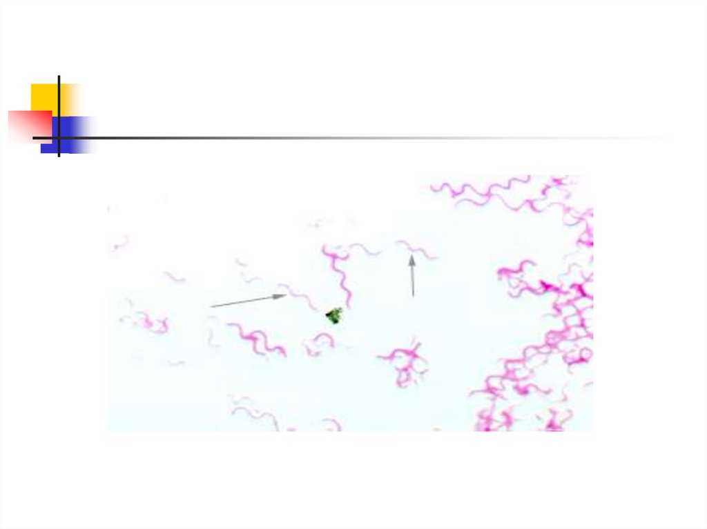
27. Morphology of the Spirochetes
The spirochetesLong
Thin
Corkscrewlike
Gram-negative
Anaerobic bacteria
There are three families are
pathogen for human:
Leptospira,Treponema, and Borrelia
28.
29.
The spirochetes - very difficult to cultureThis is due to their extreme anaerobic requirements their unique nutritional
requirements (require 1-globulin)
Over the last decade or so, some have been cultured and their characteristics
determined
But many remain uncultured
Because they were so hard to grow in culture, their differentiation was based
primarily on size and other morphological characteristics Three sizes were seen,
giving rise to the categories: small, intermediate, and large
30. Chlamydia
Chlamydia are obligate intracellular bacteria that multiply in host cellsThere are three species associated with human disease: C. psittaci, C. trachomatis, and C.
pneumoniae.
Chlamydia are small rounded organisms that vary in morphology during their replicative
cycle.
Chlamydiae are not culturable on synthetic media.
The replicative cycle of Chlamydia involves two forms, the elementary body and the
reticulate body.
The elementary body represents the infectious form, and is resistant to environmental
stresses. The elementary body is taken up by the host cells by endocytosis to form a
phagosome.
Within 8-12 hours, the elementary body reorganizes to the larger reticulate body, with
division by binary fission until the entire cell is filled with the organisms.
31.
A transmission electron microscope picture of athin section through an elementary body of C.
psittaci
Reticulate bodies of C. Psittaci
32. Mycoplasma
Mycoplasma are bacteria that lacks cell walls.Two human species are associated with disease: M. pneumoniae (pneumonia) and M.
hominus associated with genital tract infections.
The bacteria are very small (0.2
M) but pleomorphic.
They are bounded by a single triple layered membrane that contains sterols. They do not
stain well with usual stains.
Organisms can grow on enriched liquid culture medium and Mycoplasma agar to give tiny
colonies after several days, with a denser center appearance like an inverted fried egg.
33. Rickettsia
The rickettsia are bacteria which are obligate intracellular parasites.They are considered a separate group of bacteria because they have the common feature
of being spread by arthropod vectors (lice, fleas, mites and ticks).
The cells are extremely small (0.25 u in diameter) rod-shaped, coccoid and often
pleomorphic microorganisms
They have typical bacterial cell walls, no flagella (except for Rickettsia prowazekii), are
gram-negative and multiply via binary fission only inside host cells.
They occur singly, in pairs, or in strands.
Most species are found only in the cytoplasm of host cells, but those which cause spotted
fevers multiply in nuclei as well as in cytoplasm.
In the laboratory, they may be cultivated in living tissues such as embryonated chicken
eggs or vertebrate cell cultures.
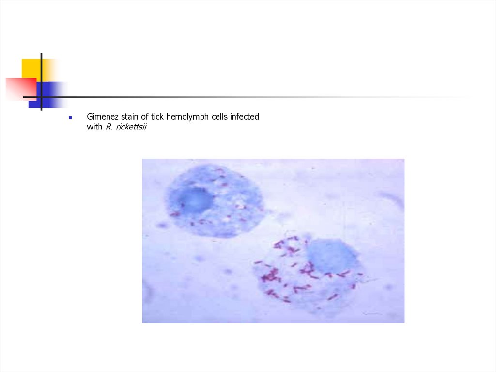
34.
Gimenez stain of tick hemolymph cells infectedwith R. rickettsii
35. Fungi
Fungi are eukaryotic organismsInclude mushrooms, molds and yeasts
They have no chlorophyll or other photosynthetic pigments
Their cell walls contain a substance called chitin
Fungal infections are mycoses
Growth in two basic forms, as yeasts and molds
They growth in synthetic mediums