


 Биология
Похожие презентации:

")


")
Introduction to Viruses. Chapter 26
1.
Chapter 26Introduction to
Viruses
© 2018 Pearson Education Ltd.
Lecture Presentations by
Nicole Tunbridge and
Kathleen Fitzpatrick
2.
A Borrowed LifeA virus is an infectious particle consisting of genes
packaged in a protein coat
Viruses are much simpler in structure than even
prokaryotic cells
Viruses cannot reproduce or carry out metabolism
outside of a host cell
Viruses exist in a shady area between life-forms and
chemicals, leading a kind of “borrowed life”
3.
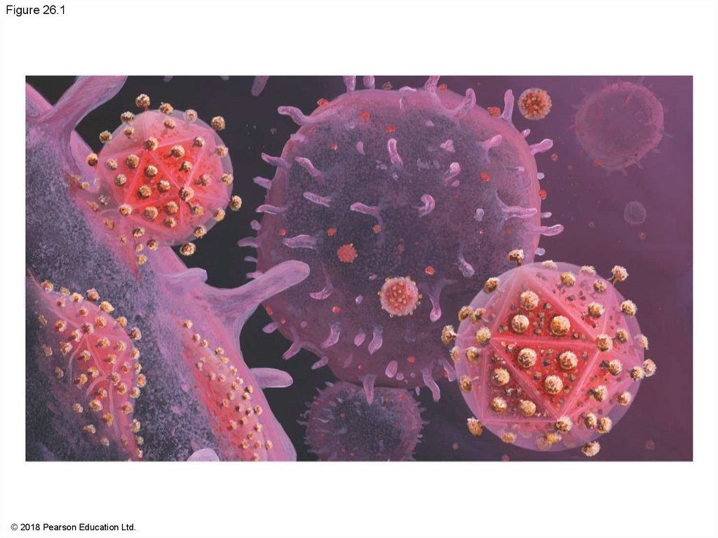
Figure 26.1© 2018 Pearson Education Ltd.
4.
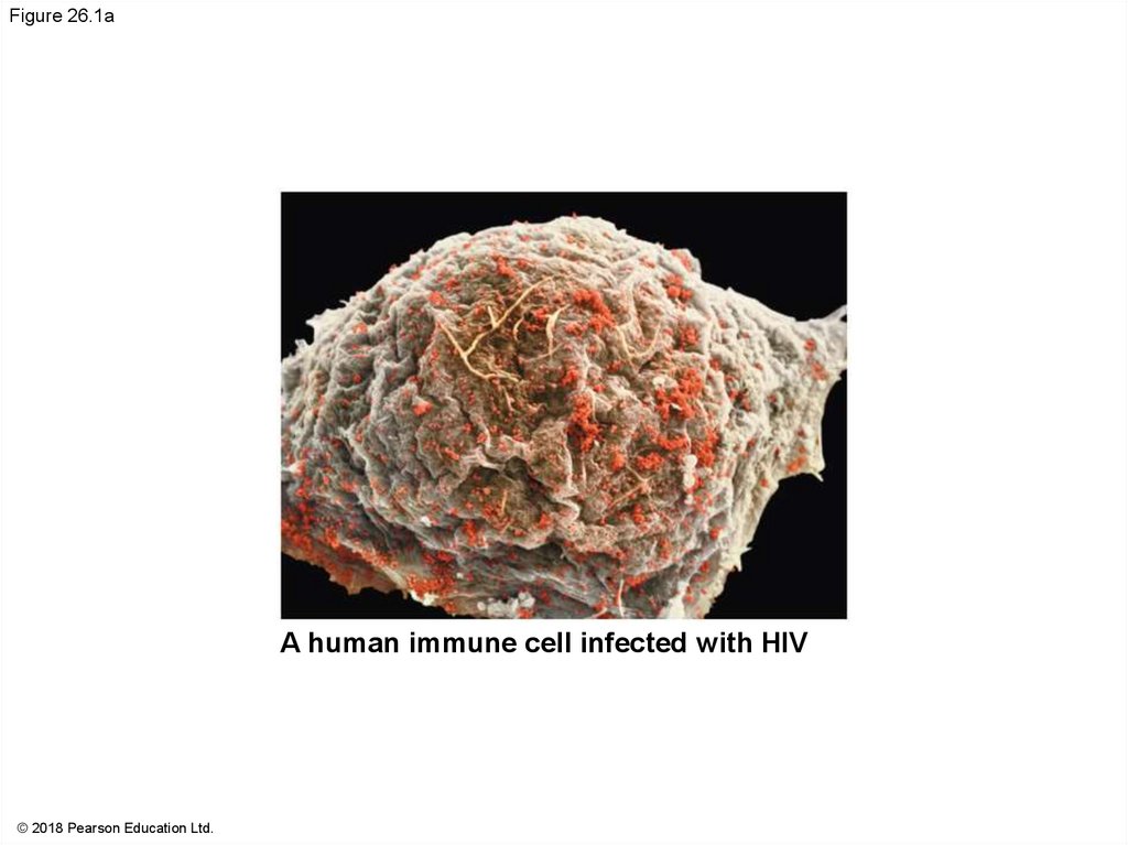
Figure 26.1aA human immune cell infected with HIV
© 2018 Pearson Education Ltd.
5.
Concept 26.1: A virus consists of a nucleic acidsurrounded by a protein coat
Viruses were detected indirectly long before they
were actually seen
6.
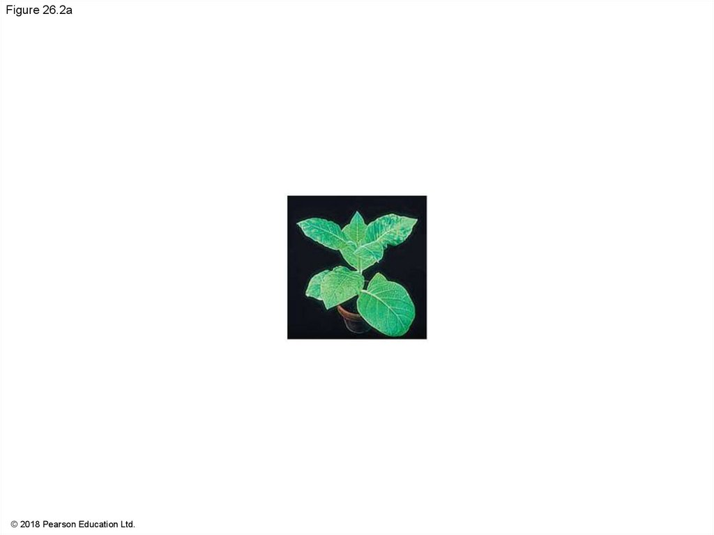
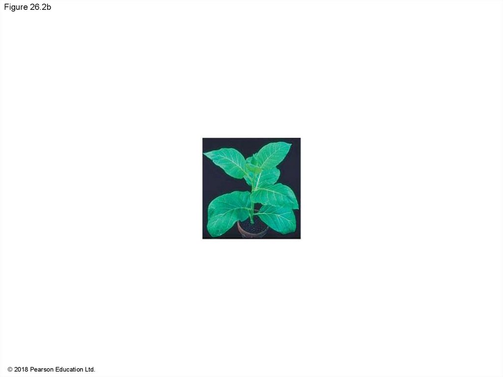
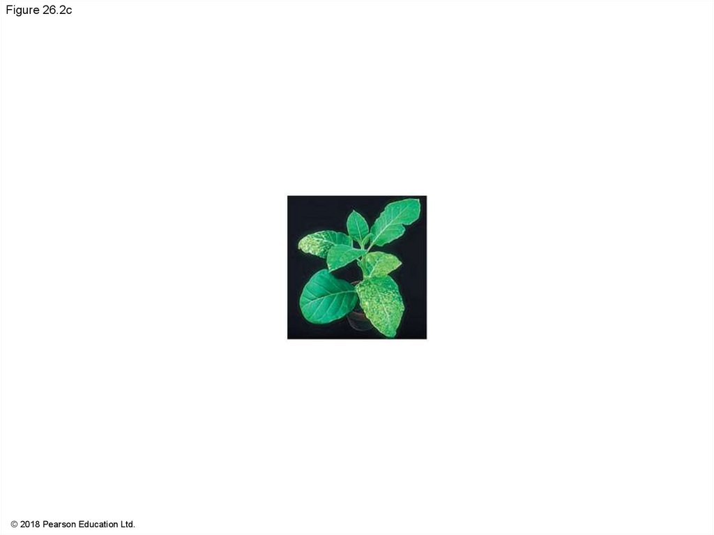
The Discovery of Viruses: Scientific InquiryTobacco mosaic disease stunts growth of tobacco
plants and gives their leaves a mosaic coloration
In the late 1800s, researchers hypothesized that
unusually small bacteria might be responsible
Later work suggested that the infectious agent did
not share features with bacteria (such as the ability
to grow on nutrient media)
In 1935, Wendell Stanley confirmed this latter
hypothesis by crystallizing the infectious particle,
now known as tobacco mosaic virus (TMV)
7.
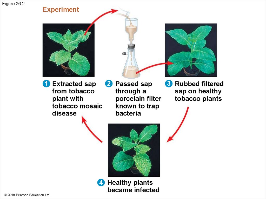
Figure 26.2Experiment
2 Passed sap
1 Extracted sap
3 Rubbed filtered
from tobacco
through a
sap on healthy
plant with
porcelain filter
tobacco plants
tobacco mosaic
known to trap
disease
bacteria
© 2018 Pearson Education Ltd.
4 Healthy plants
became infected
8.
Figure 26.2a© 2018 Pearson Education Ltd.
9.
Figure 26.2b© 2018 Pearson Education Ltd.
10.
Figure 26.2c© 2018 Pearson Education Ltd.
11.
Structure of VirusesViruses are not cells
A virus is a very small infectious particle consisting
of nucleic acid enclosed in a protein coat and, in
some cases, a membranous envelope
12.
Viral GenomesViral genomes may consist of either
double- or single-stranded DNA or
double- or single-stranded RNA
Viruses are classified as DNA viruses or RNA
viruses
The genome is either a single linear or circular
molecule of the nucleic acid
Viruses have between three and 2,000 genes in their
genome
13.
Capsids and EnvelopesA capsid is the protein shell that encloses the viral
genome
Capsids are built from protein subunits called
capsomeres
A capsid can have a variety of structures; associated
viruses may be referred to as helical or icosahedral
viruses
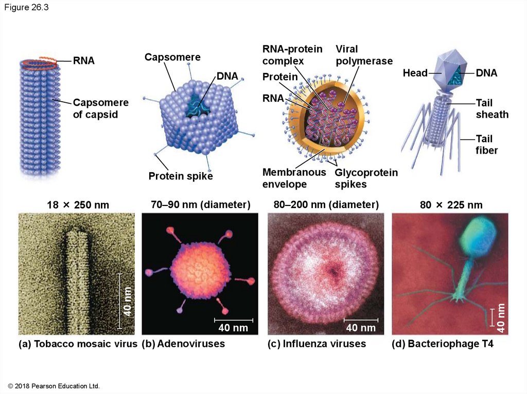
14.
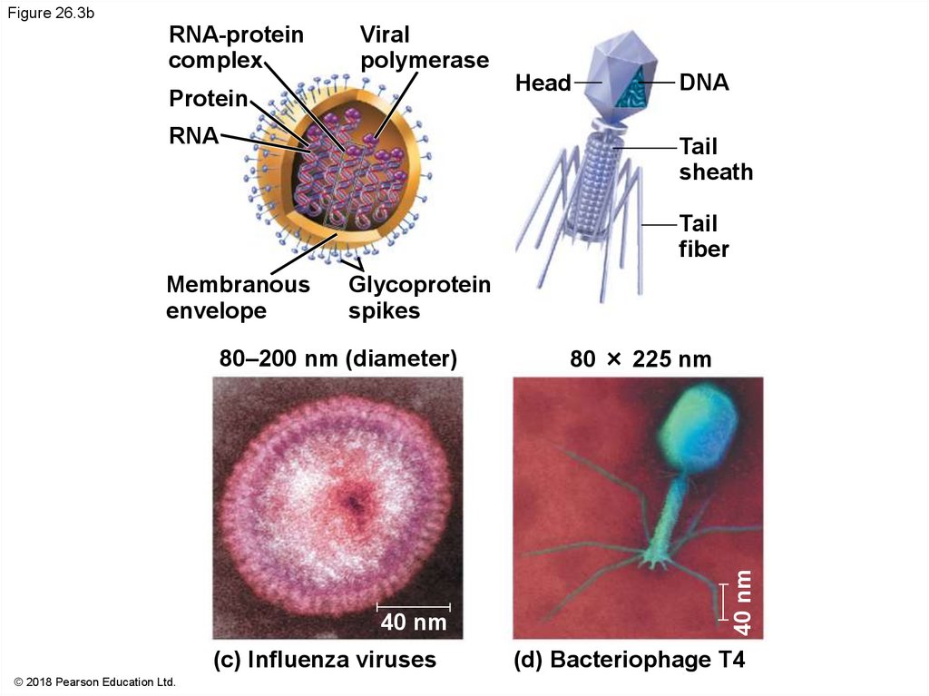
Figure 26.3RNA-protein
complex
Capsomere
RNA
DNA
Viral
polymerase
Head
Protein
RNA
Capsomere
of capsid
DNA
Tail
sheath
Tail
fiber
Membranous Glycoprotein
envelope
spikes
Protein spike
70–90 nm (diameter)
80–200 nm (diameter)
40 nm
40 nm
80 × 225 nm
(a) Tobacco mosaic virus (b) Adenoviruses
© 2018 Pearson Education Ltd.
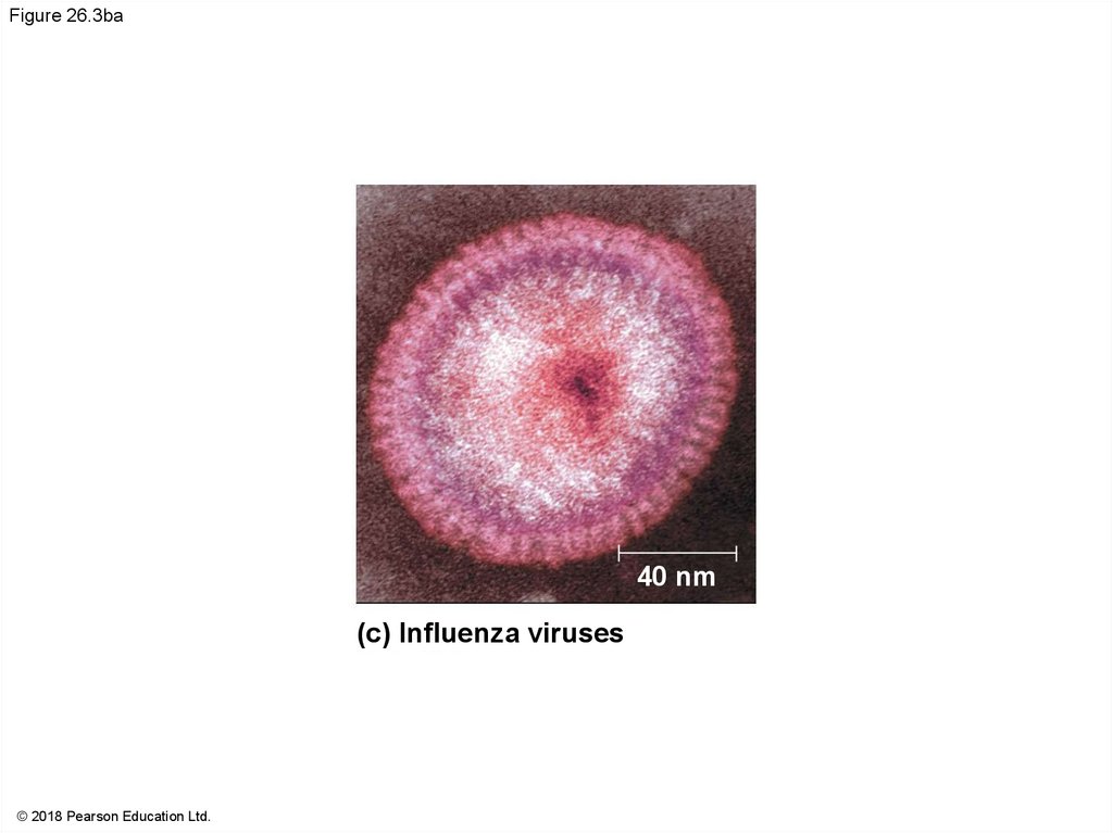
(c) Influenza viruses
40 nm
40 nm
18 × 250 nm
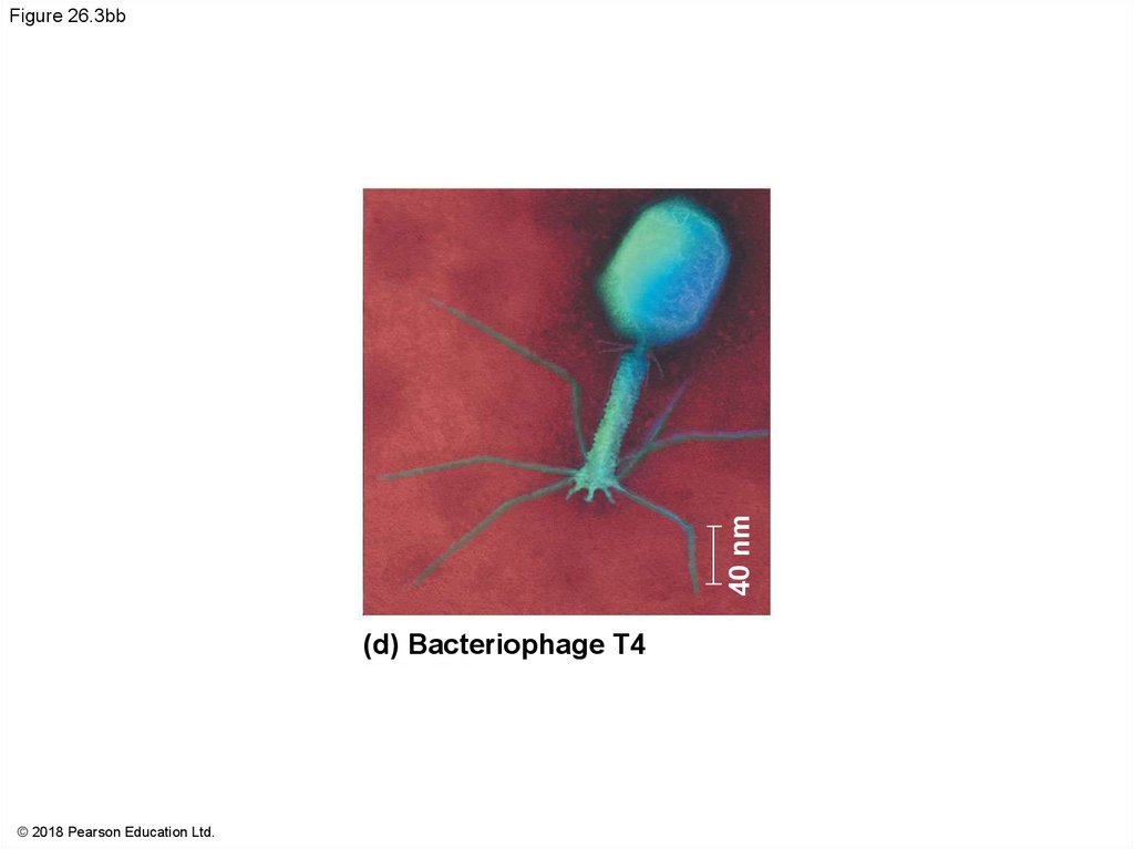
(d) Bacteriophage T4
15.
Figure 26.3aCapsomere
RNA
DNA
Capsomere
of capsid
Protein spike
18 × 250 nm
40 nm
70–90 nm (diameter)
40 nm
© 2018 Pearson Education Ltd.
(a) Tobacco mosaic
virus
(b) Adenoviruses
16.
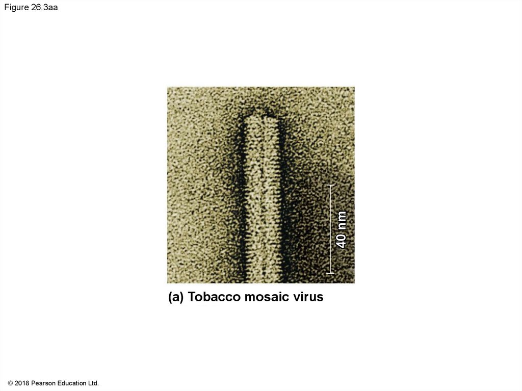
40 nmFigure 26.3aa
(a) Tobacco mosaic virus
© 2018 Pearson Education Ltd.
17.
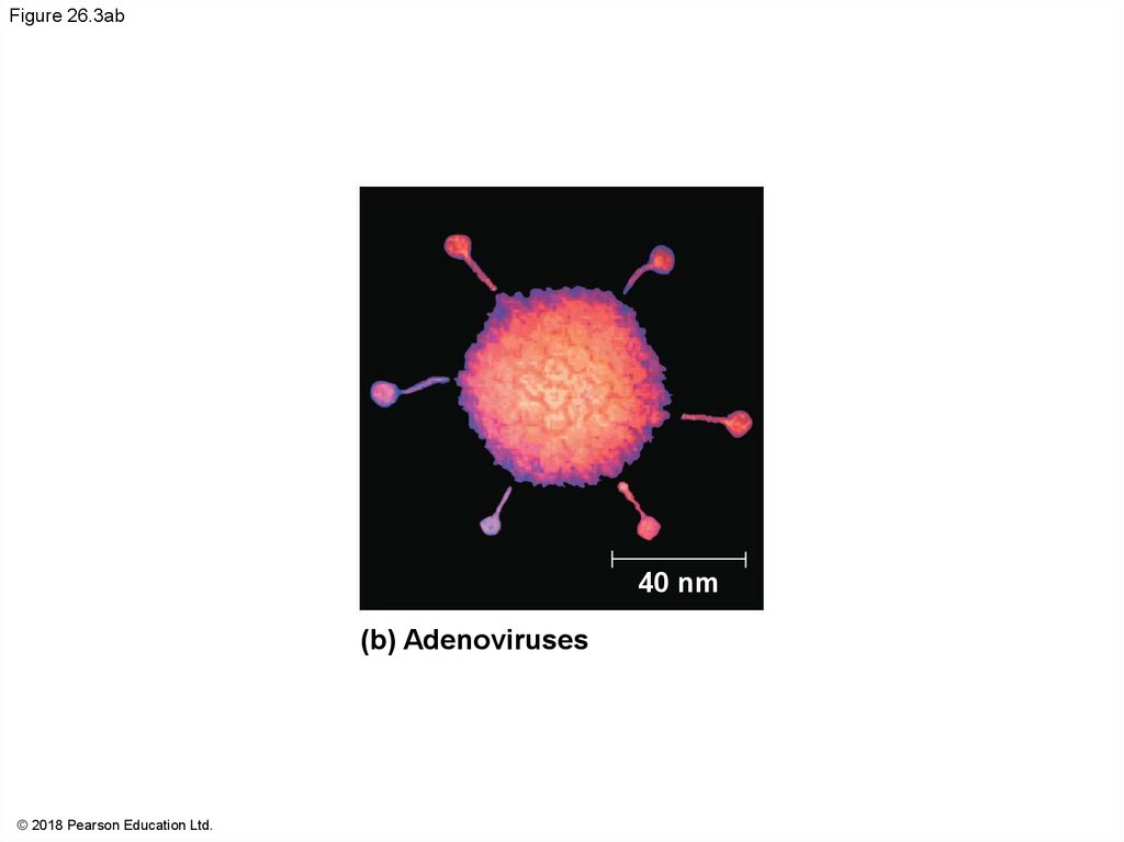
Figure 26.3ab40 nm
(b) Adenoviruses
© 2018 Pearson Education Ltd.
18.
Figure 26.3bRNA-protein
complex
Viral
polymerase
Head
Protein
RNA
DNA
Tail
sheath
Tail
fiber
Membranous
envelope
Glycoprotein
spikes
© 2018 Pearson Education Ltd.
80 × 225 nm
40 nm
40 nm
80–200 nm (diameter)
(c) Influenza viruses
(d) Bacteriophage T4
19.
Figure 26.3ba40 nm
(c) Influenza viruses
© 2018 Pearson Education Ltd.
20.
40 nmFigure 26.3bb
(d) Bacteriophage T4
© 2018 Pearson Education Ltd.
21.
Some viruses have accessory structures that helpthem infect hosts
Viral envelopes (derived from membranes of host
cells) surround the capsids of influenza viruses and
many other viruses found in animals
Viral envelopes contain a combination of viral and
host cell molecules
22.
Bacteriophages, also called phages, are virusesthat infect bacteria
They have an elongated capsid head that encloses
their DNA
A protein tail piece attaches the phage to the host
and injects the phage DNA inside
23.
Concept 26.2: Viruses replicate only in hostcells
Viruses are obligate intracellular parasites, which
means they can replicate only within a host cell
Each virus has a host range, a limited number of
host cells that it can infect
24.
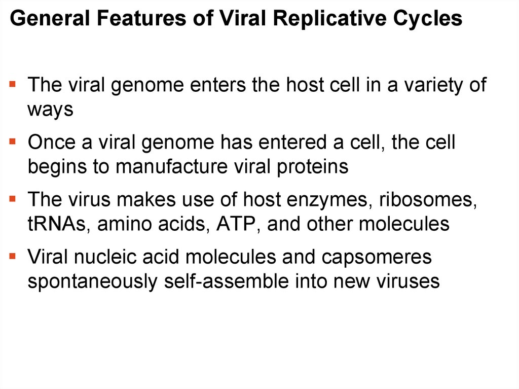
General Features of Viral Replicative CyclesThe viral genome enters the host cell in a variety of
ways
Once a viral genome has entered a cell, the cell
begins to manufacture viral proteins
The virus makes use of host enzymes, ribosomes,
tRNAs, amino acids, ATP, and other molecules
Viral nucleic acid molecules and capsomeres
spontaneously self-assemble into new viruses
25.
Figure 26.41 Entry and
uncoating
VIRUS
DNA
Capsid
2 Replication
3 Transcription and
manufacture of
capsid proteins
HOST
CELL
Viral
DNA
mRNA
Viral
DNA
© 2018 Pearson Education Ltd.
Capsid
proteins
4 Self-assembly of new
virus particles and their
exit from the cell
26.
Animation: Simplified Viral Reproductive Cycle27.
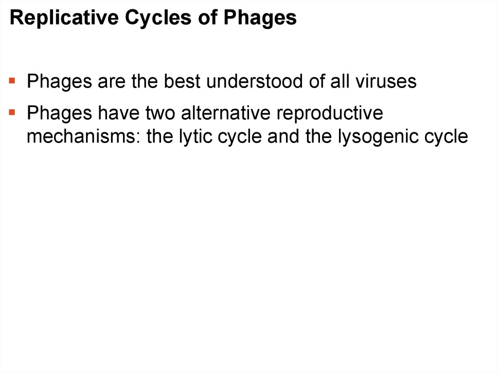
Replicative Cycles of PhagesPhages are the best understood of all viruses
Phages have two alternative reproductive
mechanisms: the lytic cycle and the lysogenic cycle
28.
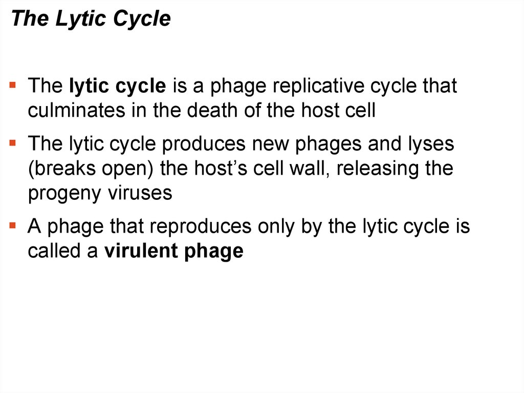
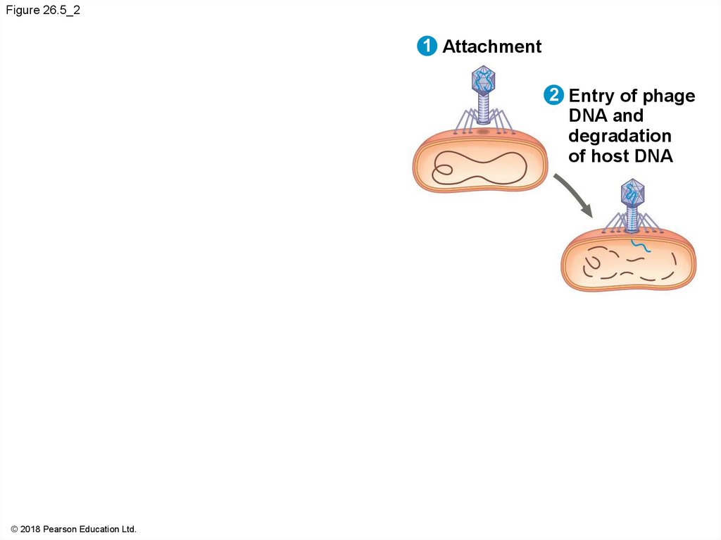
The Lytic CycleThe lytic cycle is a phage replicative cycle that
culminates in the death of the host cell
The lytic cycle produces new phages and lyses
(breaks open) the host’s cell wall, releasing the
progeny viruses
A phage that reproduces only by the lytic cycle is
called a virulent phage
29.
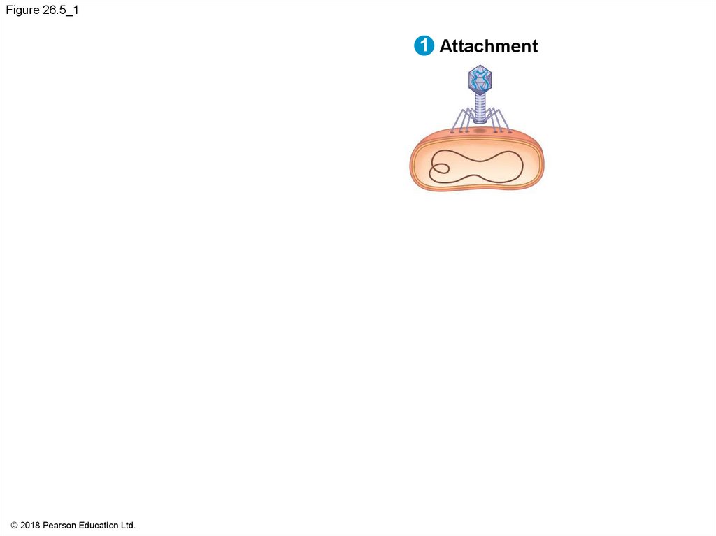
Figure 26.5_11 Attachment
© 2018 Pearson Education Ltd.
30.
Figure 26.5_21 Attachment
2 Entry of phage
DNA and
degradation
of host DNA
© 2018 Pearson Education Ltd.
31.
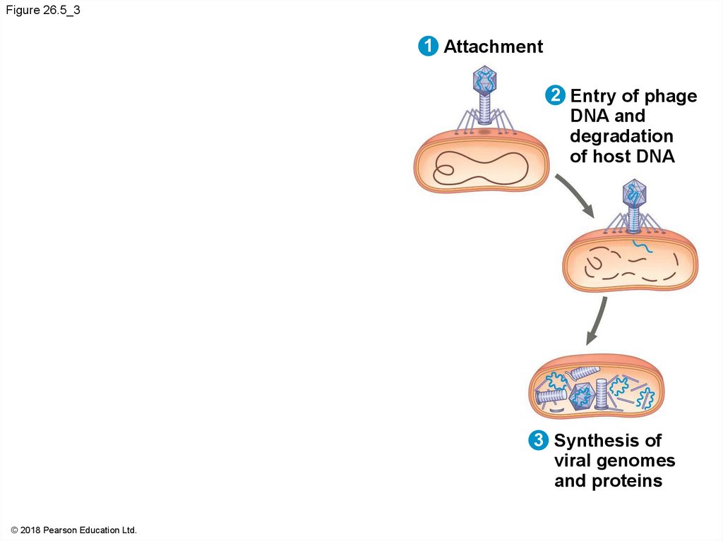
Figure 26.5_31 Attachment
2 Entry of phage
DNA and
degradation
of host DNA
3 Synthesis of
viral genomes
and proteins
© 2018 Pearson Education Ltd.
32.
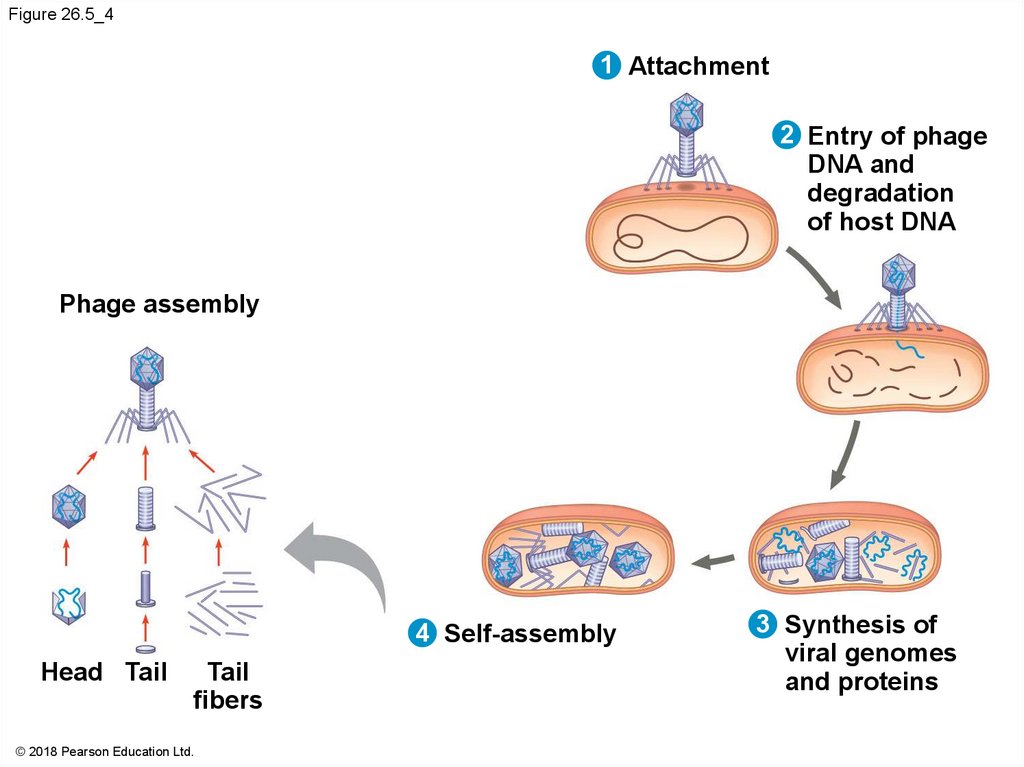
Figure 26.5_41 Attachment
2 Entry of phage
DNA and
degradation
of host DNA
Phage assembly
4 Self-assembly
Head Tail
Tail
fibers
© 2018 Pearson Education Ltd.
3 Synthesis of
viral genomes
and proteins
33.
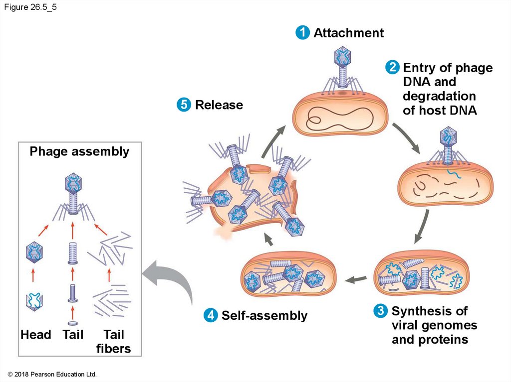
Figure 26.5_51 Attachment
5 Release
2 Entry of phage
DNA and
degradation
of host DNA
Phage assembly
4 Self-assembly
Head Tail
Tail
fibers
© 2018 Pearson Education Ltd.
3 Synthesis of
viral genomes
and proteins
34.
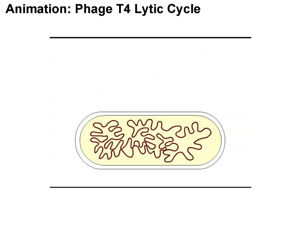
Animation: Phage T4 Lytic Cycle35.
The Lysogenic CycleThe lysogenic cycle replicates the phage genome
without destroying the host
The viral DNA molecule is incorporated into the host
cell’s chromosome
Phages that use both the lytic and lysogenic cycles
are called temperate phages
36.
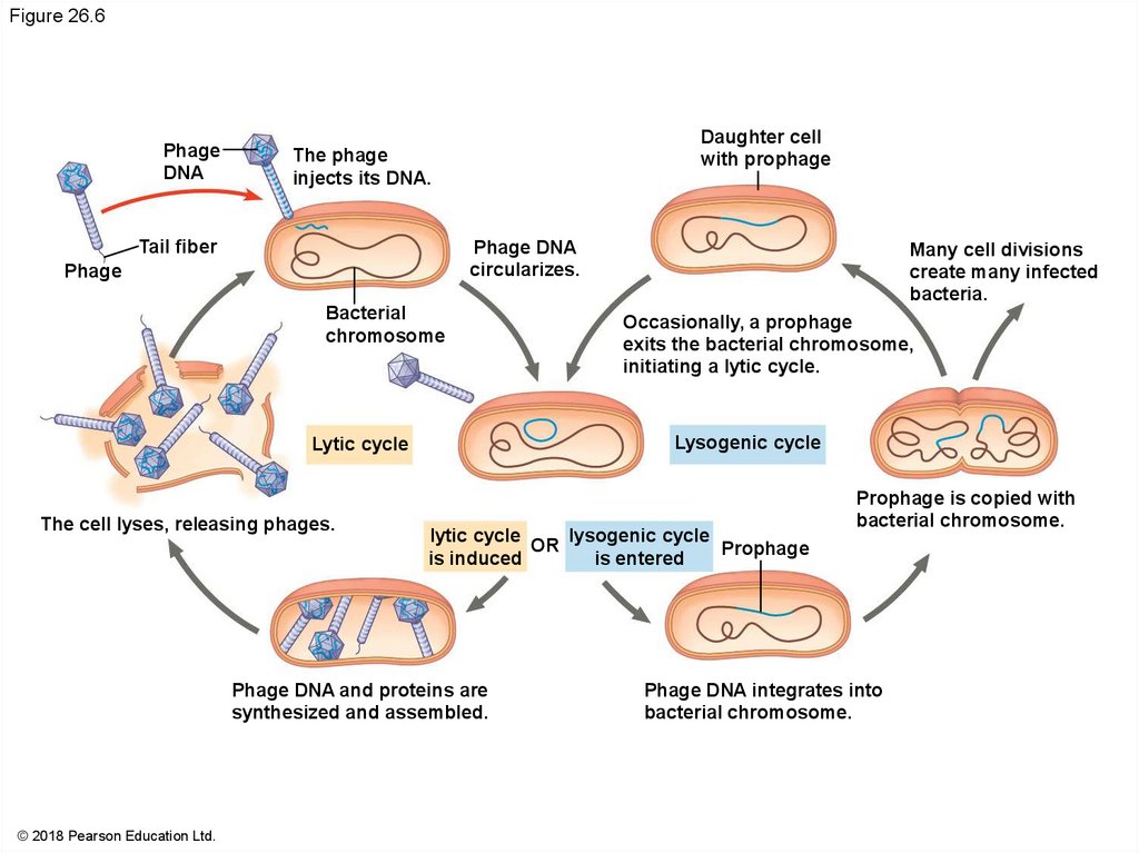
Figure 26.6Phage
DNA
Daughter cell
with prophage
The phage
injects its DNA.
Tail fiber
Phage DNA
circularizes.
Phage
Bacterial
chromosome
lytic cycle OR lysogenic cycle
Prophage
is induced
is entered
Phage DNA and proteins are
synthesized and assembled.
© 2018 Pearson Education Ltd.
Occasionally, a prophage
exits the bacterial chromosome,
initiating a lytic cycle.
Lysogenic cycle
Lytic cycle
The cell lyses, releasing phages.
Many cell divisions
create many infected
bacteria.
Prophage is copied with
bacterial chromosome.
Phage DNA integrates into
bacterial chromosome.
37.
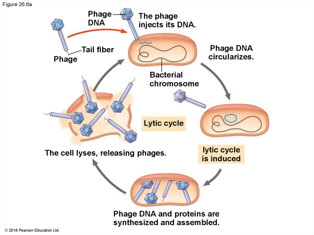
Figure 26.6aPhage
DNA
The phage
injects its DNA.
Phage DNA
circularizes.
Tail fiber
Phage
Bacterial
chromosome
Lytic cycle
The cell lyses, releasing phages.
lytic cycle
is induced
Phage DNA and proteins are
synthesized and assembled.
© 2018 Pearson Education Ltd.
38.
Figure 26.6bDaughter cell
with prophage
Many cell divisions
create many infected
bacteria.
Occasionally, a prophage
exits the bacterial chromosome,
initiating a lytic cycle.
Lysogenic cycle
lysogenic cycle
is entered
Prophage is copied with
bacterial chromosome.
Prophage
Phage DNA integrates into
bacterial chromosome.
© 2018 Pearson Education Ltd.
39.
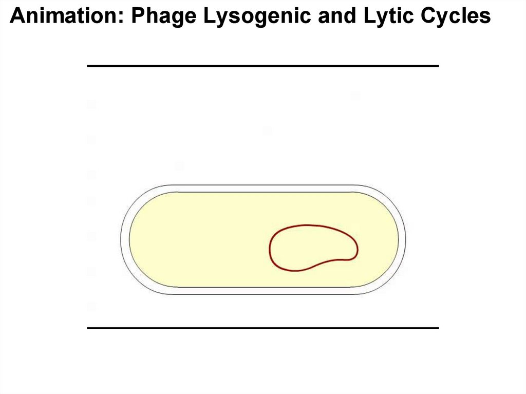
Animation: Phage Lysogenic and Lytic Cycles40.
The integrated viral DNA is known as a prophageEvery time the host divides, it copies the phage DNA
and passes the copies to daughter cells
An environmental signal can trigger the virus
genome to exit the bacterial chromosome and switch
to the lytic mode
41.
Bacterial Defenses Against PhagesBacteria have their own defenses against phages
Natural selection favors bacterial mutants with
surface proteins that cannot be recognized as
receptors by a particular type of phage
Foreign DNA can be identified as such and cut up by
cellular enzymes called restriction enzymes
The bacterium’s own DNA is protected from the
restriction enzymes by being methylated
42.
Both bacteria and archaea can protect themselvesfrom viral infection with the CRISPR-Cas system
It is based on sequences called clustered regularly
interspaced short palindromic repeats (CRISPRs)
Each “spacer” sequence between the repeats
corresponds to DNA from a phage that had infected
the cell
Particular nuclease proteins interact with the
CRISPR region; these are called CRISPRassociated (Cas) proteins
43.
When a phage infects a bacterial cell that has theCRISPR-Cas system, the phage DNA is integrated
between two repeat sequences
If the cell survives the infection, it can block any
attempt of the same type of phage to reinfect it
The attempt of the phage to infect the cell triggers
transcription of the CRISPR region
The resulting RNAs are cut into pieces and bound by
Cas proteins
44.
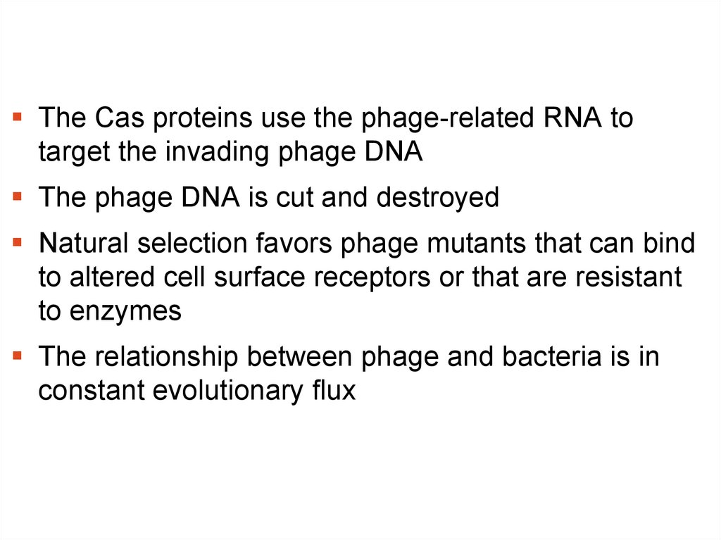
The Cas proteins use the phage-related RNA totarget the invading phage DNA
The phage DNA is cut and destroyed
Natural selection favors phage mutants that can bind
to altered cell surface receptors or that are resistant
to enzymes
The relationship between phage and bacteria is in
constant evolutionary flux
45.
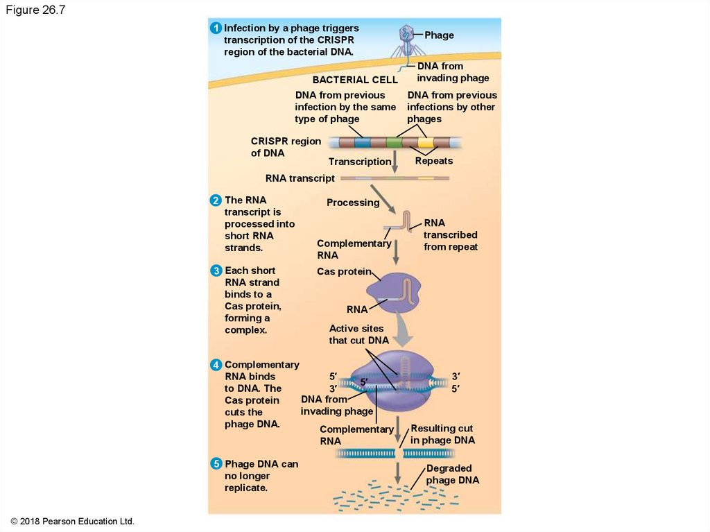
Figure 26.71 Infection by a phage triggers
transcription of the CRISPR
region of the bacterial DNA.
BACTERIAL CELL
DNA from previous
infection by the same
type of phage
CRISPR region
of DNA
Transcription
Phage
DNA from
invading phage
DNA from previous
infections by other
phages
Repeats
RNA transcript
2 The RNA
transcript is
processed into
short RNA
strands.
3 Each short
RNA strand
binds to a
Cas protein,
forming a
complex.
Processing
Complementary
RNA
Cas protein
RNA
Active sites
that cut DNA
4 Complementary
RNA binds
5′
5′
to DNA. The
3′
DNA from
Cas protein
invading phage
cuts the
phage DNA.
Complementary
RNA
5 Phage DNA can
no longer
replicate.
© 2018 Pearson Education Ltd.
RNA
transcribed
from repeat
3′
5′
Resulting cut
in phage DNA
Degraded
phage DNA
46.
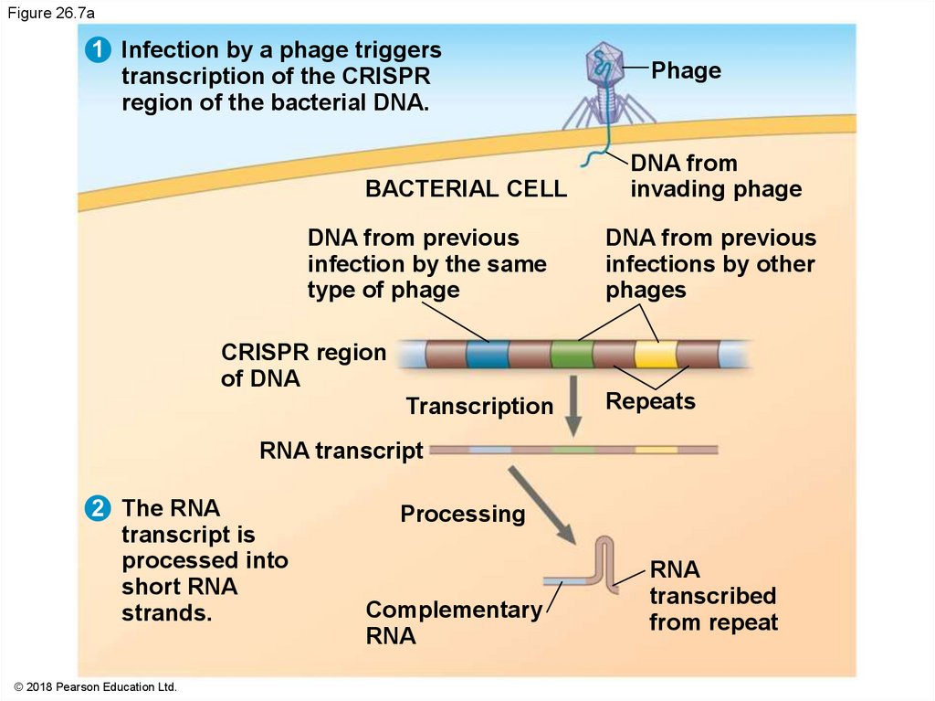
Figure 26.7a1 Infection by a phage triggers
transcription of the CRISPR
region of the bacterial DNA.
BACTERIAL CELL
DNA from previous
infection by the same
type of phage
Phage
DNA from
invading phage
DNA from previous
infections by other
phages
CRISPR region
of DNA
Transcription
Repeats
RNA transcript
2 The RNA
transcript is
processed into
short RNA
strands.
© 2018 Pearson Education Ltd.
Processing
Complementary
RNA
RNA
transcribed
from repeat
47.
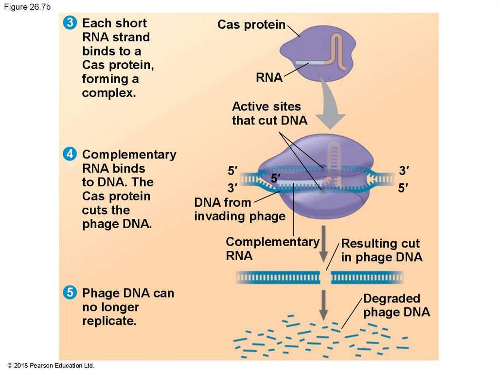
Figure 26.7b3 Each short
RNA strand
binds to a
Cas protein,
forming a
complex.
Cas protein
RNA
Active sites
that cut DNA
4 Complementary
RNA binds
to DNA. The
Cas protein
cuts the
phage DNA.
5′
5′
3′
DNA from
invading phage
Complementary
RNA
5 Phage DNA can
no longer
replicate.
© 2018 Pearson Education Ltd.
3′
5′
Resulting cut
in phage DNA
Degraded
phage DNA
48.
Figure 26.7c© 2018 Pearson Education Ltd.
49.
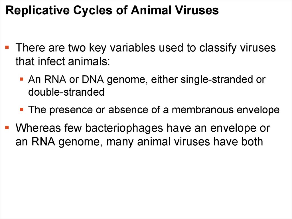
Replicative Cycles of Animal VirusesThere are two key variables used to classify viruses
that infect animals:
An RNA or DNA genome, either single-stranded or
double-stranded
The presence or absence of a membranous envelope
Whereas few bacteriophages have an envelope or
an RNA genome, many animal viruses have both
50.
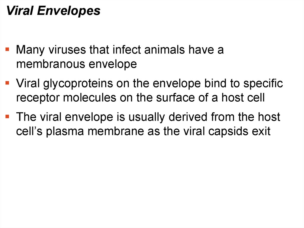
Viral EnvelopesMany viruses that infect animals have a
membranous envelope
Viral glycoproteins on the envelope bind to specific
receptor molecules on the surface of a host cell
The viral envelope is usually derived from the host
cell’s plasma membrane as the viral capsids exit
51.
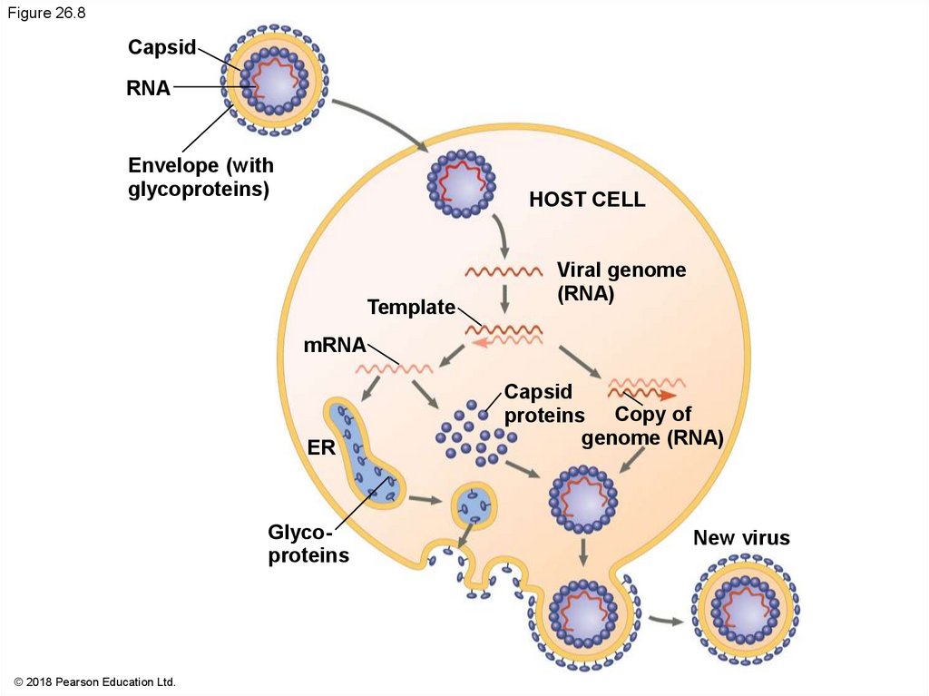
Figure 26.8Capsid
RNA
Envelope (with
glycoproteins)
HOST CELL
Template
Viral genome
(RNA)
mRNA
ER
Glycoproteins
© 2018 Pearson Education Ltd.
Capsid
proteins Copy of
genome (RNA)
New virus
52.
Other viral membranes form from the host’s nuclearenvelope and are then replaced by an envelope
made from Golgi apparatus membrane
53.
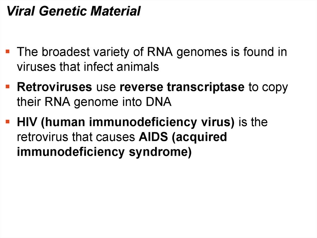
Viral Genetic MaterialThe broadest variety of RNA genomes is found in
viruses that infect animals
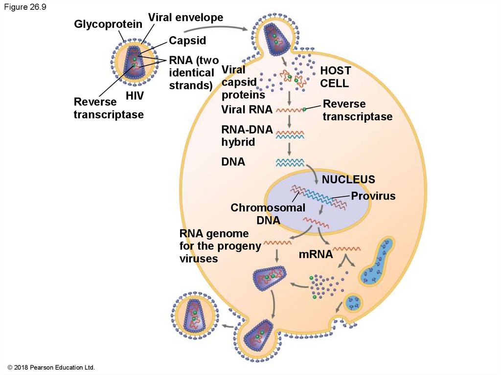
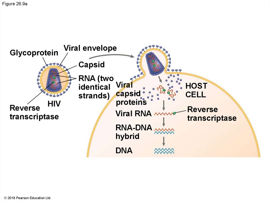
Retroviruses use reverse transcriptase to copy
their RNA genome into DNA
HIV (human immunodeficiency virus) is the
retrovirus that causes AIDS (acquired
immunodeficiency syndrome)
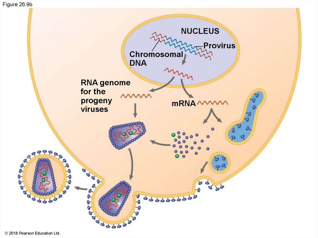
54.
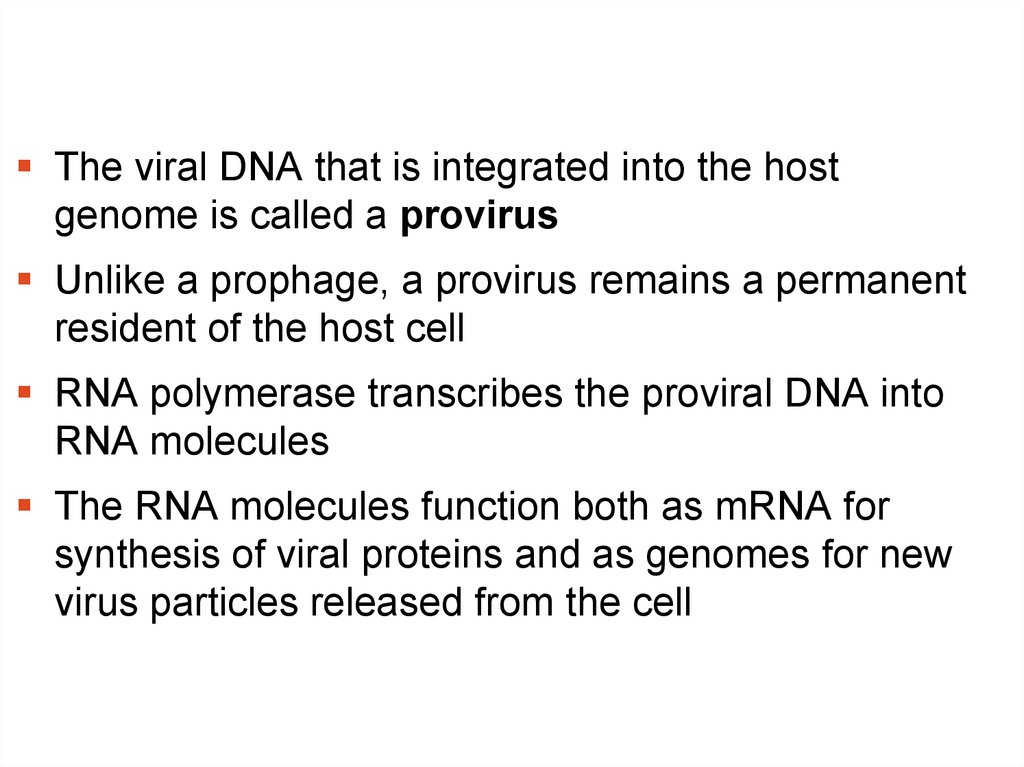
The viral DNA that is integrated into the hostgenome is called a provirus
Unlike a prophage, a provirus remains a permanent
resident of the host cell
RNA polymerase transcribes the proviral DNA into
RNA molecules
The RNA molecules function both as mRNA for
synthesis of viral proteins and as genomes for new
virus particles released from the cell
55.
Table 26.1© 2018 Pearson Education Ltd.
56.
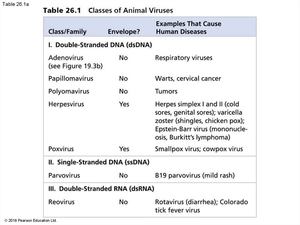
Table 26.1a© 2018 Pearson Education Ltd.
57.
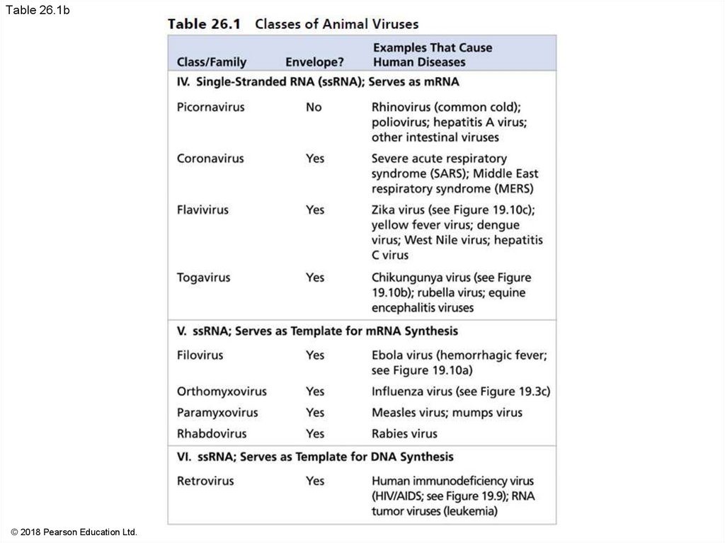
Table 26.1b© 2018 Pearson Education Ltd.
58.
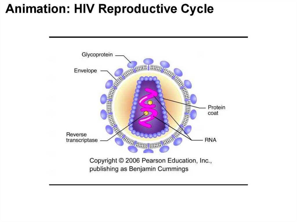
Figure 26.9Glycoprotein
Viral envelope
Capsid
HIV
Reverse
transcriptase
RNA (two
identical Viral
strands) capsid
proteins
Viral RNA
HOST
CELL
Reverse
transcriptase
RNA-DNA
hybrid
DNA
NUCLEUS
Provirus
Chromosomal
DNA
RNA genome
for the progeny
mRNA
viruses
© 2018 Pearson Education Ltd.
59.
Figure 26.9aGlycoprotein
Viral envelope
Capsid
HIV
Reverse
transcriptase
RNA (two
identical Viral
strands) capsid
proteins
Viral RNA
RNA-DNA
hybrid
DNA
© 2018 Pearson Education Ltd.
HOST
CELL
Reverse
transcriptase
60.
Figure 26.9bNUCLEUS
Provirus
Chromosomal
DNA
RNA genome
for the
progeny
viruses
© 2018 Pearson Education Ltd.
mRNA
61.
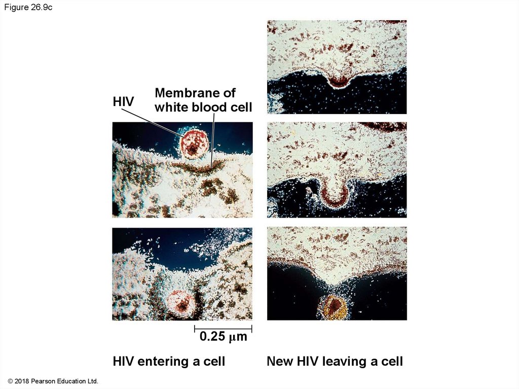
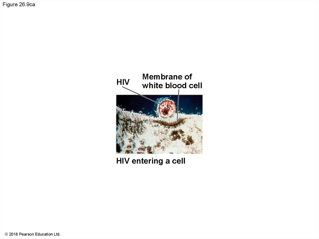
Figure 26.9cHIV
Membrane of
white blood cell
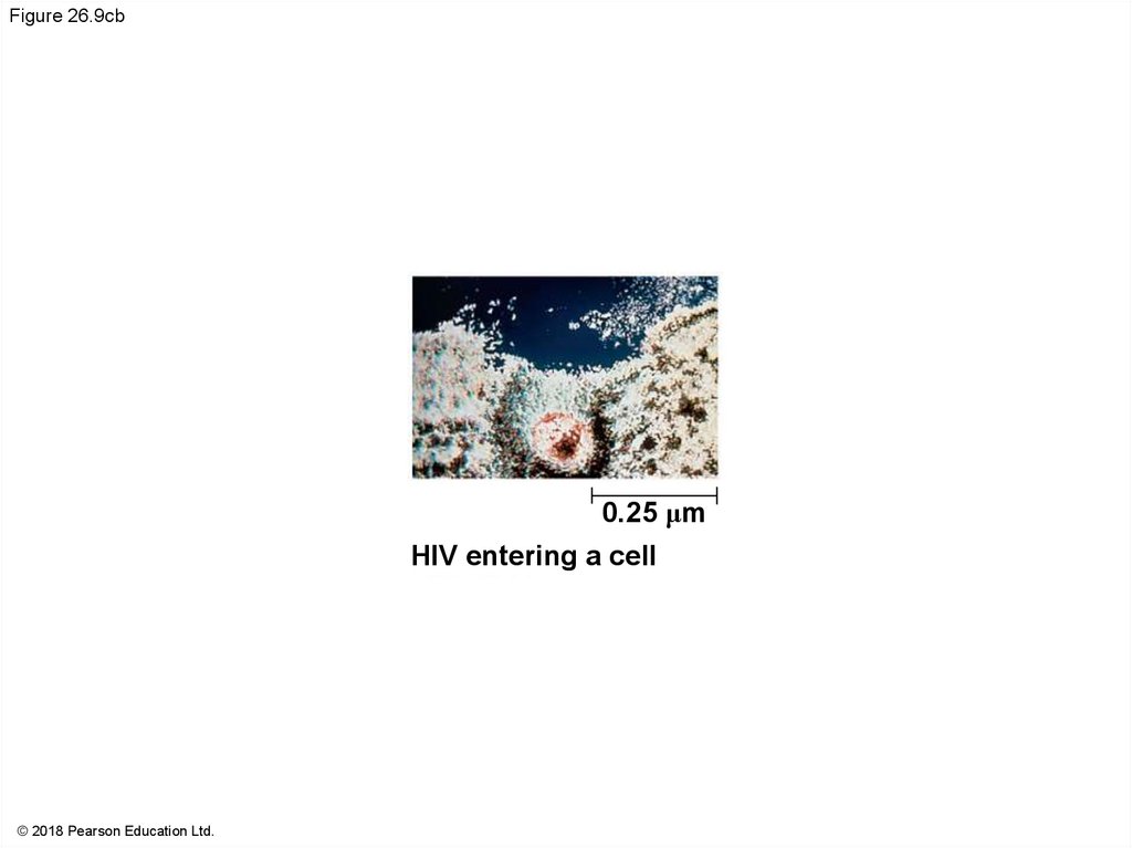
0.25 μm
HIV entering a cell
© 2018 Pearson Education Ltd.
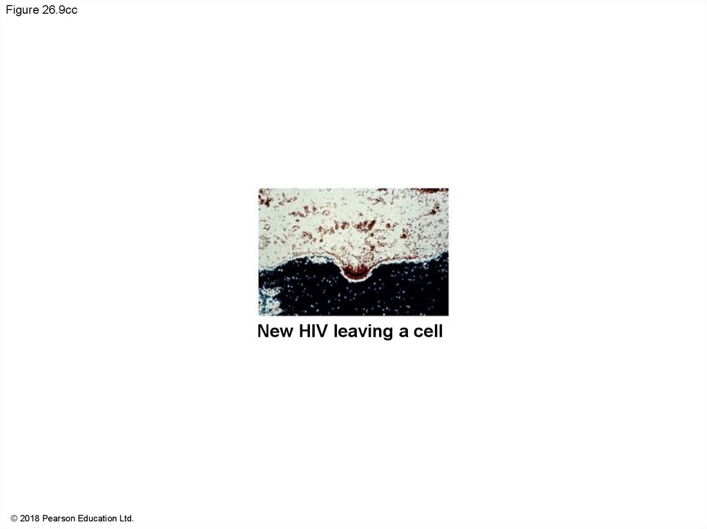
New HIV leaving a cell
62.
Figure 26.9caHIV
Membrane of
white blood cell
HIV entering a cell
© 2018 Pearson Education Ltd.
63.
Figure 26.9cb0.25 μm
HIV entering a cell
© 2018 Pearson Education Ltd.
64.
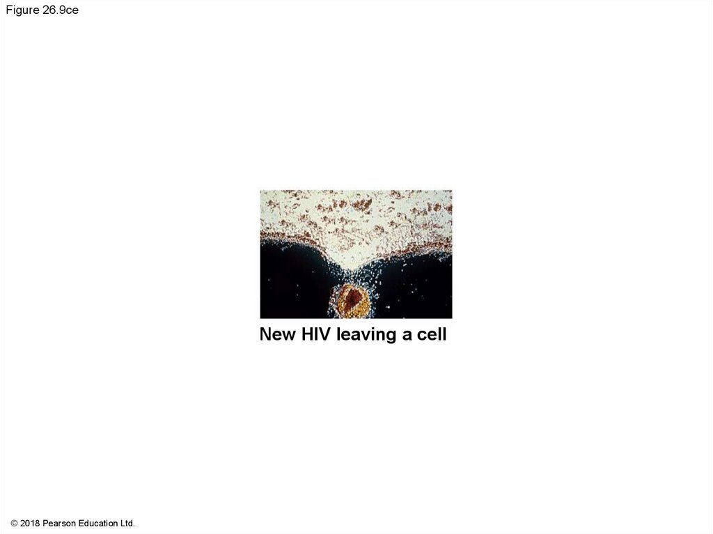
Figure 26.9ccNew HIV leaving a cell
© 2018 Pearson Education Ltd.
65.
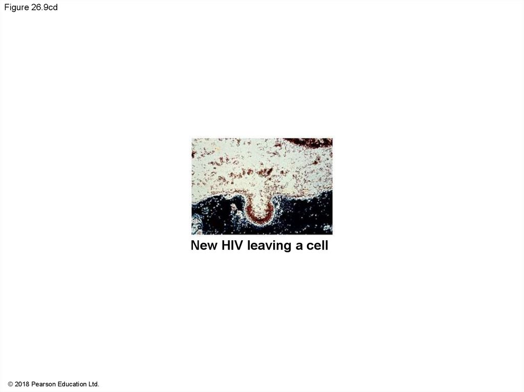
Figure 26.9cdNew HIV leaving a cell
© 2018 Pearson Education Ltd.
66.
Figure 26.9ceNew HIV leaving a cell
© 2018 Pearson Education Ltd.
67.
Animation: HIV Reproductive Cycle68.
Evolution of VirusesViruses do not fit our definition of living organisms
Since viruses can replicate only within cells, they
probably evolved as bits of cellular nucleic acid
Candidates for the source of viral genomes include
plasmids and transposons
Plasmids, transposons, and viruses are all mobile
genetic elements
69.
The largest virus yet discovered is the size of a smallbacterium
Its genome encodes proteins involved in translation,
DNA repair, protein folding, and polysaccharide
synthesis
There is controversy about whether this virus
evolved before or after cells
70.
Concept 26.3: Viruses and prions areformidable pathogens in animals and plants
Diseases caused by viral infections affect humans,
agricultural crops, and livestock worldwide
Smaller, less complex entities called prions also
cause disease in plants and animals, respectively
71.
Viral Diseases in AnimalsViruses may damage or kill cells by causing the
release of hydrolytic enzymes from lysosomes
Some viruses cause infected cells to produce toxins
that lead to disease symptoms
Others have molecular components such as
envelope proteins that are toxic
72.
A vaccine is a harmless derivatives of pathogenicmicrobes that stimulate the immune system to mount
defenses against the harmful pathogen
Vaccines can prevent certain viral illnesses
Viral infections cannot be treated by antibiotics
Antiviral drugs can help to treat, not cure, viral
infections by inhibiting synthesis of viral DNA and by
interfering with viral assembly
73.
Emerging VirusesEmerging viruses are those that suddenly become
apparent
The Ebola virus is one of several emerging viruses
that cause hemorrhagic fever, an often fatal illness
Other examples include the chikungunya virus and
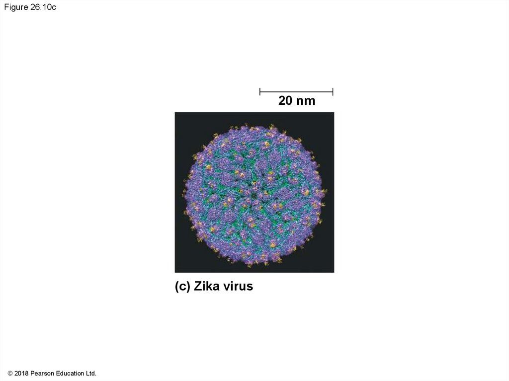
the recently emerging Zika virus (2015)
74.
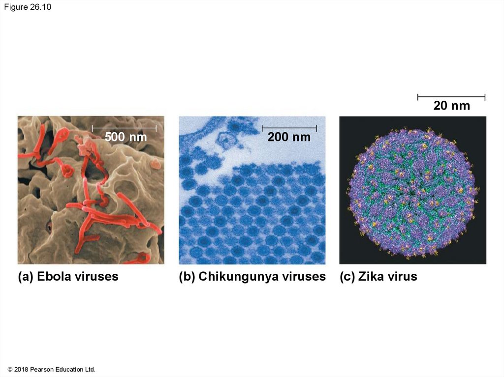
Figure 26.1020 nm
500 nm
(a) Ebola viruses
© 2018 Pearson Education Ltd.
200 nm
(b) Chikungunya viruses
(c) Zika virus
75.
Figure 26.10a500 nm
(a) Ebola viruses
© 2018 Pearson Education Ltd.
76.
Figure 26.10b200 nm
(b) Chikungunya viruses
© 2018 Pearson Education Ltd.
77.
Figure 26.10c20 nm
(c) Zika virus
© 2018 Pearson Education Ltd.
78.
In 2009, a general outbreak (epidemic) of a flu-likeillness appeared in Mexico and the United States,
caused by an influenza virus named H1N1
A global epidemic is called a pandemic
79.
Three processes contribute to the emergence of newviral diseases:
RNA viruses have an unusually high rate of mutation
The disease can be disseminated from a small,
isolated human population and can eventually spread
around the world
About three-quarters of new human diseases
originate by spreading to humans from animals
80.
Flu epidemics are caused by type A influenzaviruses; these infect a wide variety of animals
including birds, pigs, horses, and humans
Strains of influenza A are given standardized names
based on the viral surface proteins hemagglutinin
(HA) and neuraminidase (NA)
H1N1 is the strain that caused the 2009 flu
pandemic
81.
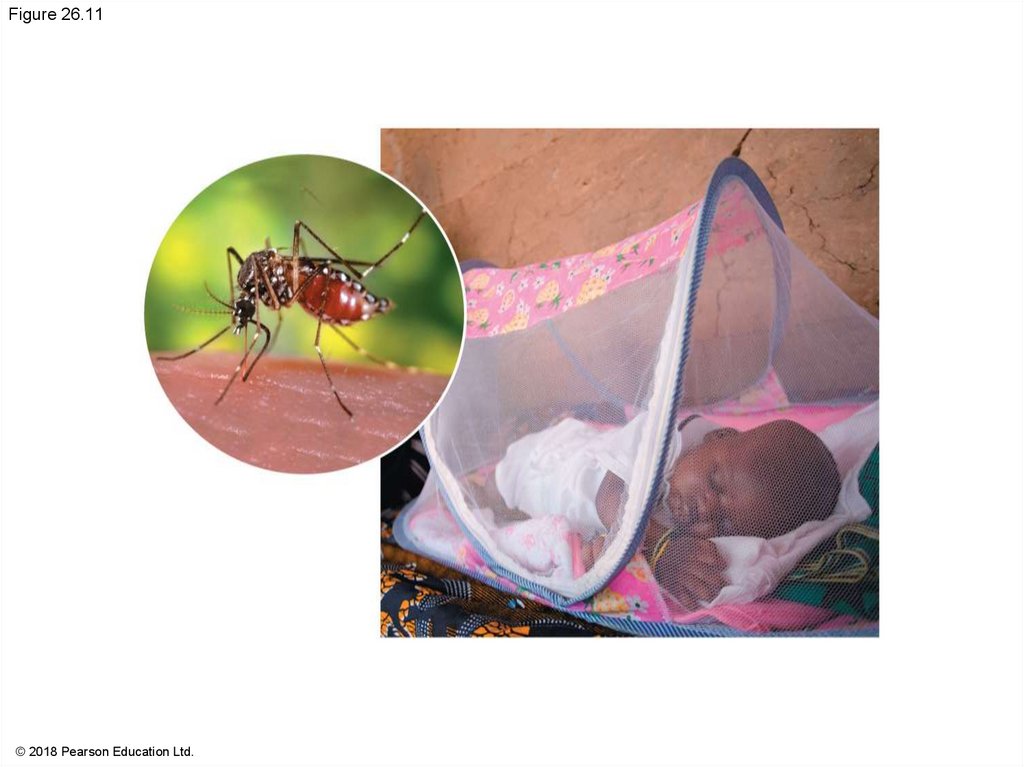
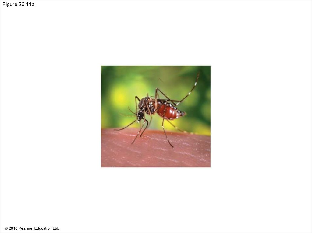
Changes in host behavior or the environment canincrease the spread of viruses responsible for
emerging diseases
New roads into a remote area may increase spread
of viral diseases
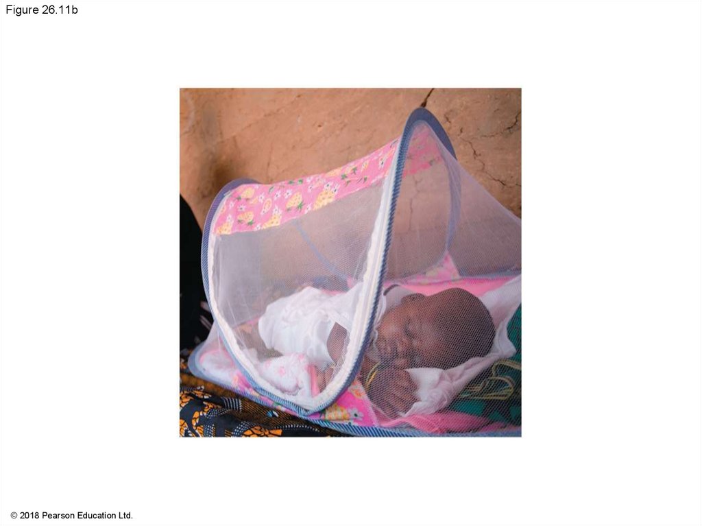
The use of insecticides and mosquito nets may help
prevent the spread
It is possible that global climate change may allow
mosquitoes that carry viruses to expand their range
82.
Figure 26.11© 2018 Pearson Education Ltd.
83.
Figure 26.11a© 2018 Pearson Education Ltd.
84.
Figure 26.11b© 2018 Pearson Education Ltd.
85.
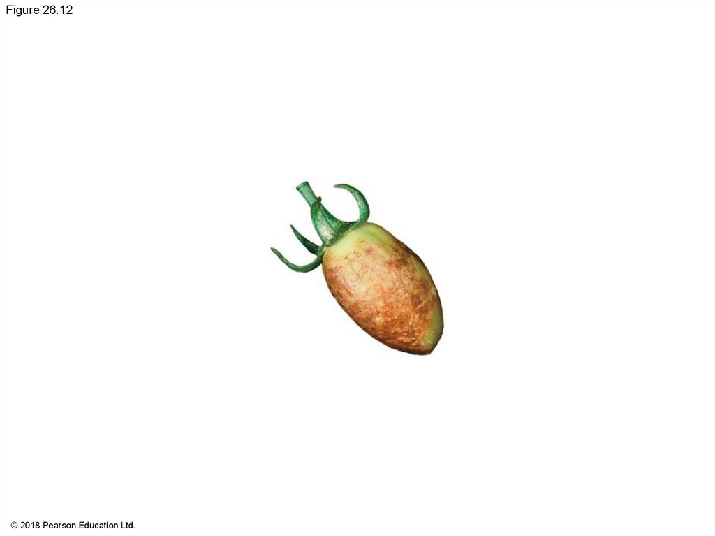
Viral Diseases in PlantsMore than 2,000 types of viral diseases of plants are
known and cause spots on leaves and fruits, stunted
growth, and damaged flowers or roots
Most plant viruses have an RNA genome
Many have a helical capsid, while others have an
icosahedral capsid
86.
Figure 26.12© 2018 Pearson Education Ltd.
87.
Plant viruses spread disease by two major routes:Horizontal transmission, entering through damaged
cell walls
Vertical transmission, inheriting the virus from a
parent
88.
Prions: Proteins as Infectious AgentsPrions are infectious proteins that appear to cause
degenerative brain diseases in animals
Scrapie in sheep, mad cow disease, and CreutzfeldtJakob disease in humans are all caused by prions
Prions are incorrectly folded proteins, can be
transmitted in food, act slowly, and are virtually
indestructible
89.
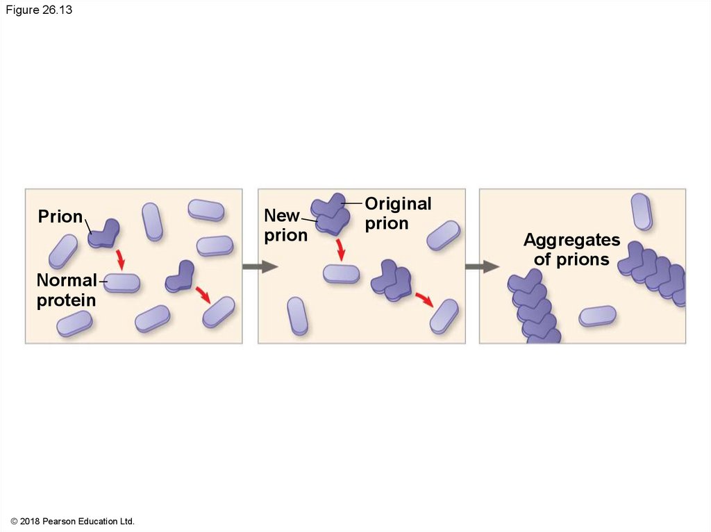
Prions are somehow able to convert a normal formof the protein into the misfolded version
Then several prions aggregate into a complex that
can convert more proteins to prions
Prions might also be involved in diseases such as
Alzheimer’s and Parkinson’s disease
90.
Figure 26.13Prion
Normal
protein
© 2018 Pearson Education Ltd.
New
prion
Original
prion
Aggregates
of prions
91.
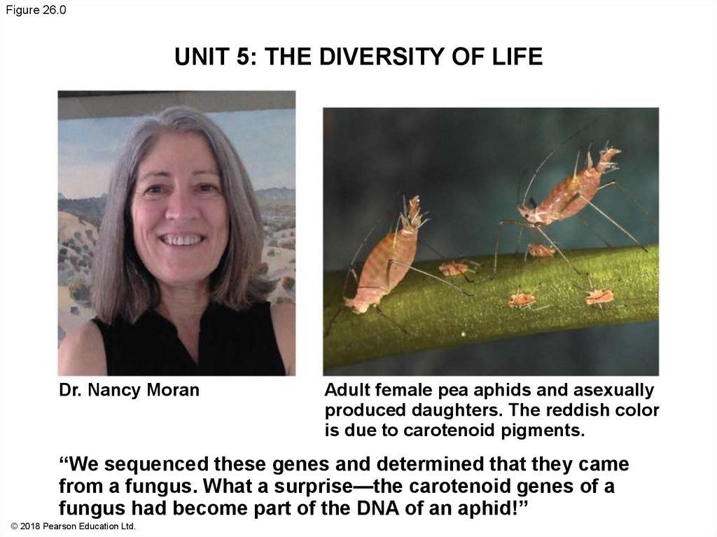
Figure 26.0UNIT 5: THE DIVERSITY OF LIFE
Dr. Nancy Moran
Adult female pea aphids and asexually
produced daughters. The reddish color
is due to carotenoid pigments.
“We sequenced these genes and determined that they came
from a fungus. What a surprise—the carotenoid genes of a
fungus had become part of the DNA of an aphid!”
© 2018 Pearson Education Ltd.
92.
Figure 26.0aDr. Nancy Moran
© 2018 Pearson Education Ltd.
93.
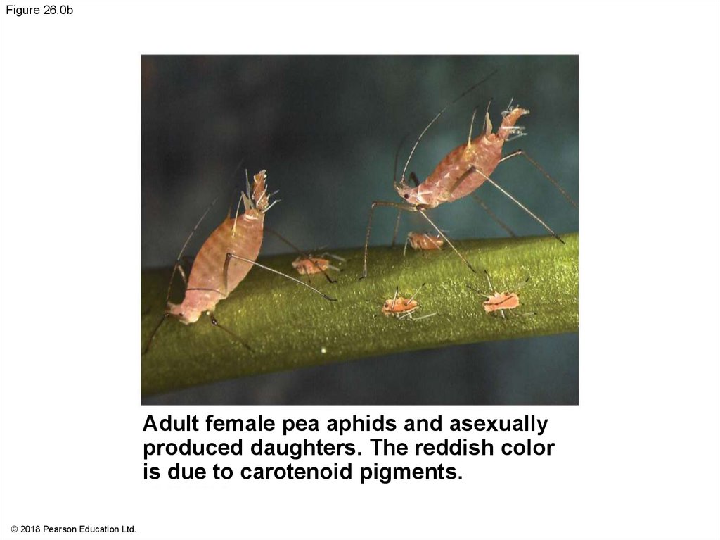
Figure 26.0bAdult female pea aphids and asexually
produced daughters. The reddish color
is due to carotenoid pigments.
© 2018 Pearson Education Ltd.
94.
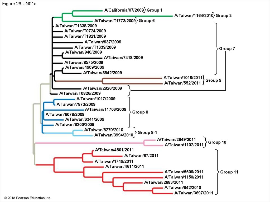
Figure 26.UN01aA/California/07/2009 Group 1
A/Taiwan/1164/2010 Group 3
A/Taiwan/T1773/2009 Group 6
A/Taiwan/T1338/2009
A/Taiwan/T0724/2009
A/Taiwan/T1821/2009
A/Taiwan/937/2009
A/Taiwan/T1339/2009
A/Taiwan/940/2009
A/Taiwan/7418/2009
A/Taiwan/8575/2009
A/Taiwan/4909/2009
A/Taiwan/8542/2009
Group 7
A/Taiwan/1018/2011
Group 9
A/Taiwan/552/2011
A/Taiwan/2826/2009
A/Taiwan/T0826/2009
A/Taiwan/1017/2009
A/Taiwan/7873/2009
A/Taiwan/11706/2009
Group 8
A/Taiwan/6078/2009
A/Taiwan/6341/2009
A/Taiwan/6200/2009
A/Taiwan/5270/2010
Group 8-1
A/Taiwan/3994/2010
A/Taiwan/2649/2011
Group 10
A/Taiwan/1102/2011
A/Taiwan/4501/2011
A/Taiwan/67/2011
A/Taiwan/1749/2011
A/Taiwan/4611/2011
A/Taiwan/5506/2011
Group 11
A/Taiwan/1150/2011
A/Taiwan/2883/2011
A/Taiwan/842/2010
A/Taiwan/3697/2011
© 2018 Pearson Education Ltd.
95.
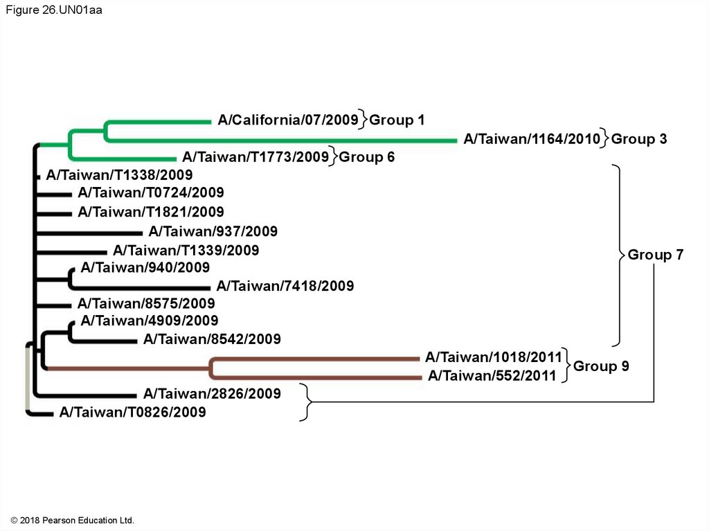
Figure 26.UN01aaA/California/07/2009 Group 1
A/Taiwan/1164/2010 Group 3
A/Taiwan/T1773/2009 Group 6
A/Taiwan/T1338/2009
A/Taiwan/T0724/2009
A/Taiwan/T1821/2009
A/Taiwan/937/2009
A/Taiwan/T1339/2009
A/Taiwan/940/2009
A/Taiwan/7418/2009
A/Taiwan/8575/2009
A/Taiwan/4909/2009
A/Taiwan/8542/2009
Group 7
A/Taiwan/1018/2011
Group 9
A/Taiwan/552/2011
A/Taiwan/2826/2009
A/Taiwan/T0826/2009
© 2018 Pearson Education Ltd.
96.
Figure 26.UN01abA/Taiwan/1017/2009
A/Taiwan/7873/2009
A/Taiwan/11706/2009
Group 8
A/Taiwan/6078/2009
A/Taiwan/6341/2009
A/Taiwan/6200/2009
A/Taiwan/5270/2010
Group 8-1
A/Taiwan/3994/2010
A/Taiwan/2649/2011
A/Taiwan/1102/2011
Group 10
A/Taiwan/4501/2011
A/Taiwan/67/2011
A/Taiwan/1749/2011
A/Taiwan/4611/2011
A/Taiwan/5506/2011
A/Taiwan/1150/2011
A/Taiwan/2883/2011
A/Taiwan/842/2010
A/Taiwan/3697/2011
© 2018 Pearson Education Ltd.
Group 11
97.
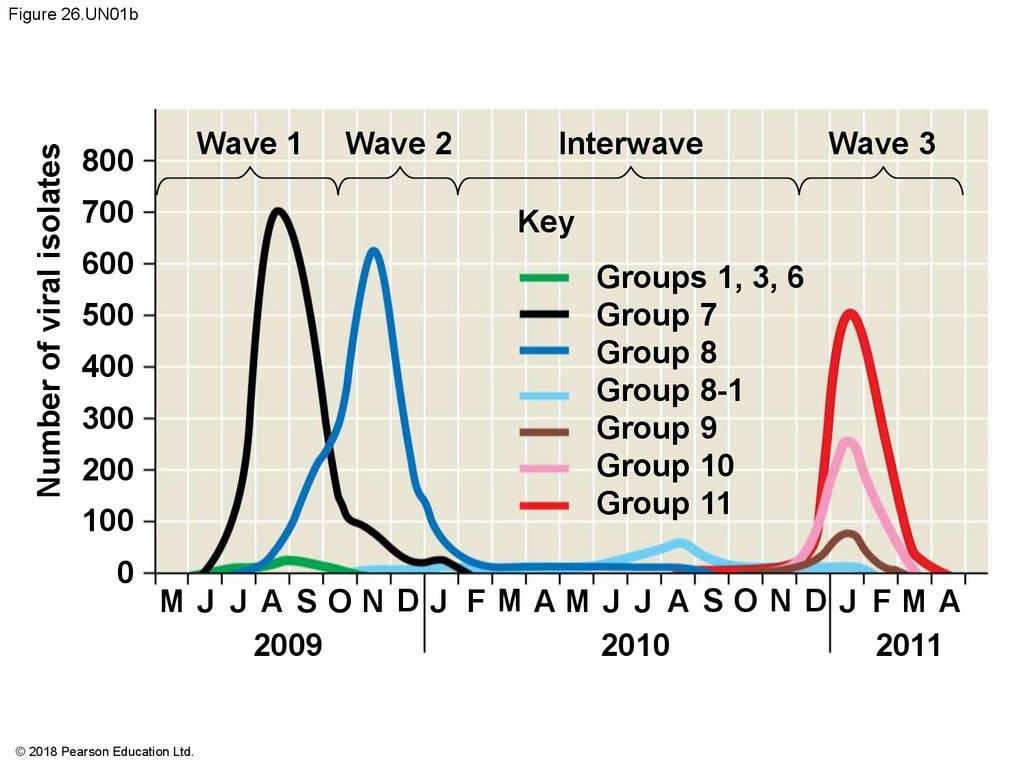
Number of viral isolatesFigure 26.UN01b
Wave 1
800
700
Interwave
Wave 3
Key
600
Groups 1, 3, 6
Group 7
Group 8
Group 8-1
Group 9
Group 10
Group 11
500
400
300
200
100
0
Wave 2
M J J A SON DJ FMAM J J A SO N D J FM A
2009
2010
2011
© 2018 Pearson Education Ltd.
98.
Figure 26.UN01c© 2018 Pearson Education Ltd.
99.
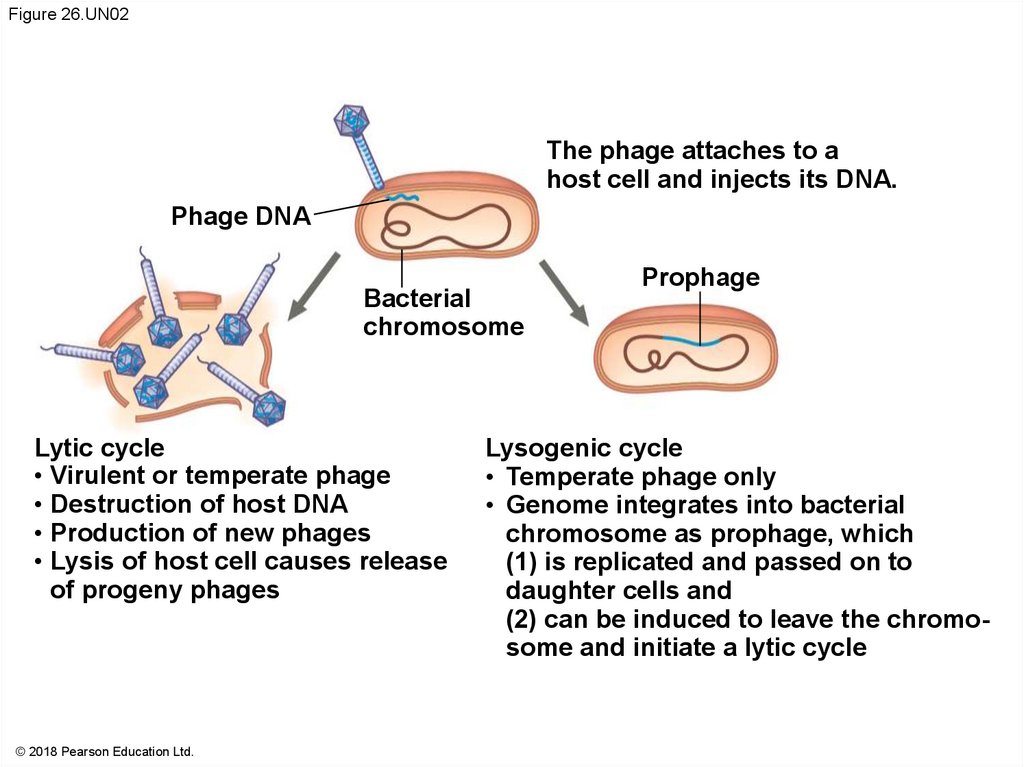
Figure 26.UN02The phage attaches to a
host cell and injects its DNA.
Phage DNA
Bacterial
chromosome
Lytic cycle
• Virulent or temperate phage
• Destruction of host DNA
• Production of new phages
• Lysis of host cell causes release
of progeny phages
© 2018 Pearson Education Ltd.
Prophage
Lysogenic cycle
• Temperate phage only
• Genome integrates into bacterial
chromosome as prophage, which
(1) is replicated and passed on to
daughter cells and
(2) can be induced to leave the chromosome and initiate a lytic cycle
100.
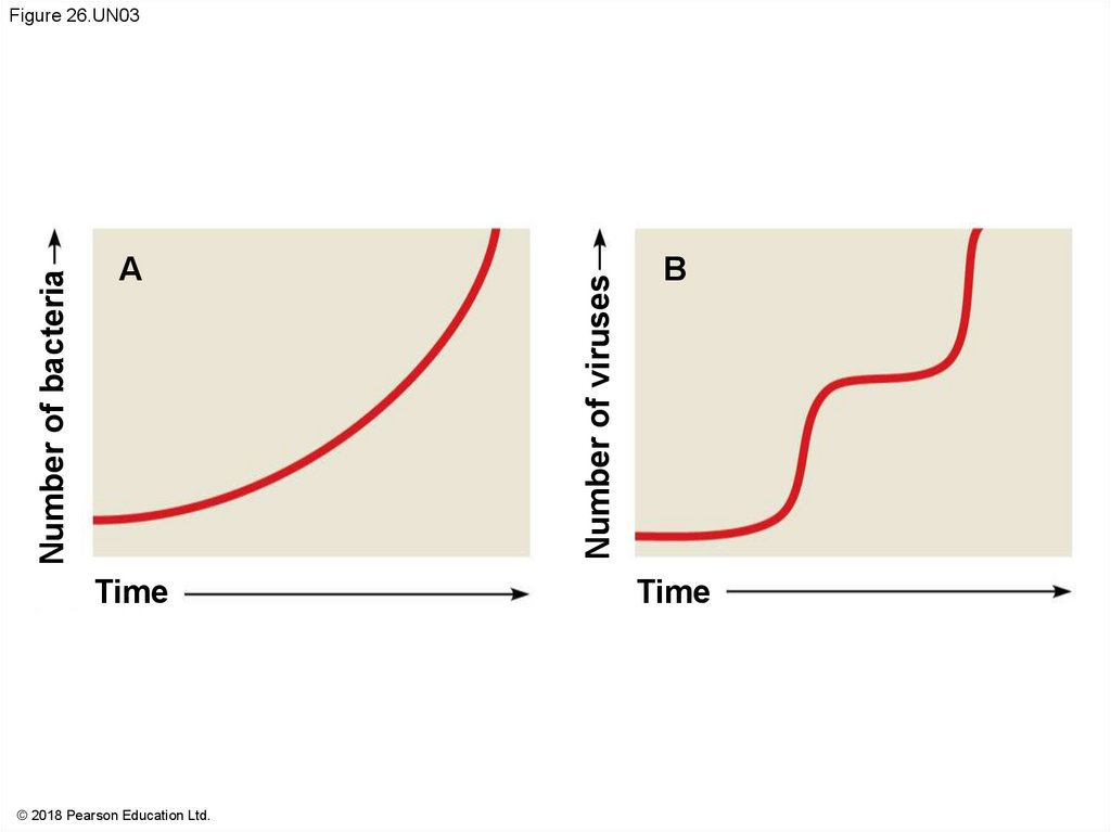
ATime
© 2018 Pearson Education Ltd.
Number of viruses
Number of bacteria
Figure 26.UN03
B
Time
101.
Figure 26.UN04© 2018 Pearson Education Ltd.