Похожие презентации:
")


Identification of natural transparent pure ruby by inclusions visible only in a microscope
1.
Identification of naturaltransparent pure ruby by
inclusions visible only in a
microscope (final
presentation)
2.
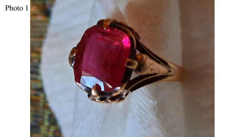
Photo 13.
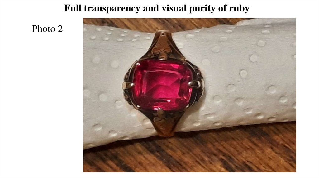
Full transparency and visual purity of rubyPhoto 2
4.
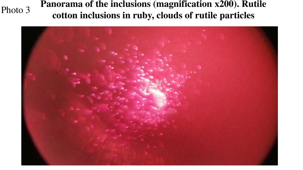
Photo 3Panorama of the inclusions (magnification x200). Rutile
cotton inclusions in ruby, clouds of rutile particles
5.
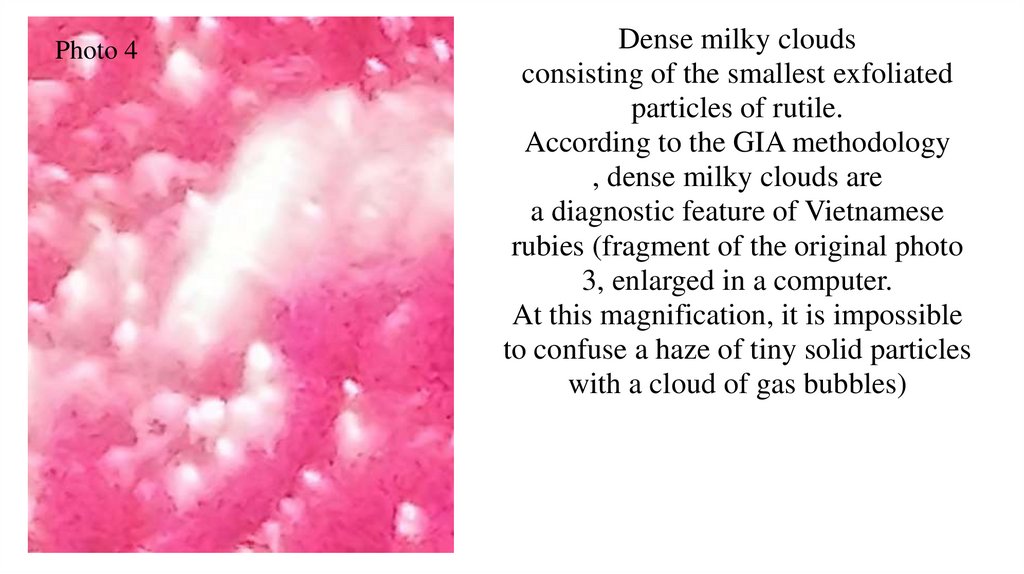
Photo 4Dense milky clouds
consisting of the smallest exfoliated
particles of rutile.
According to the GIA methodology
, dense milky clouds are
a diagnostic feature of Vietnamese
rubies (fragment of the original photo
3, enlarged in a computer.
At this magnification, it is impossible
to confuse a haze of tiny solid particles
with a cloud of gas bubbles)
6.
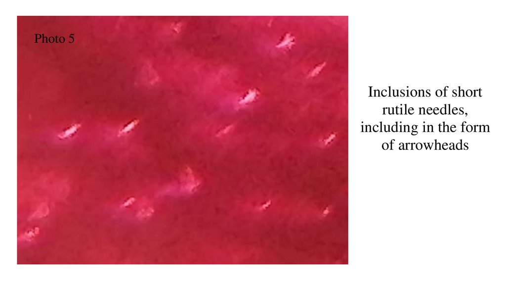
Photo 5Inclusions of short
rutile needles,
including in the form
of arrowheads
7.
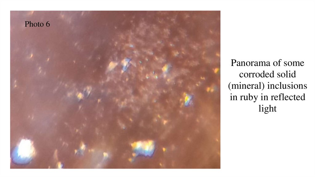
Photo 6Panorama of some
corroded solid
(mineral) inclusions
in ruby in reflected
light
8.
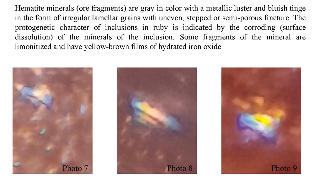
Hematite minerals (ore fragments) are gray in color with a metallic luster and bluish tingein the form of irregular lamellar grains with uneven, stepped or semi-porous fracture. The
protogenetic character of inclusions in ruby is indicated by the corroding (surface
dissolution) of the minerals of the inclusion. Some fragments of the mineral are
limonitized and have yellow-brown films of hydrated iron oxide
Photo 7
Photo 8
Photo 9
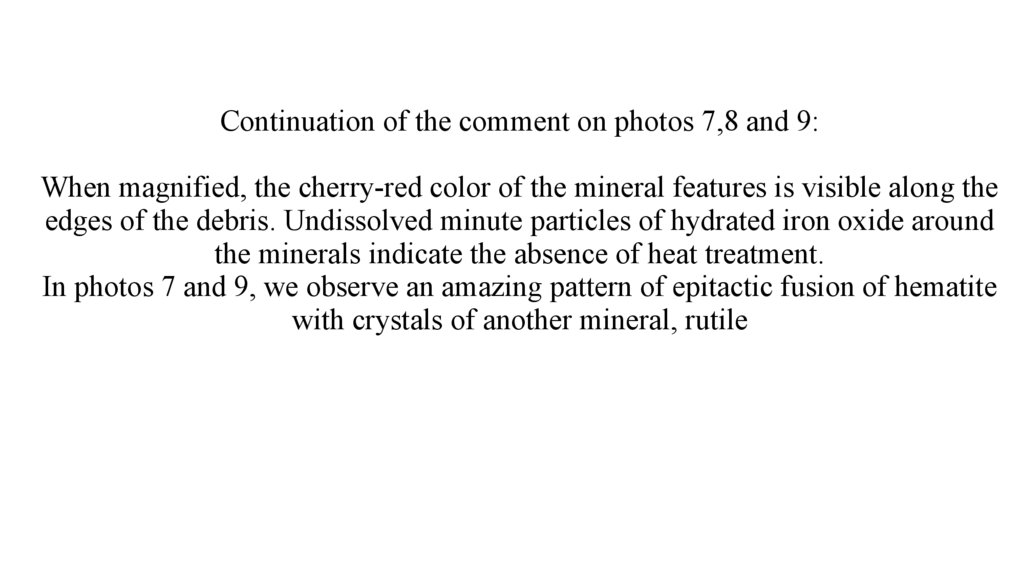
9.
Continuation of the comment on photos 7,8 and 9:s visible along the edges of the debris. Undissolved minute particles of hydrated iron o
9, we observe an amazing pattern of epitactic fusion of hematite with crystals of anoth
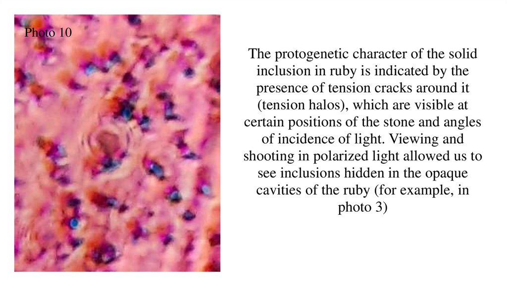
10.
Photo 10The protogenetic character of the solid
inclusion in ruby is indicated by the
presence of tension cracks around it
(tension halos), which are visible at
certain positions of the stone and angles
of incidence of light. Viewing and
shooting in polarized light allowed us to
see inclusions hidden in the opaque
cavities of the ruby (for example, in
photo 3)
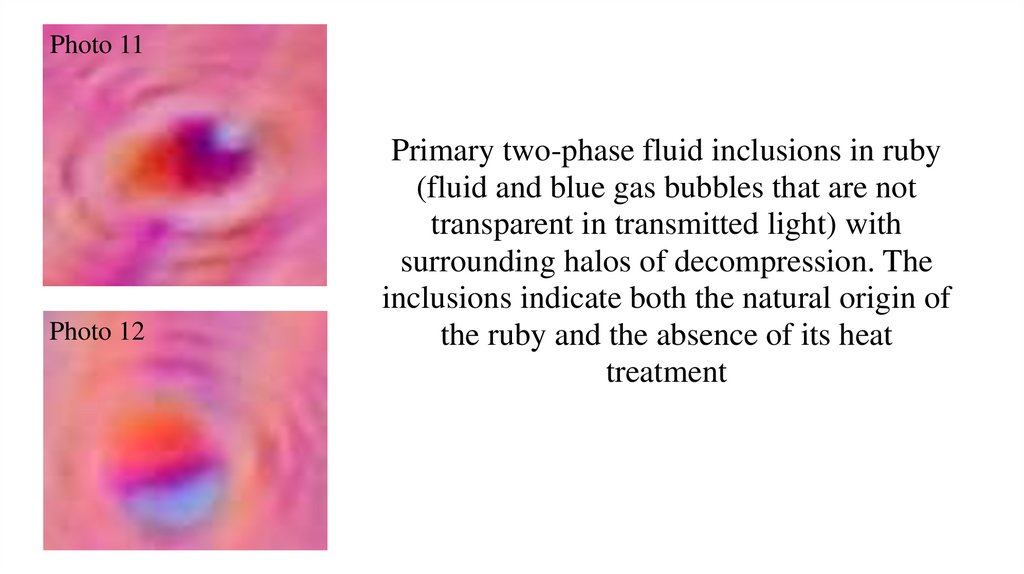
11.
Photo 11Photo 12
Primary two-phase fluid inclusions in ruby
(fluid and blue gas bubbles that are not
transparent in transmitted light) with
surrounding halos of decompression. The
inclusions indicate both the natural origin of
the ruby and the absence of its heat
treatment
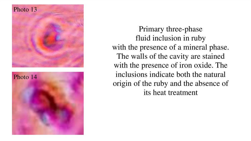
12.
Photo 13Photo 14
Primary three-phase
fluid inclusion in ruby
with the presence of a mineral phase.
The walls of the cavity are stained
with the presence of iron oxide. The
inclusions indicate both the natural
origin of the ruby and the absence of
its heat treatment
13.
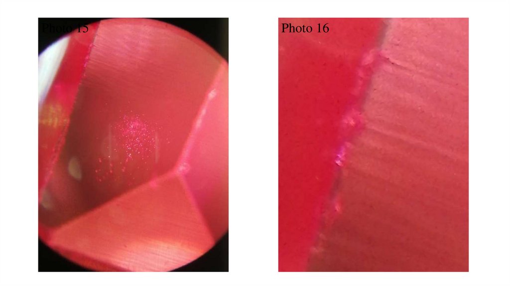
Photo 15Photo 16
14.
Comment on photos 15 and 16:The lines (stripes) visible in the photographs are on the surface of the ruby (the
results of manual cutting and polishing), in no way can they be mistaken for
ruby growth lines.
Some of the lines are a continuation of the clearly visible chips on the ribs. The
stripes do not cross faceted borders, the distances between them are different,
the stripes themselves are of different thicknesses, do not contrast and are not
concentric.
Photo 15 with a magnification of x100, photo 16 is a fragment of photo 15,
enlarged in a computer
15.
The dimensions of the stone (approximately): length and width 10x9 mm, thickness 5-6 mm, bThere are no inclusions (turbidities) visible to the naked eye, the stone is visually completely cl
However, when examining the stone under a microscope, inclusions were found concentrated i
The author of the study is Sergey Korshunov, s-777@rambler.ru