Похожие презентации:


Аллопатрическое видообразование. Симпатрическое видообразование
1.
1. Аллопатрическое видообразование.Причины дискретности видов с точки зрения
Биологической концепции вида
2. Симпатрическое видообразование
Причины дискретности видов с точки зрения
Морфологической концепции вида
2.
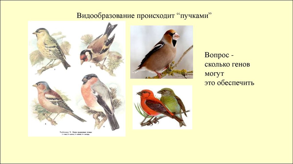
Видообразование происходит “пучками”Вопрос сколько генов
могут
это обеспечить
3.
4.
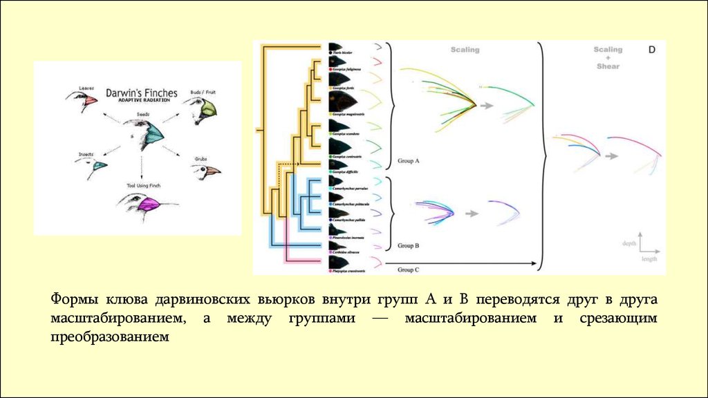
Формы клюва дарвиновских вьюрков внутри групп A и B переводятся друг в другамасштабированием, а между группами — масштабированием и срезающим
преобразованием
5.
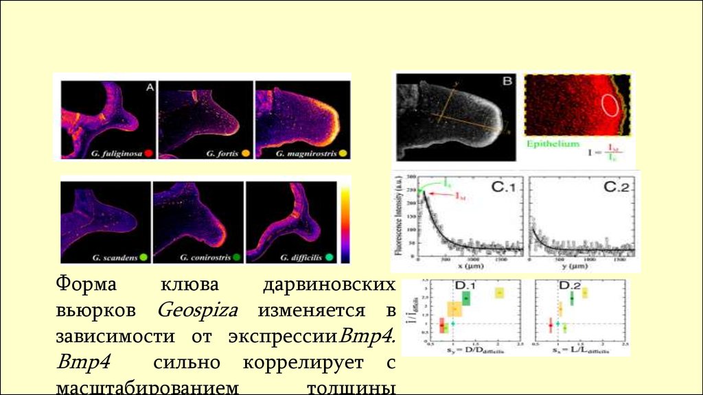
Формаклюва
дарвиновских
вьюрков Geospiza изменяется в
зависимости от экспрессииBmp4.
Bmp4
сильно коррелирует с
масштабированием
толщины
6.
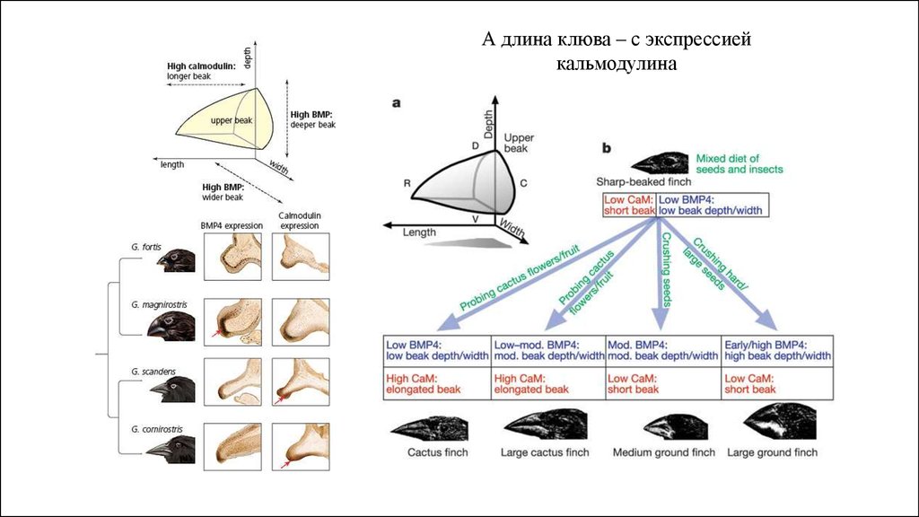
А длина клюва – с экспрессиейкальмодулина
7.
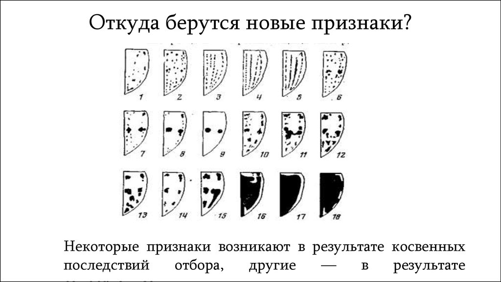
Откуда берутся новые признаки?Некоторые признаки возникают в результате косвенных
последствий
отбора,
другие
—
в
результате
8.
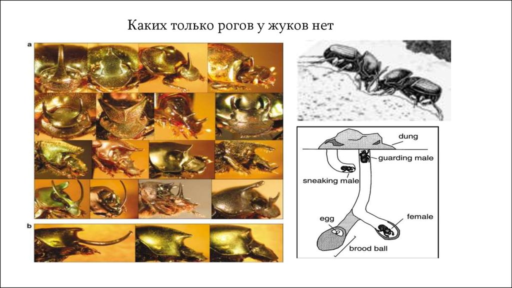
Каких только рогов у жуков нет9.
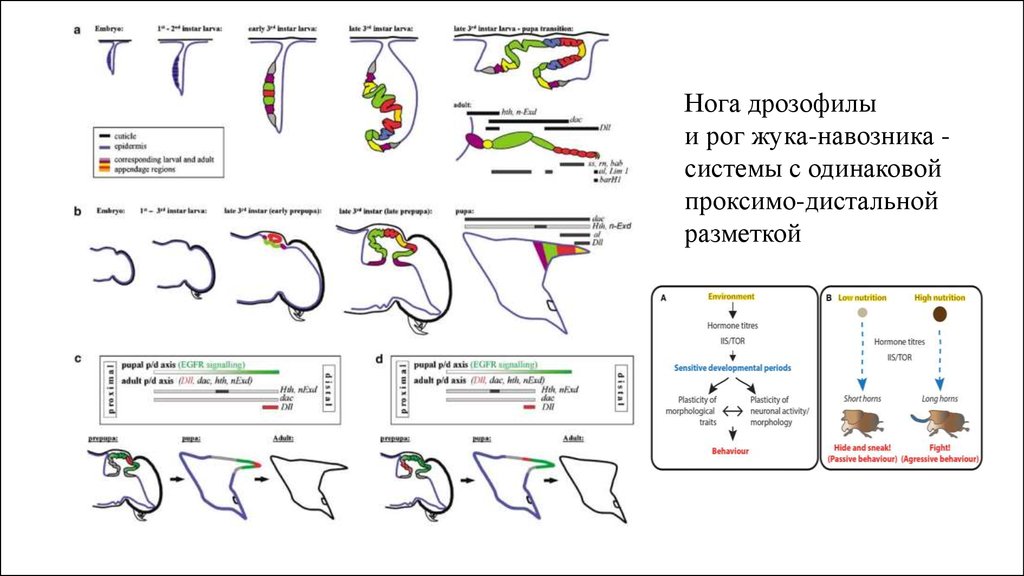
Нога дрозофилыи рог жука-навозника системы с одинаковой
проксимо-дистальной
разметкой
10.
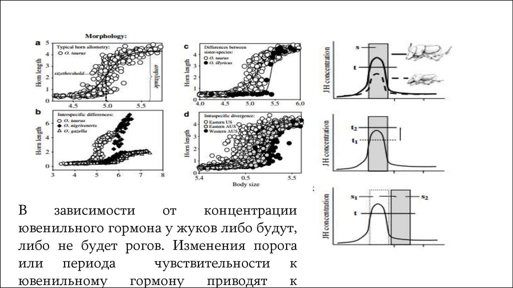
Взависимости
от
концентрации
ювенильного гормона у жуков либо будут,
либо не будет рогов. Изменения порога
или периода
чувствительности к
ювенильному
гормону
приводят
к
11.
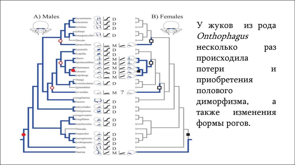
У жуковиз рода
Onthophagus
несколько
раз
происходила
потери
и
приобретения
полового
диморфизма,
а
также изменения
формы рогов.
12.
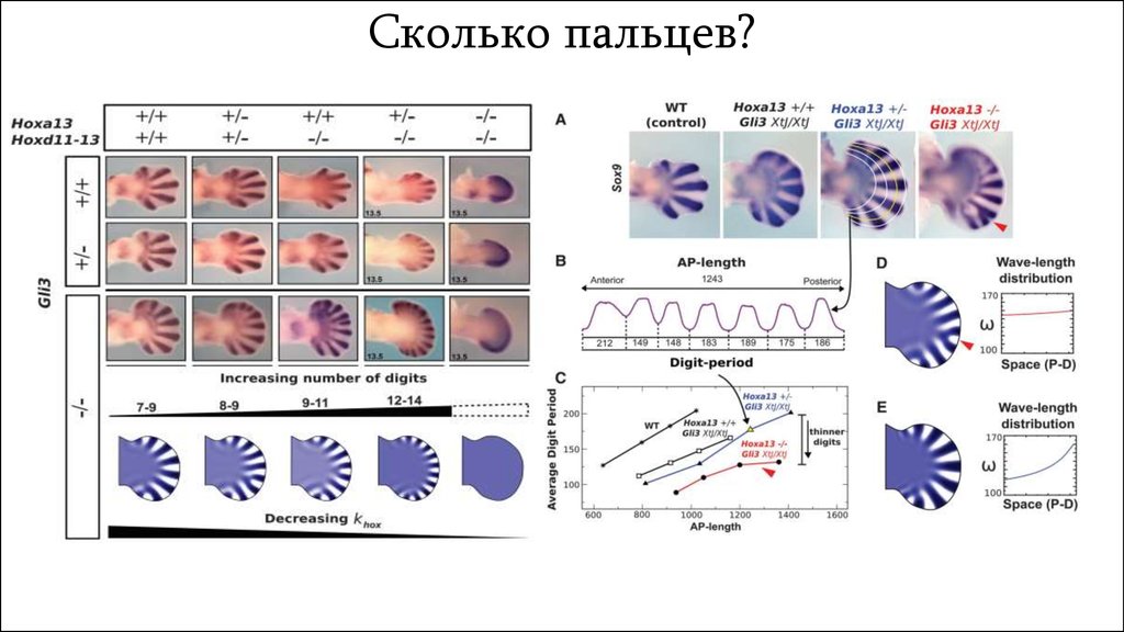
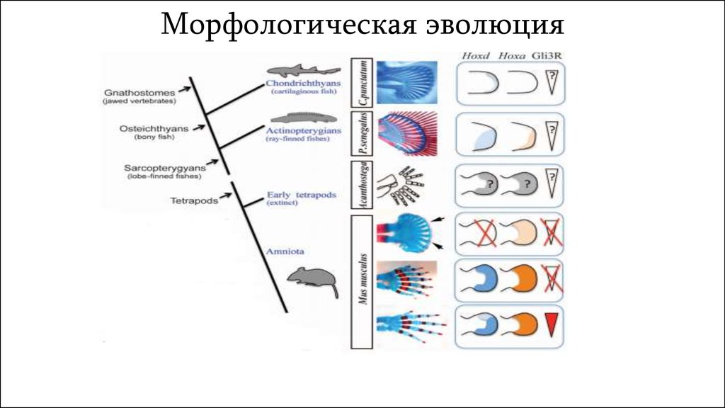
Сколько пальцев?13.
Морфологическая эволюция14.
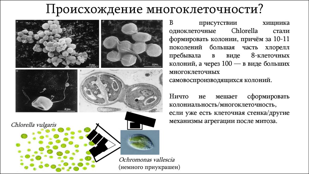
Происхождение многоклеточности?В
присутствии
хищника
одноклеточные
Chlorella
стали
формировать колонии, причём за 10-11
поколений большая часть хлорелл
пребывала
в
виде
8-клеточных
колоний, а через 100 — в виде больших
многоклеточных
самовоспроизводящихся колоний.
Chlorella vulgaris
Ничто не мешает сформировать
колониальность/многоклеточность,
если уже есть клеточная стенка/другие
механизмы агрегации после митоза.
Ochromonas vallescia
(немного приукрашен)
15.
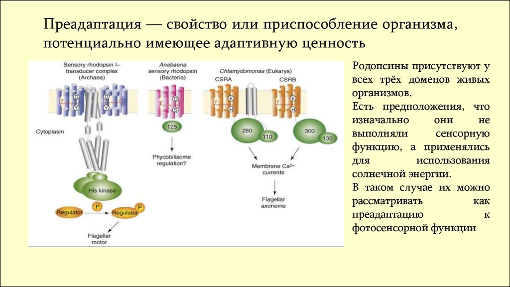
Преадаптация — свойство или приспособление организма,потенциально имеющее адаптивную ценность
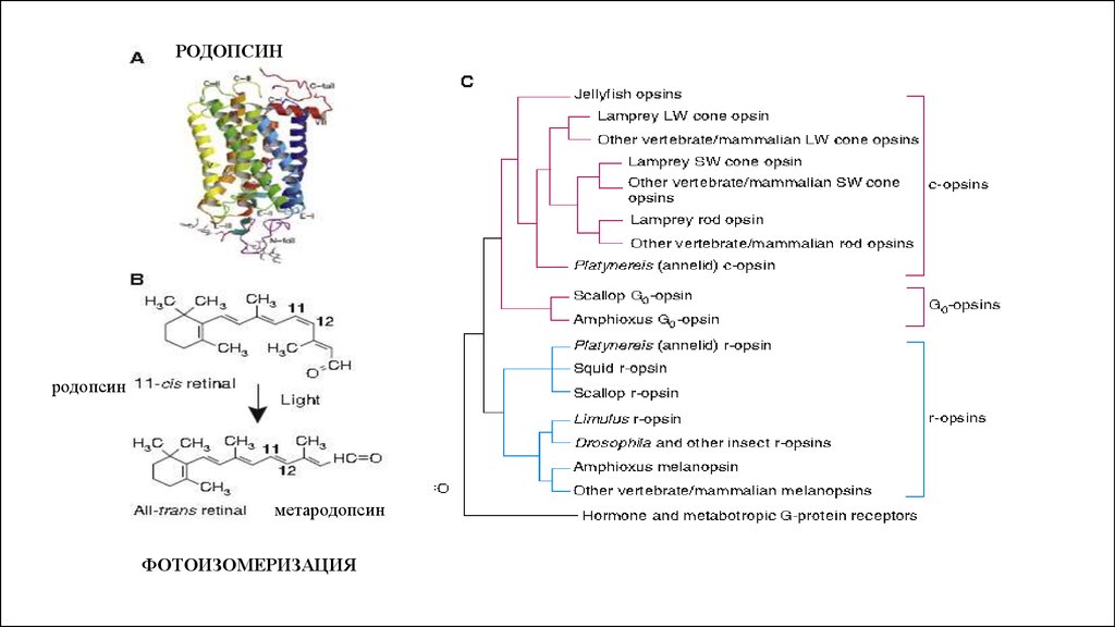
Родопсины присутствуют у
всех трёх доменов живых
организмов.
Есть предположения, что
изначально
они
не
выполняли
сенсорную
функцию, а применялись
для
использования
солнечной энергии.
В таком случае их можно
рассматривать
как
преадаптацию
к
фотосенсорной функции
16.
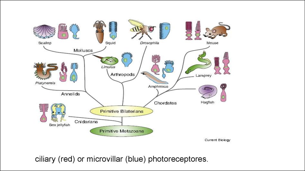
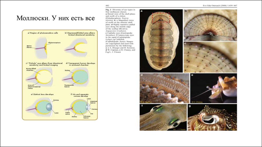
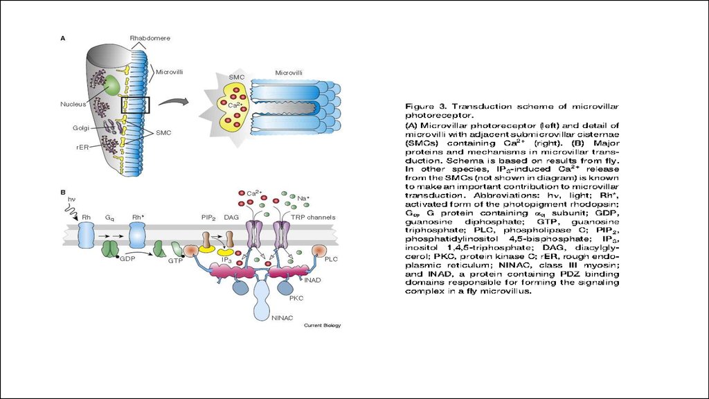
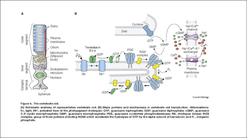
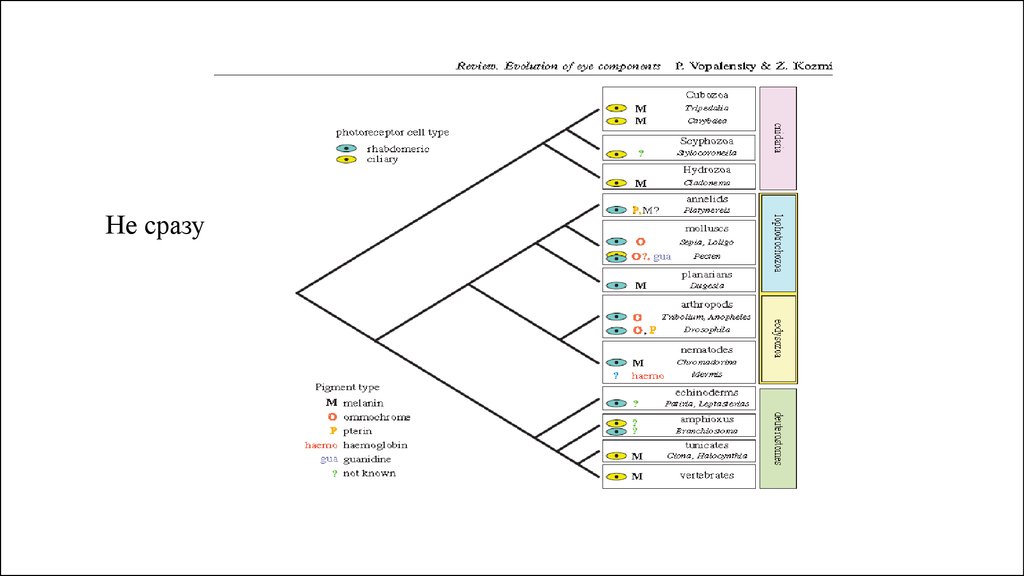
ciliary (red) or microvillar (blue) photoreceptores.17.
Моллюски. У них есть все18.
РОДОПСИНродопсин
метародопсин
ФОТОИЗОМЕРИЗАЦИЯ
19.
20.
21.
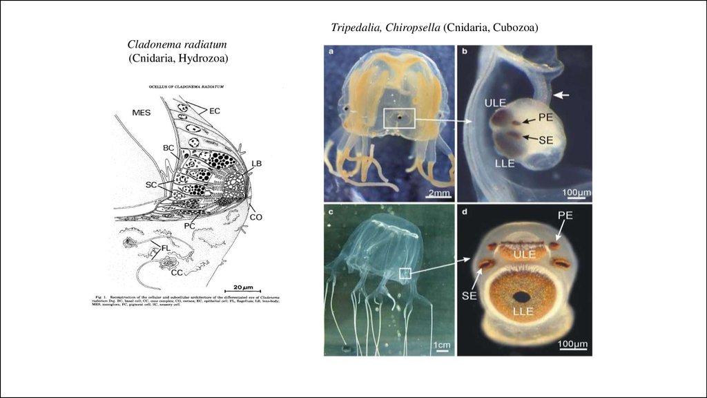
Tripedalia, Chiropsella (Cnidaria, Cubozoa)Cladonema radiatum
(Cnidaria, Hydrozoa)
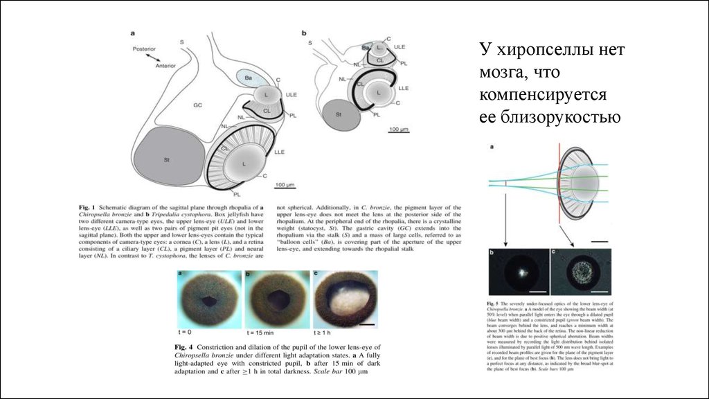
22.
У хиропселлы нетмозга, что
компенсируется
ее близорукостью
23.
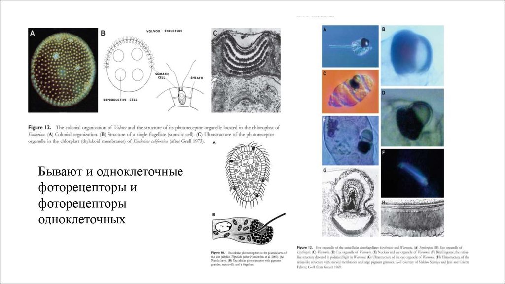
Бывают и одноклеточныефоторецепторы и
фоторецепторы
одноклеточных
24.
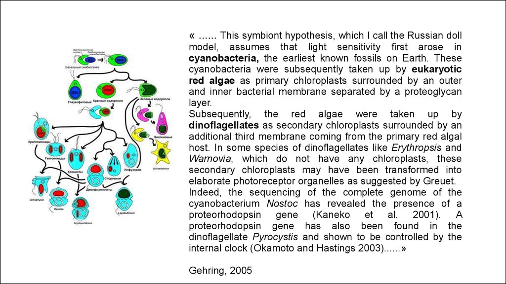
« ...... This symbiont hypothesis, which I call the Russian dollmodel, assumes that light sensitivity first arose in
cyanobacteria, the earliest known fossils on Earth. These
cyanobacteria were subsequently taken up by eukaryotic
red algae as primary chloroplasts surrounded by an outer
and inner bacterial membrane separated by a proteoglycan
layer.
Subsequently, the red algae were taken up by
dinoflagellates as secondary chloroplasts surrounded by an
additional third membrane coming from the primary red algal
host. In some species of dinoflagellates like Erythropsis and
Warnovia, which do not have any chloroplasts, these
secondary chloroplasts may have been transformed into
elaborate photoreceptor organelles as suggested by Greuet.
Indeed, the sequencing of the complete genome of the
cyanobacterium Nostoc has revealed the presence of a
proteorhodopsin gene (Kaneko et al. 2001). A
proteorhodopsin gene has also been found in the
dinoflagellate Pyrocystis and shown to be controlled by the
internal clock (Okamoto and Hastings 2003)......»
Gehring, 2005
25.
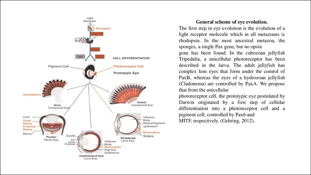
General scheme of eye evolution.The first step in eye evolution is the evolution of a
light receptor molecule which in all metazoans is
rhodopsin. In the most ancestral metazoa, the
sponges, a single Pax gene, but no opsin
gene has been found. In the cubozoan jellyfish
Tripedalia, a unicellular photoreceptor has been
described in the larva. The adult jellyfish has
complex lens eyes that form under the control of
PaxB, whereas the eyes of a hydrozoan jellyfish
(Cladonema) are controlled by PaxA. We propose
that from the unicellular
photoreceptor cell, the prototypic eye postulated by
Darwin originated by a first step of cellular
differentiation into a photoreceptor cell and a
pigment cell, controlled by Pax6 and
MITF, respectively. (Gehring, 2012).
26.
Не сразу27.
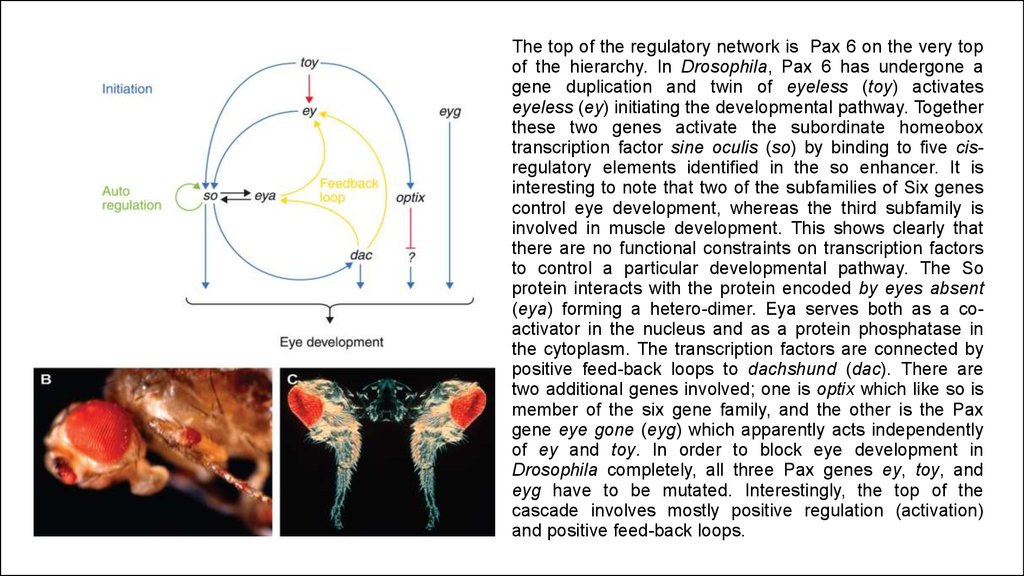
The top of the regulatory network is Pax 6 on the very topof the hierarchy. In Drosophila, Pax 6 has undergone a
gene duplication and twin of eyeless (toy) activates
eyeless (ey) initiating the developmental pathway. Together
these two genes activate the subordinate homeobox
transcription factor sine oculis (so) by binding to five cisregulatory elements identified in the so enhancer. It is
interesting to note that two of the subfamilies of Six genes
control eye development, whereas the third subfamily is
involved in muscle development. This shows clearly that
there are no functional constraints on transcription factors
to control a particular developmental pathway. The So
protein interacts with the protein encoded by eyes absent
(eya) forming a hetero-dimer. Eya serves both as a coactivator in the nucleus and as a protein phosphatase in
the cytoplasm. The transcription factors are connected by
positive feed-back loops to dachshund (dac). There are
two additional genes involved; one is optix which like so is
member of the six gene family, and the other is the Pax
gene eye gone (eyg) which apparently acts independently
of ey and toy. In order to block eye development in
Drosophila completely, all three Pax genes ey, toy, and
eyg have to be mutated. Interestingly, the top of the
cascade involves mostly positive regulation (activation)
and positive feed-back loops.
28.
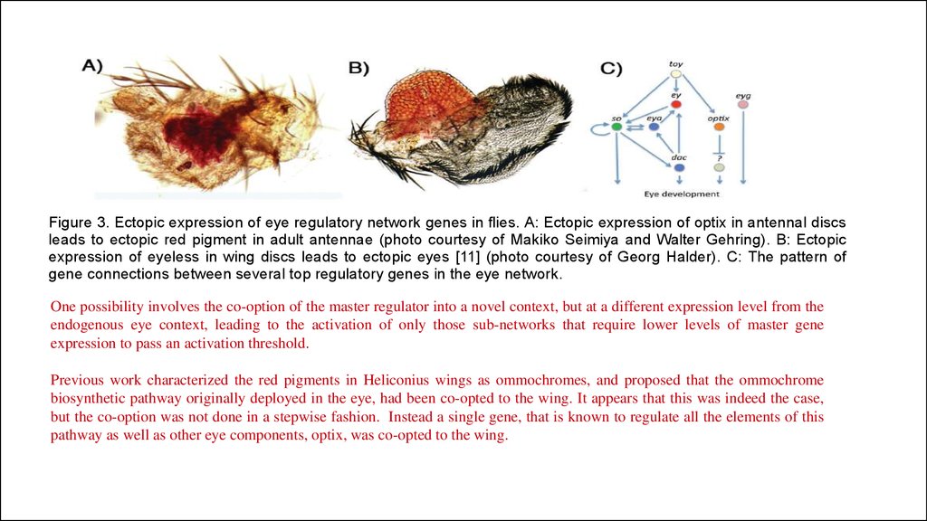
Figure 3. Ectopic expression of eye regulatory network genes in flies. A: Ectopic expression of optix in antennal discsleads to ectopic red pigment in adult antennae (photo courtesy of Makiko Seimiya and Walter Gehring). B: Ectopic
expression of eyeless in wing discs leads to ectopic eyes [11] (photo courtesy of Georg Halder). C: The pattern of
gene connections between several top regulatory genes in the eye network.
One possibility involves the co-option of the master regulator into a novel context, but at a different expression level from the
endogenous eye context, leading to the activation of only those sub-networks that require lower levels of master gene
expression to pass an activation threshold.
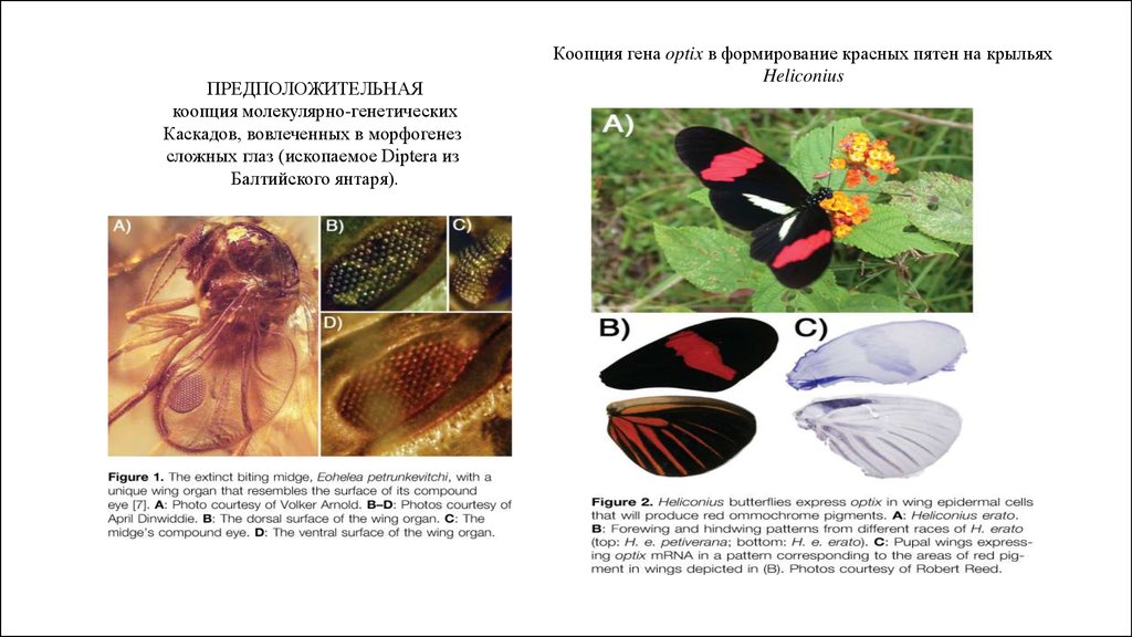
Previous work characterized the red pigments in Heliconius wings as ommochromes, and proposed that the ommochrome
biosynthetic pathway originally deployed in the eye, had been co-opted to the wing. It appears that this was indeed the case,
but the co-option was not done in a stepwise fashion. Instead a single gene, that is known to regulate all the elements of this
pathway as well as other eye components, optix, was co-opted to the wing.
29.
Monteiro, 2011ПРЕДПОЛОЖИТЕЛЬНАЯ
коопция молекулярно-генетических
Каскадов, вовлеченных в морфогенез
сложных глаз (ископаемое Diptera из
Балтийского янтаря).
Коопция гена optix в формирование красных пятен на крыльях
Heliconius
30.
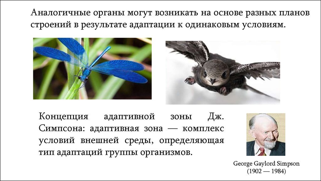
Аналогичные органы могут возникать на основе разных плановстроений в результате адаптации к одинаковым условиям.
Концепция
адаптивной
зоны
Дж.
Симпсона: адаптивная зона — комплекс
условий внешней среды, определяющая
тип адаптаций группы организмов.
George Gaylord Simpson
(1902 — 1984)
31.
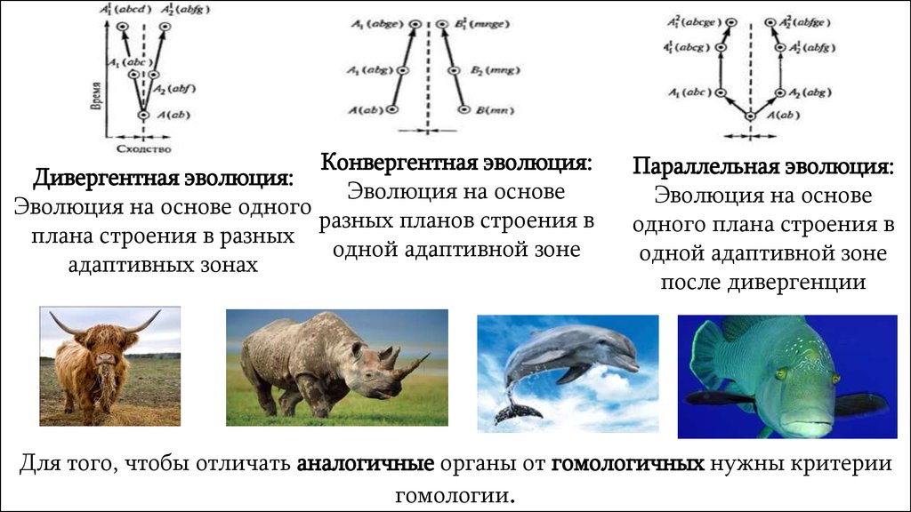
Конвергентная эволюция:Дивергентная эволюция:
Эволюция на основе
Эволюция на основе одного
разных планов строения в
плана строения в разных
одной адаптивной зоне
адаптивных зонах
Параллельная эволюция:
Эволюция на основе
одного плана строения в
одной адаптивной зоне
после дивергенции
Для того, чтобы отличать аналогичные органы от гомологичных нужны критерии
гомологии.