


 Биология
Похожие презентации:


")


The Structure and Function of Large Biological Molecules
1. Chapter 5
The Structure and Function ofLarge Biological Molecules
PowerPoint® Lecture Presentations for
Biology
Eighth Edition
Neil Campbell and Jane Reece
Lectures by Chris Romero, updated by Erin Barley with contributions from Joan Sharp
Copyright © 2008 Pearson Education, Inc., publishing as Pearson Benjamin Cummings
2. Overview: The Molecules of Life
• All living things are made up of four classes oflarge biological molecules: carbohydrates,
lipids, proteins, and nucleic acids
• Within cells, small organic molecules are joined
together to form larger molecules
• Macromolecules are large molecules
composed of thousands of covalently
connected atoms
• Molecular structure and function are
inseparable
Copyright © 2008 Pearson Education, Inc., publishing as Pearson Benjamin Cummings
3.
Fig. 5-14. Concept 5.1: Macromolecules are polymers, built from monomers
• A polymer is a long molecule consisting ofmany similar building blocks
• These small building-block molecules are
called monomers
• Three of the four classes of life’s organic
molecules are polymers:
– Carbohydrates
– Proteins
– Nucleic acids
Copyright © 2008 Pearson Education, Inc., publishing as Pearson Benjamin Cummings
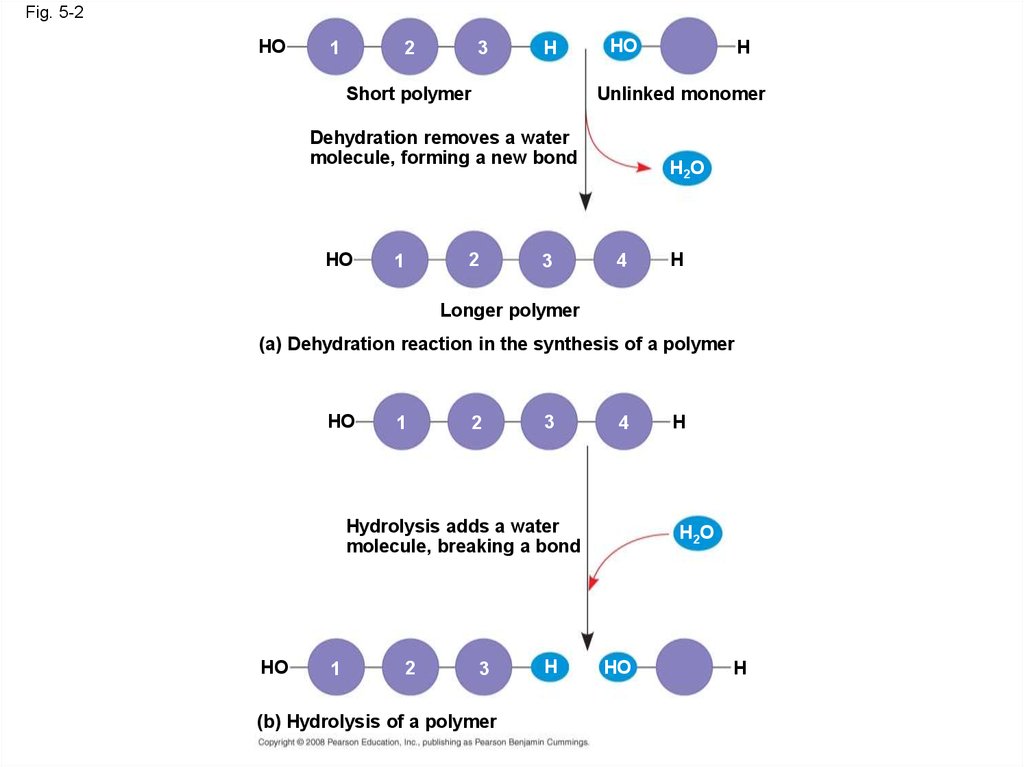
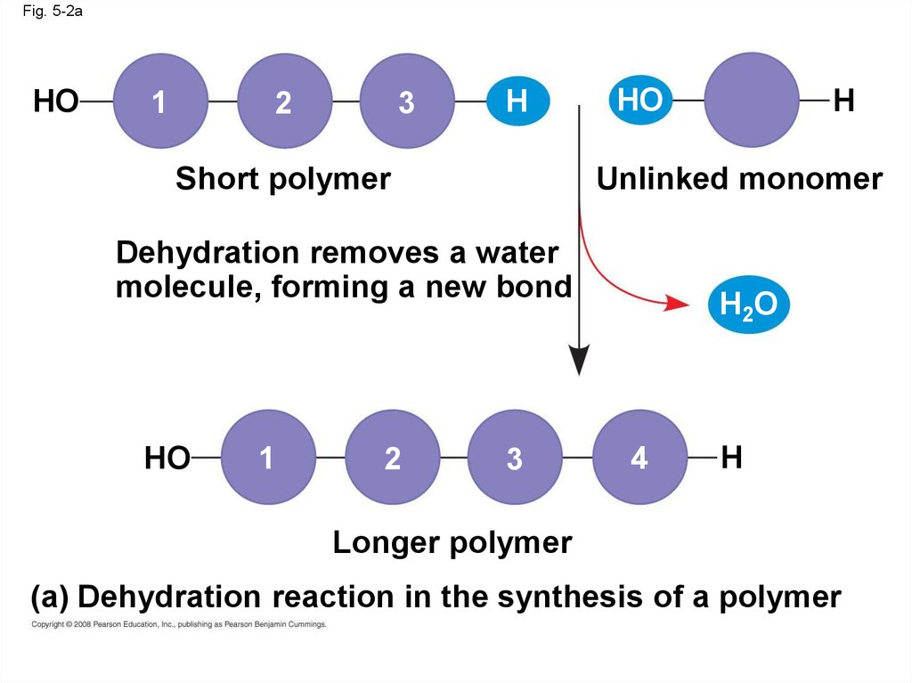
5. The Synthesis and Breakdown of Polymers
• A condensation reaction or more specificallya dehydration reaction occurs when two
monomers bond together through the loss of a
water molecule
• Enzymes are macromolecules that speed up
the dehydration process
• Polymers are disassembled to monomers by
hydrolysis, a reaction that is essentially the
reverse of the dehydration reaction
Animation: Polymers
Copyright © 2008 Pearson Education, Inc., publishing as Pearson Benjamin Cummings
6.
Fig. 5-2HO
1
2
3
H
Short polymer
HO
Unlinked monomer
Dehydration removes a water
molecule, forming a new bond
HO
2
1
H
3
H2O
4
H
Longer polymer
(a) Dehydration reaction in the synthesis of a polymer
HO
1
2
3
4
Hydrolysis adds a water
molecule, breaking a bond
HO
1
2
3
(b) Hydrolysis of a polymer
H
H
H2O
HO
H
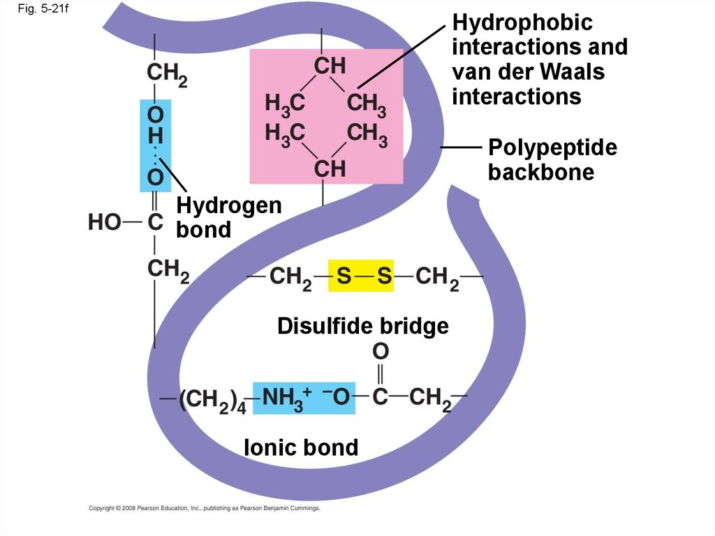
7.
Fig. 5-2aHO
1
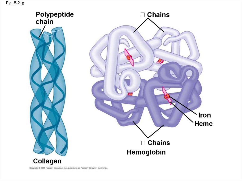
2
3
H
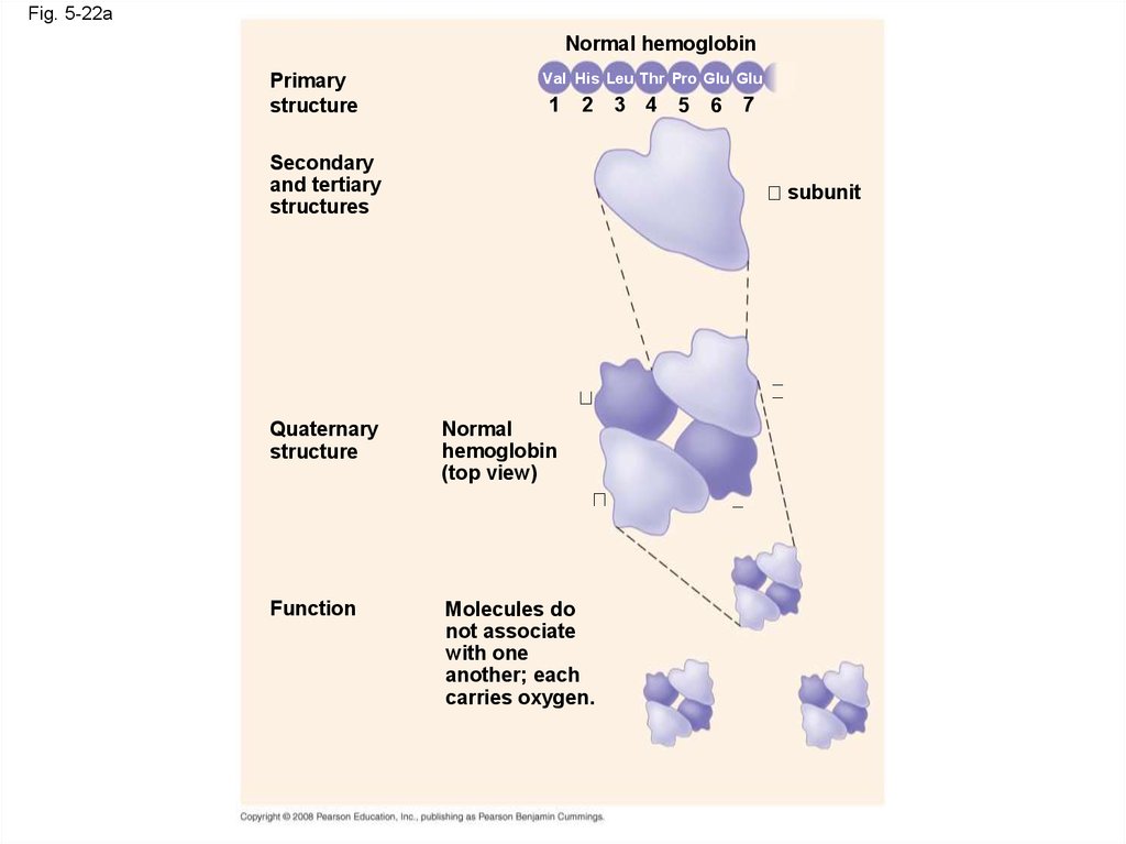
Short polymer
HO
Unlinked monomer
Dehydration removes a water
molecule, forming a new bond
HO
1
2
H
3
H2O
4
H
Longer polymer
(a) Dehydration reaction in the synthesis of a polymer
8.
Fig. 5-2bHO
1
2
3
4
Hydrolysis adds a water
molecule, breaking a bond
HO
1
2
3
(b) Hydrolysis of a polymer
H
H
H2O
HO
H
9. The Diversity of Polymers
• Each cell has thousands of different kinds ofmacromolecules2 3
H
HO
• Macromolecules vary among cells of an
organism, vary more within a species, and vary
even more between species
• An immense variety of polymers can be built
from a small set of monomers
Copyright © 2008 Pearson Education, Inc., publishing as Pearson Benjamin Cummings
10. Concept 5.2: Carbohydrates serve as fuel and building material
• Carbohydrates include sugars and thepolymers of sugars
• The simplest carbohydrates are
monosaccharides, or single sugars
• Carbohydrate macromolecules are
polysaccharides, polymers composed of many
sugar building blocks
Copyright © 2008 Pearson Education, Inc., publishing as Pearson Benjamin Cummings
11. Sugars
• Monosaccharides have molecular formulasthat are usually multiples of CH2O
• Glucose (C6H12O6) is the most common
monosaccharide
• Monosaccharides are classified by
– The location of the carbonyl group (as aldose
or ketose)
– The number of carbons in the carbon skeleton
Copyright © 2008 Pearson Education, Inc., publishing as Pearson Benjamin Cummings
12.
Fig. 5-3Trioses (C3H6O3)
Pentoses (C5H10O5)
Hexoses (C6H12O6)
Glyceraldehyde
Ribose
Glucose
Galactose
Dihydroxyacetone
Ribulose
Fructose
13.
Fig. 5-3aTrioses (C3H6O3)
Pentoses (C5H10O5)
Hexoses (C6H12O6)
Glyceraldehyde
Ribose
Glucose
Galactose
14.
Fig. 5-3bTrioses (C3H6O3)
Pentoses (C5H10O5)
Hexoses (C6H12O6)
Dihydroxyacetone
Ribulose
Fructose
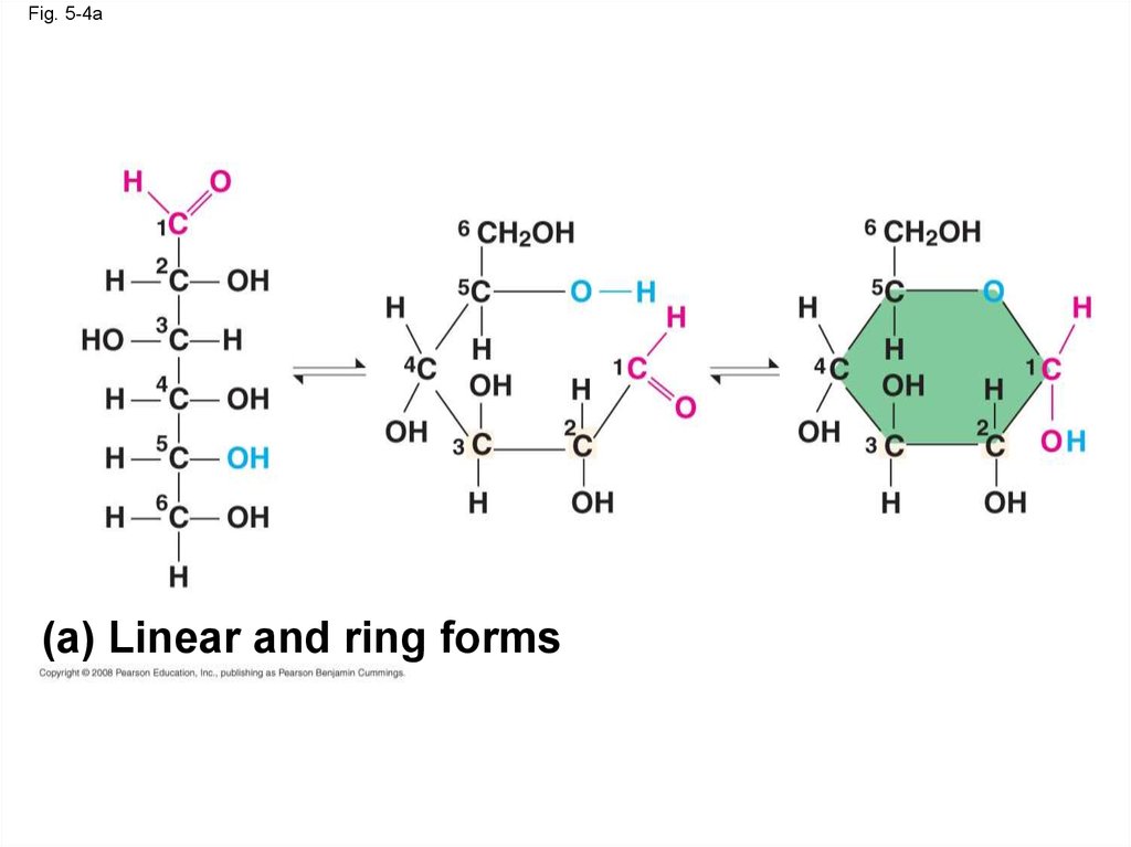
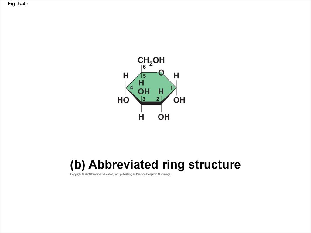
15.
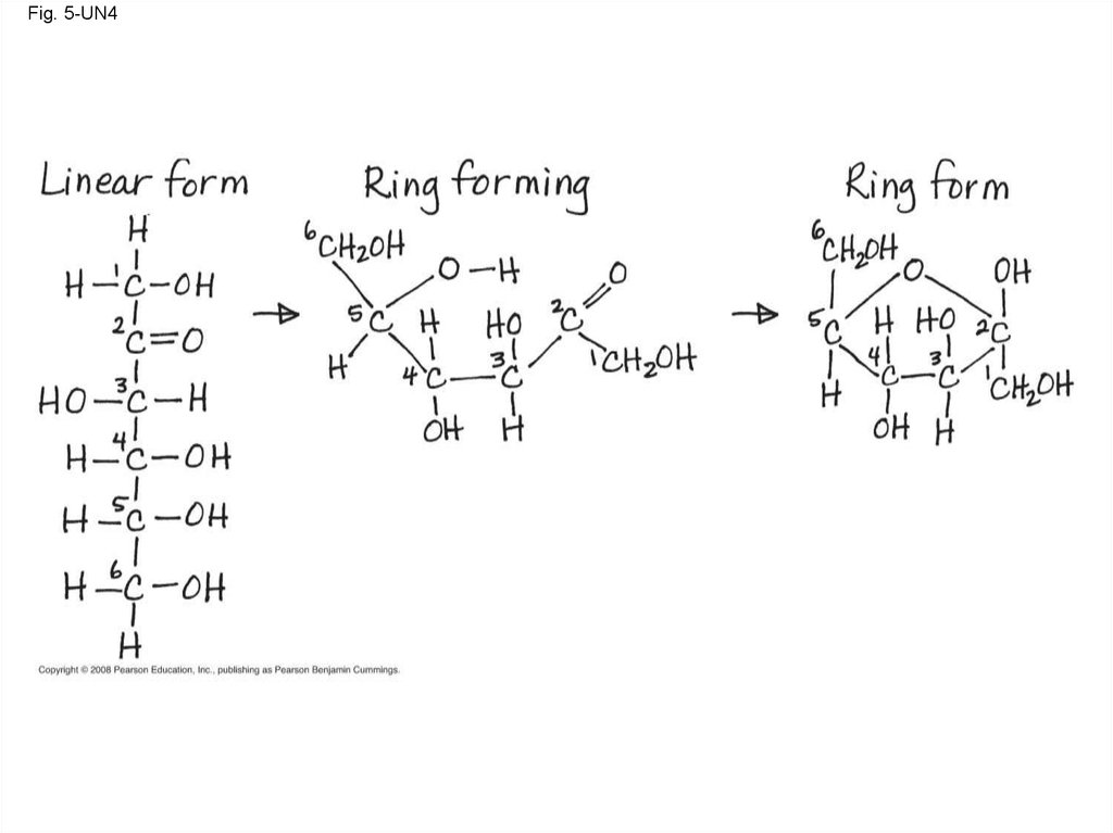
• Though often drawn as linear skeletons, inaqueous solutions many sugars form rings
• Monosaccharides serve as a major fuel for
cells and as raw material for building molecules
Copyright © 2008 Pearson Education, Inc., publishing as Pearson Benjamin Cummings
16.
Fig. 5-4(a) Linear and ring forms
(b) Abbreviated ring structure
17.
Fig. 5-4a(a) Linear and ring forms
18.
Fig. 5-4b(b) Abbreviated ring structure
19.
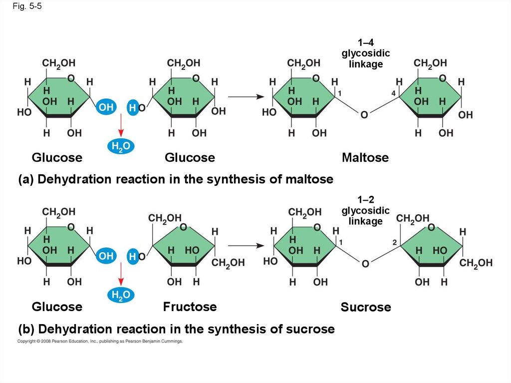
• A disaccharide is formed when a dehydrationreaction joins two monosaccharides
• This covalent bond is called a glycosidic
linkage
Animation: Disaccharides
Copyright © 2008 Pearson Education, Inc., publishing as Pearson Benjamin Cummings
20.
Fig. 5-51–4
glycosidic
linkage
Glucose
Glucose
Maltose
(a) Dehydration reaction in the synthesis of maltose
1–2
glycosidic
linkage
Glucose
Fructose
(b) Dehydration reaction in the synthesis of sucrose
Sucrose
21. Polysaccharides
• Polysaccharides, the polymers of sugars,have storage and structural roles
• The structure and function of a polysaccharide
are determined by its sugar monomers and the
positions of glycosidic linkages
Copyright © 2008 Pearson Education, Inc., publishing as Pearson Benjamin Cummings
22. Storage Polysaccharides
• Starch, a storage polysaccharide of plants,consists entirely of glucose monomers
• Plants store surplus starch as granules within
chloroplasts and other plastids
Copyright © 2008 Pearson Education, Inc., publishing as Pearson Benjamin Cummings
23.
Fig. 5-6Chloroplast
Mitochondria Glycogen granules
Starch
0.5 µm
1 µm
Glycogen
Amylose
Amylopectin
(a) Starch: a plant polysaccharide
(b) Glycogen: an animal polysaccharide
24.
• Glycogen is a storage polysaccharide inanimals
• Humans and other vertebrates store glycogen
mainly in liver and muscle cells
Copyright © 2008 Pearson Education, Inc., publishing as Pearson Benjamin Cummings
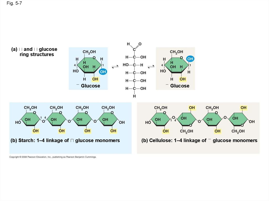
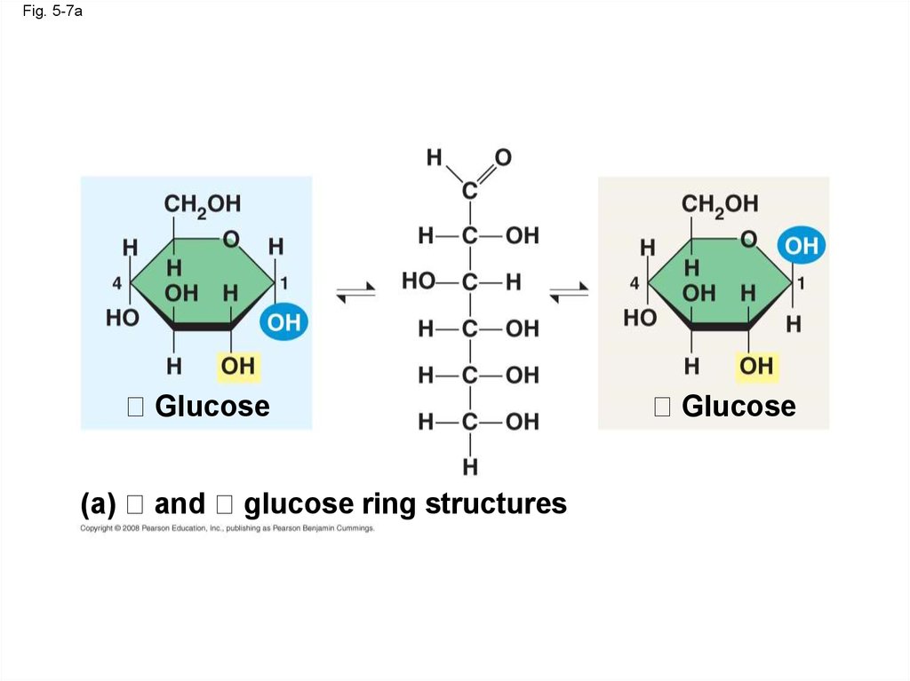
25. Structural Polysaccharides
• The polysaccharide cellulose is a majorcomponent of the tough wall of plant cells
• Like starch, cellulose is a polymer of glucose,
but the glycosidic linkages differ
• The difference is based on two ring forms for
glucose: alpha ( ) and beta ( )
Animation: Polysaccharides
Copyright © 2008 Pearson Education, Inc., publishing as Pearson Benjamin Cummings
26.
Fig. 5-7(a)
and glucose
ring structures
Glucose
(b) Starch: 1–4 linkage of
glucose monomers
Glucose
(b) Cellulose: 1–4 linkage of
glucose monomers
27.
Fig. 5-7aGlucose
(a)
and
glucose ring structures
Glucose
28.
Fig. 5-7bc(b) Starch: 1–4 linkage of
glucose monomers
(c) Cellulose: 1–4 linkage of
glucose monomers
29.
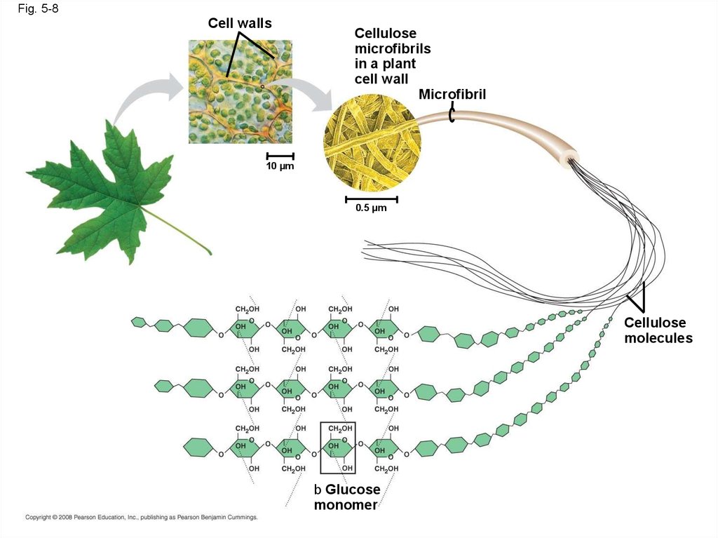
• Polymers with glucose are helical• Polymers with glucose are straight
• In straight structures, H atoms on one
strand can bond with OH groups on other
strands
• Parallel cellulose molecules held together
this way are grouped into microfibrils, which
form strong building materials for plants
Copyright © 2008 Pearson Education, Inc., publishing as Pearson Benjamin Cummings
30.
Fig. 5-8Cell walls
Cellulose
microfibrils
in a plant
cell wall
Microfibril
10 µm
0.5 µm
Cellulose
molecules
b Glucose
monomer
31.
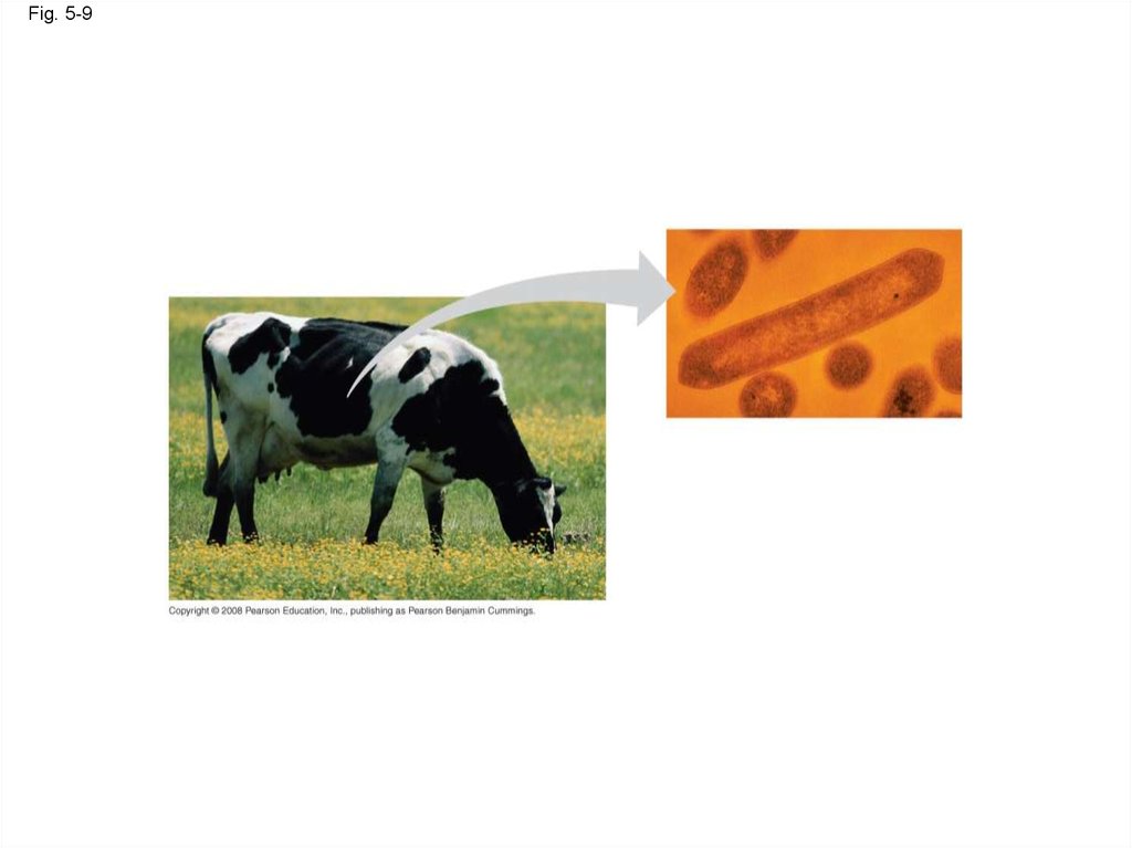
• Enzymes that digest starch by hydrolyzinglinkages can’t hydrolyze linkages in cellulose
• Cellulose in human food passes through the
digestive tract as insoluble fiber
• Some microbes use enzymes to digest
cellulose
• Many herbivores, from cows to termites, have
symbiotic relationships with these microbes
Copyright © 2008 Pearson Education, Inc., publishing as Pearson Benjamin Cummings
32.
Fig. 5-933.
• Chitin, another structural polysaccharide, isfound in the exoskeleton of arthropods
• Chitin also provides structural support for the
cell walls of many fungi
Copyright © 2008 Pearson Education, Inc., publishing as Pearson Benjamin Cummings
34.
Fig. 5-10(a) The structure
of the chitin
monomer.
(b) Chitin forms the
exoskeleton of
arthropods.
(c) Chitin is used to make
a strong and flexible
surgical thread.
35. Concept 5.3: Lipids are a diverse group of hydrophobic molecules
• Lipids are the one class of large biologicalmolecules that do not form polymers
• The unifying feature of lipids is having little or
no affinity for water
• Lipids are hydrophobic because they consist
mostly of hydrocarbons, which form nonpolar
covalent bonds
• The most biologically important lipids are fats,
phospholipids, and steroids
Copyright © 2008 Pearson Education, Inc., publishing as Pearson Benjamin Cummings
36. Fats
• Fats are constructed from two types of smallermolecules: glycerol and fatty acids
• Glycerol is a three-carbon alcohol with a
hydroxyl group attached to each carbon
• A fatty acid consists of a carboxyl group
attached to a long carbon skeleton
Copyright © 2008 Pearson Education, Inc., publishing as Pearson Benjamin Cummings
37.
Fig. 5-11Fatty acid
(palmitic acid)
Glycerol
(a) Dehydration reaction in the synthesis of a fat
Ester linkage
(b) Fat molecule (triacylglycerol)
38.
Fig. 5-11aFatty acid
(palmitic acid)
Glycerol
(a) Dehydration reaction in the synthesis of a fat
39.
Fig. 5-11bEster linkage
(b) Fat molecule (triacylglycerol)
40.
• Fats separate from water becausewater molecules form hydrogen bonds
with each other and exclude the fats
• In a fat, three fatty acids are joined to
glycerol by an ester linkage, creating a
triacylglycerol, or triglyceride
Copyright © 2008 Pearson Education, Inc., publishing as Pearson Benjamin Cummings
41.
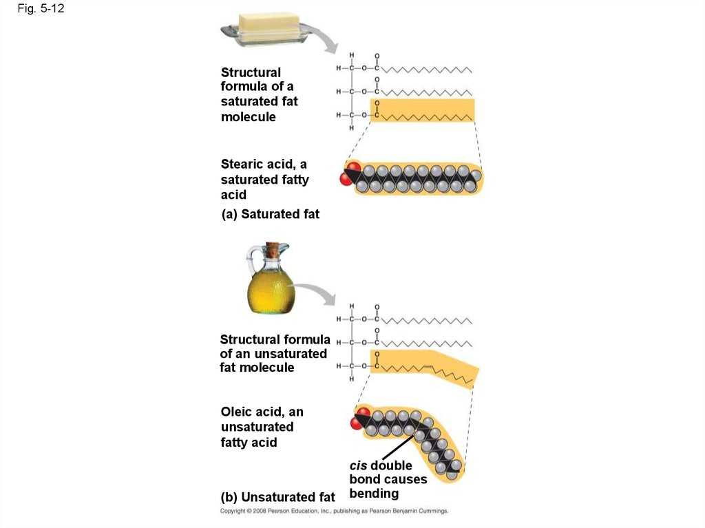
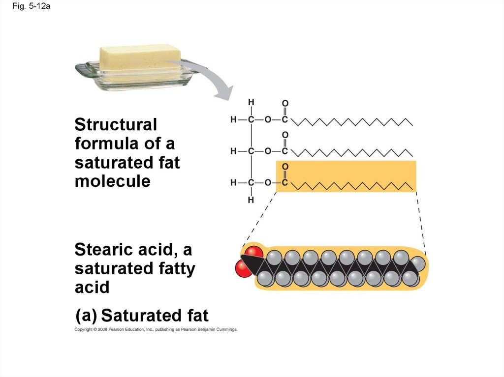
• Fatty acids vary in length (number of carbons)and in the number and locations of double
bonds
• Saturated fatty acids have the maximum
number of hydrogen atoms possible and no
double bonds
• Unsaturated fatty acids have one or more
double bonds
Animation: Fats
Copyright © 2008 Pearson Education, Inc., publishing as Pearson Benjamin Cummings
42.
Fig. 5-12Structural
formula of a
saturated fat
molecule
Stearic acid, a
saturated fatty
acid
(a) Saturated fat
Structural formula
of an unsaturated
fat molecule
Oleic acid, an
unsaturated
fatty acid
(b) Unsaturated fat
cis double
bond causes
bending
43.
Fig. 5-12aStructural
formula of a
saturated fat
molecule
Stearic acid, a
saturated fatty
acid
(a) Saturated fat
44.
Fig. 5-12bStructural formula
of an unsaturated
fat molecule
Oleic acid, an
unsaturated
fatty acid
(b) Unsaturated fat
cis double
bond causes
bending
45.
• Fats made from saturated fatty acids are calledsaturated fats, and are solid at room
temperature
• Most animal fats are saturated
• Fats made from unsaturated fatty acids are
called unsaturated fats or oils, and are liquid at
room temperature
• Plant fats and fish fats are usually unsaturated
Copyright © 2008 Pearson Education, Inc., publishing as Pearson Benjamin Cummings
46.
• A diet rich in saturated fats may contribute tocardiovascular disease through plaque deposits
• Hydrogenation is the process of converting
unsaturated fats to saturated fats by adding
hydrogen
• Hydrogenating vegetable oils also creates
unsaturated fats with trans double bonds
• These trans fats may contribute more than
saturated fats to cardiovascular disease
Copyright © 2008 Pearson Education, Inc., publishing as Pearson Benjamin Cummings
47.
• The major function of fats is energy storage• Humans and other mammals store their fat in
adipose cells
• Adipose tissue also cushions vital organs and
insulates the body
Copyright © 2008 Pearson Education, Inc., publishing as Pearson Benjamin Cummings
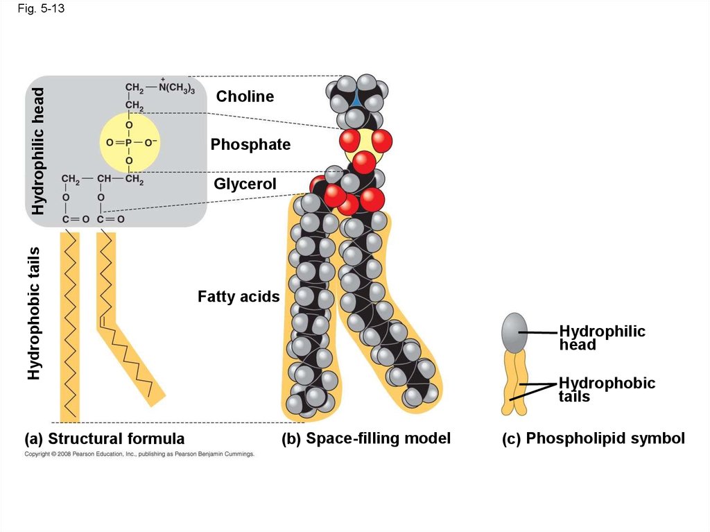
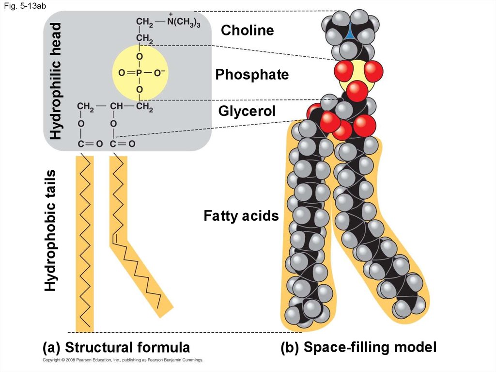
48. Phospholipids
• In a phospholipid, two fatty acids and aphosphate group are attached to glycerol
• The two fatty acid tails are hydrophobic, but the
phosphate group and its attachments form a
hydrophilic head
Copyright © 2008 Pearson Education, Inc., publishing as Pearson Benjamin Cummings
49.
Hydrophobic tailsHydrophilic head
Fig. 5-13
(a) Structural formula
Choline
Phosphate
Glycerol
Fatty acids
Hydrophilic
head
Hydrophobic
tails
(b) Space-filling model
(c) Phospholipid symbol
50.
Hydrophobic tailsHydrophilic head
Fig. 5-13ab
(a) Structural formula
Choline
Phosphate
Glycerol
Fatty acids
(b) Space-filling model
51.
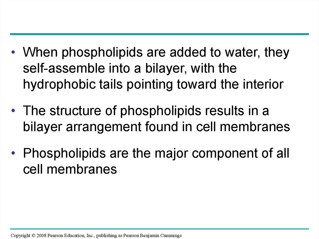
• When phospholipids are added to water, theyself-assemble into a bilayer, with the
hydrophobic tails pointing toward the interior
• The structure of phospholipids results in a
bilayer arrangement found in cell membranes
• Phospholipids are the major component of all
cell membranes
Copyright © 2008 Pearson Education, Inc., publishing as Pearson Benjamin Cummings
52.
Fig. 5-14Hydrophilic
head
Hydrophobic
tail
WATER
WATER
53. Steroids
• Steroids are lipids characterized by a carbonskeleton consisting of four fused rings
• Cholesterol, an important steroid, is a
component in animal cell membranes
• Although cholesterol is essential in animals,
high levels in the blood may contribute to
cardiovascular disease
Copyright © 2008 Pearson Education, Inc., publishing as Pearson Benjamin Cummings
54.
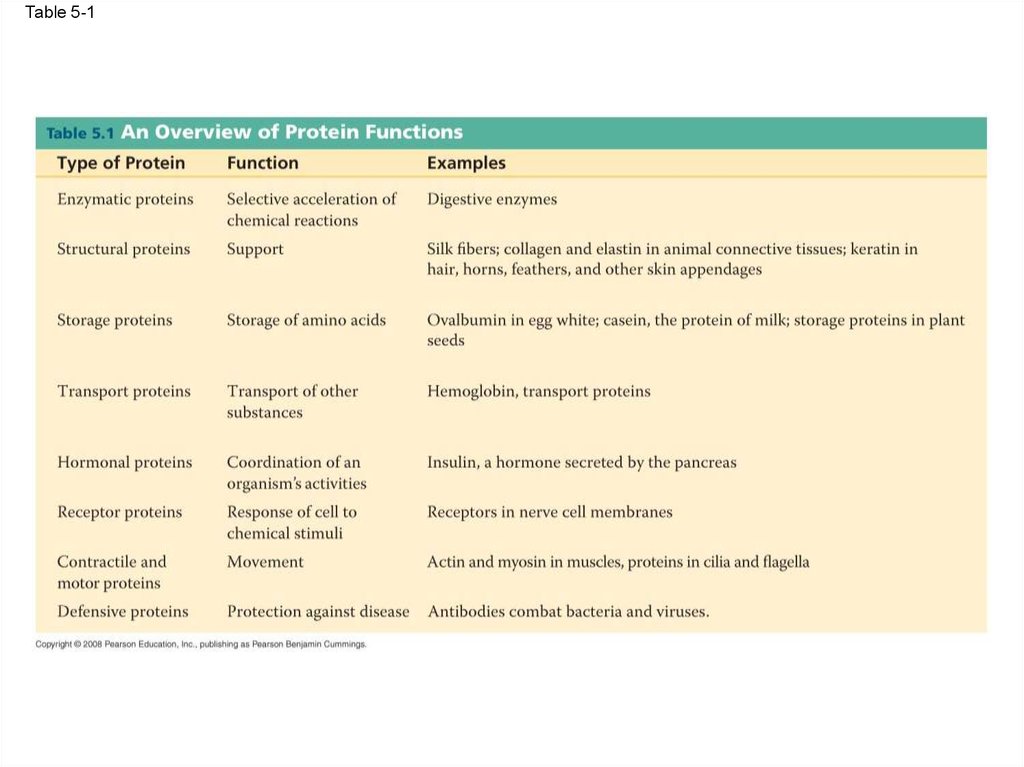
Fig. 5-1555. Concept 5.4: Proteins have many structures, resulting in a wide range of functions
• Proteins account for more than 50% of the drymass of most cells
• Protein functions include structural support,
storage, transport, cellular communications,
movement, and defense against foreign
substances
Copyright © 2008 Pearson Education, Inc., publishing as Pearson Benjamin Cummings
56.
Table 5-157.
Animation: Structural ProteinsAnimation: Storage Proteins
Animation: Transport Proteins
Animation: Receptor Proteins
Animation: Contractile Proteins
Animation: Defensive Proteins
Animation: Hormonal Proteins
Animation: Sensory Proteins
Animation: Gene Regulatory Proteins
Copyright © 2008 Pearson Education, Inc., publishing as Pearson Benjamin Cummings
58.
• Enzymes are a type of protein that acts as acatalyst to speed up chemical reactions
• Enzymes can perform their functions
repeatedly, functioning as workhorses that
carry out the processes of life
Animation: Enzymes
Copyright © 2008 Pearson Education, Inc., publishing as Pearson Benjamin Cummings
59.
Fig. 5-16Substrate
(sucrose)
Glucose
OH
Fructose
HO
Enzyme
(sucrase)
H2O
60. Polypeptides
• Polypeptides are polymers built from thesame set of 20 amino acids
• A protein consists of one or more polypeptides
Copyright © 2008 Pearson Education, Inc., publishing as Pearson Benjamin Cummings
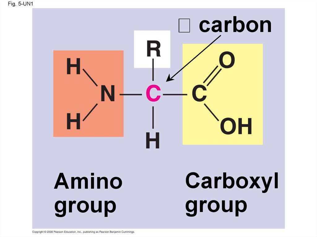
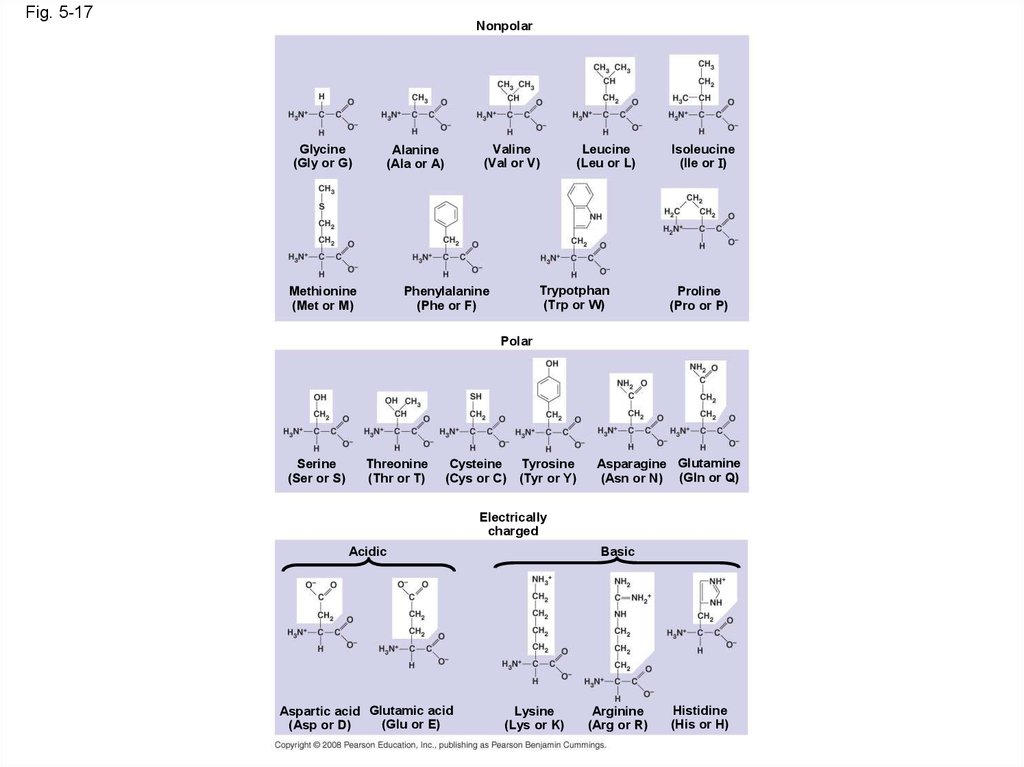
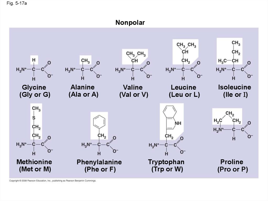
61. Amino Acid Monomers
• Amino acids are organic molecules withcarboxyl and amino groups
• Amino acids differ in their properties due to
differing side chains, called R groups
Copyright © 2008 Pearson Education, Inc., publishing as Pearson Benjamin Cummings
62.
Fig. 5-UN1carbon
Amino
group
Carboxyl
group
63.
Fig. 5-17Nonpolar
Glycine
(Gly or G)
Valine
(Val or V)
Alanine
(Ala or A)
Methionine
(Met or M)
Leucine
(Leu or L)
Trypotphan
(Trp or W)
Phenylalanine
(Phe or F)
Isoleucine
(Ile or I)
Proline
(Pro or P)
Polar
Serine
(Ser or S)
Threonine
(Thr or T)
Cysteine Tyrosine
(Cys or C) (Tyr or Y)
Asparagine Glutamine
(Asn or N) (Gln or Q)
Electrically
charged
Acidic
Aspartic acid Glutamic acid
(Glu or E)
(Asp or D)
Basic
Lysine
(Lys or K)
Arginine
(Arg or R)
Histidine
(His or H)
64.
Fig. 5-17aNonpolar
Glycine
(Gly or G)
Methionine
(Met or M)
Alanine
(Ala or A)
Valine
(Val or V)
Phenylalanine
(Phe or F)
Leucine
(Leu or L)
Tryptophan
(Trp or W)
Isoleucine
(Ile or I)
Proline
(Pro or P)
65.
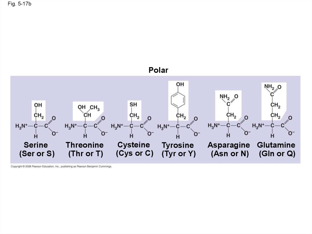
Fig. 5-17bPolar
Serine
(Ser or S)
Threonine
(Thr or T)
Cysteine
(Cys or C)
Tyrosine
(Tyr or Y)
Asparagine Glutamine
(Asn or N) (Gln or Q)
66.
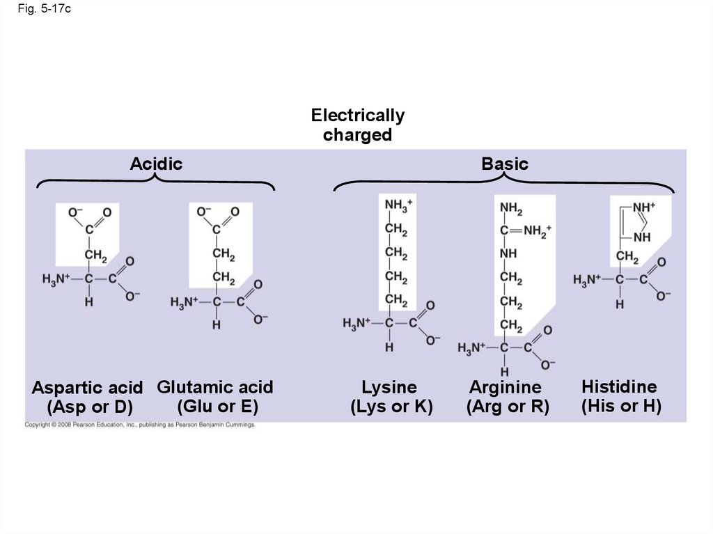
Fig. 5-17cElectrically
charged
Acidic
Aspartic acid Glutamic acid
(Glu or E)
(Asp or D)
Basic
Lysine
(Lys or K)
Arginine
(Arg or R)
Histidine
(His or H)
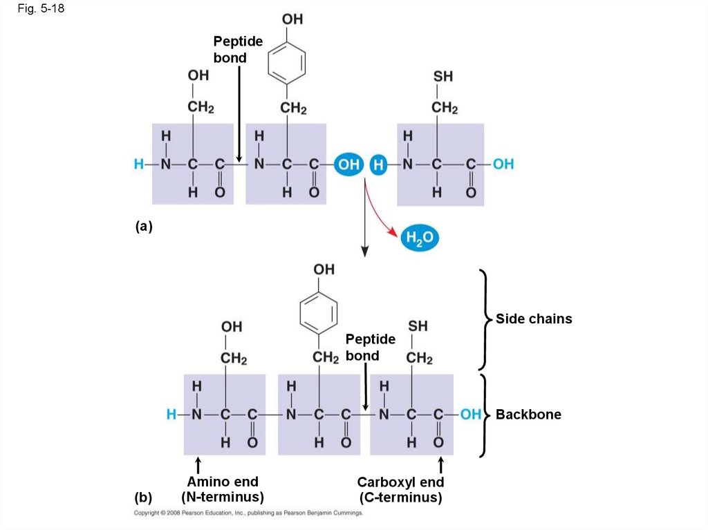
67. Amino Acid Polymers
• Amino acids are linked by peptide bonds• A polypeptide is a polymer of amino acids
• Polypeptides range in length from a few to
more than a thousand monomers
• Each polypeptide has a unique linear sequence
of amino acids
Copyright © 2008 Pearson Education, Inc., publishing as Pearson Benjamin Cummings
68.
Fig. 5-18Peptide
bond
(a)
Side chains
Peptide
bond
Backbone
(b)
Amino end
(N-terminus)
Carboxyl end
(C-terminus)
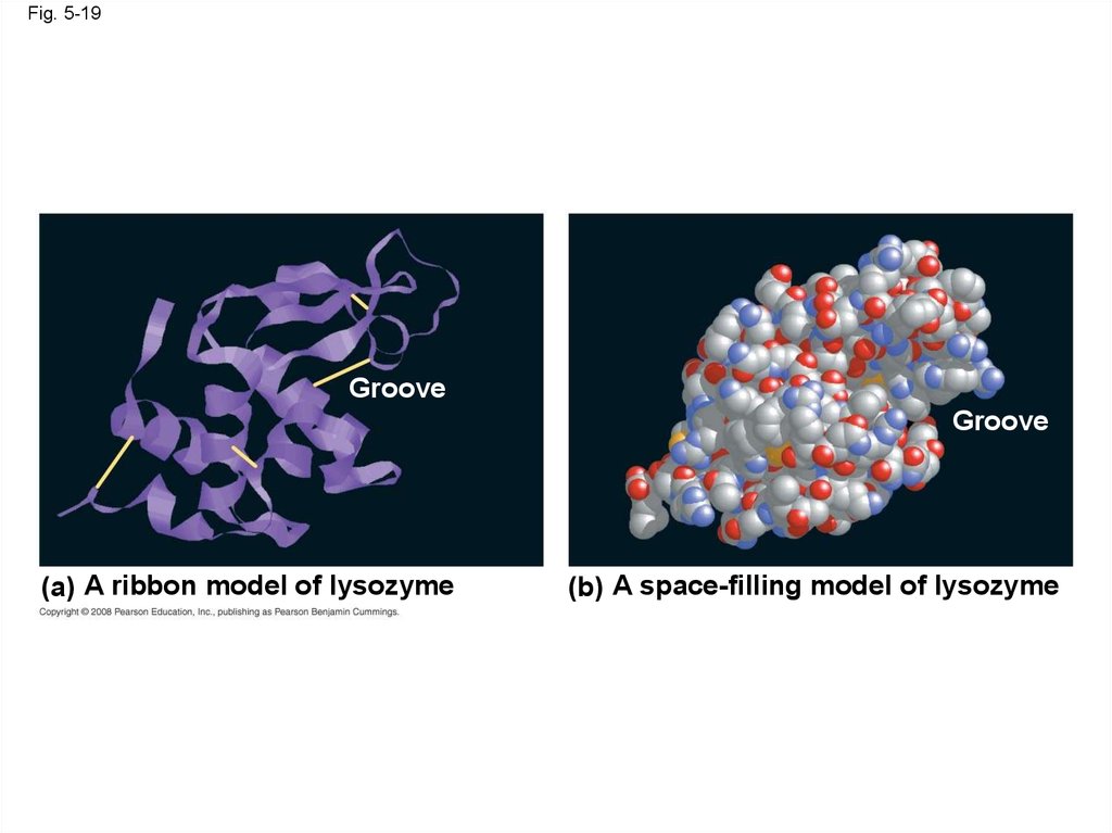
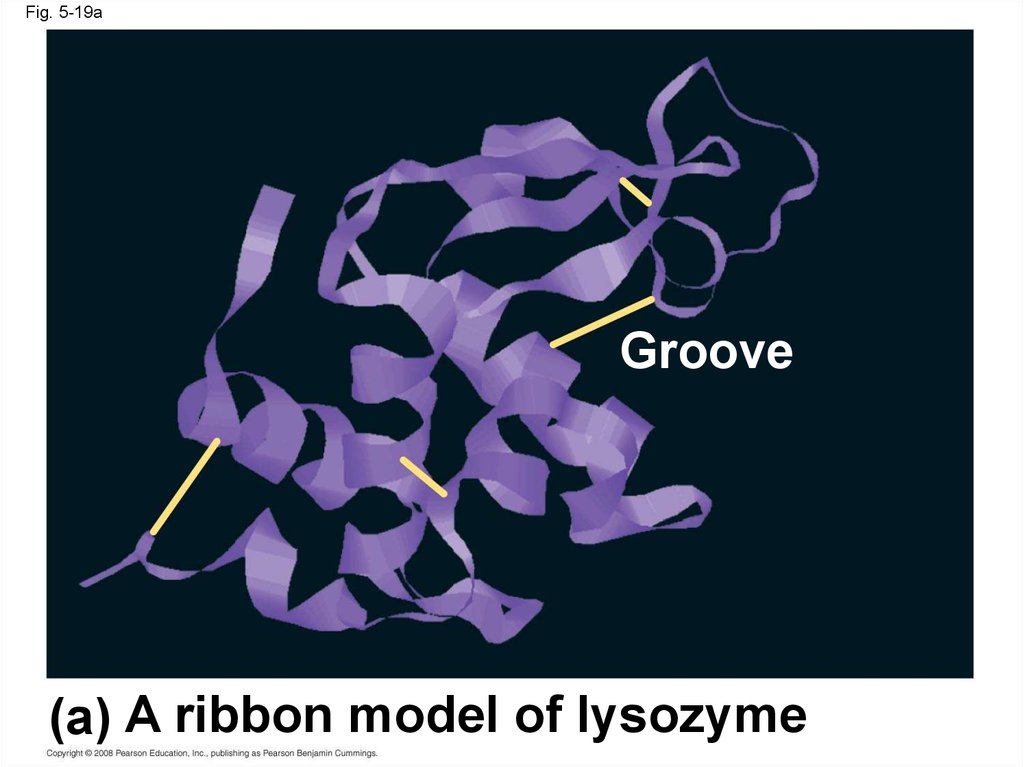
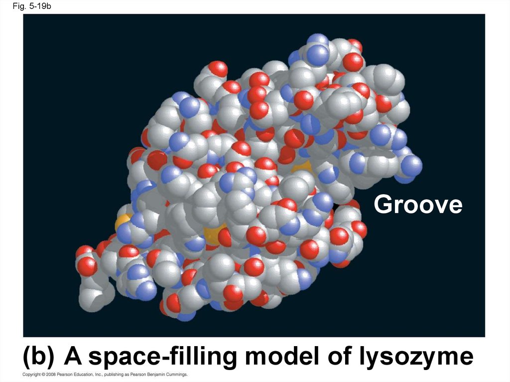
69. Protein Structure and Function
• A functional protein consists of one or morepolypeptides twisted, folded, and coiled into a
unique shape
Copyright © 2008 Pearson Education, Inc., publishing as Pearson Benjamin Cummings
70.
Fig. 5-19Groove
Groove
(a) A ribbon model of lysozyme
(b) A space-filling model of lysozyme
71.
Fig. 5-19aGroove
(a) A ribbon model of lysozyme
72.
Fig. 5-19bGroove
(b) A space-filling model of lysozyme
73.
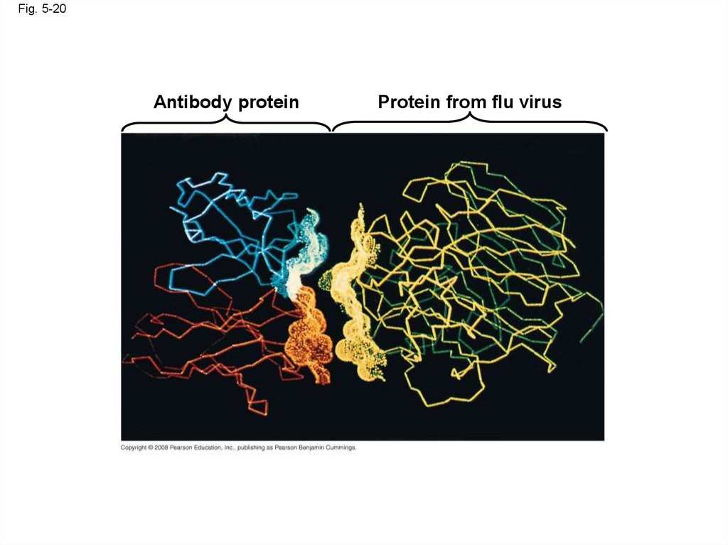
• The sequence of amino acids determines aprotein’s three-dimensional structure
• A protein’s structure determines its function
Copyright © 2008 Pearson Education, Inc., publishing as Pearson Benjamin Cummings
74.
Fig. 5-20Antibody protein
Protein from flu virus
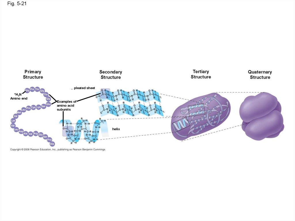
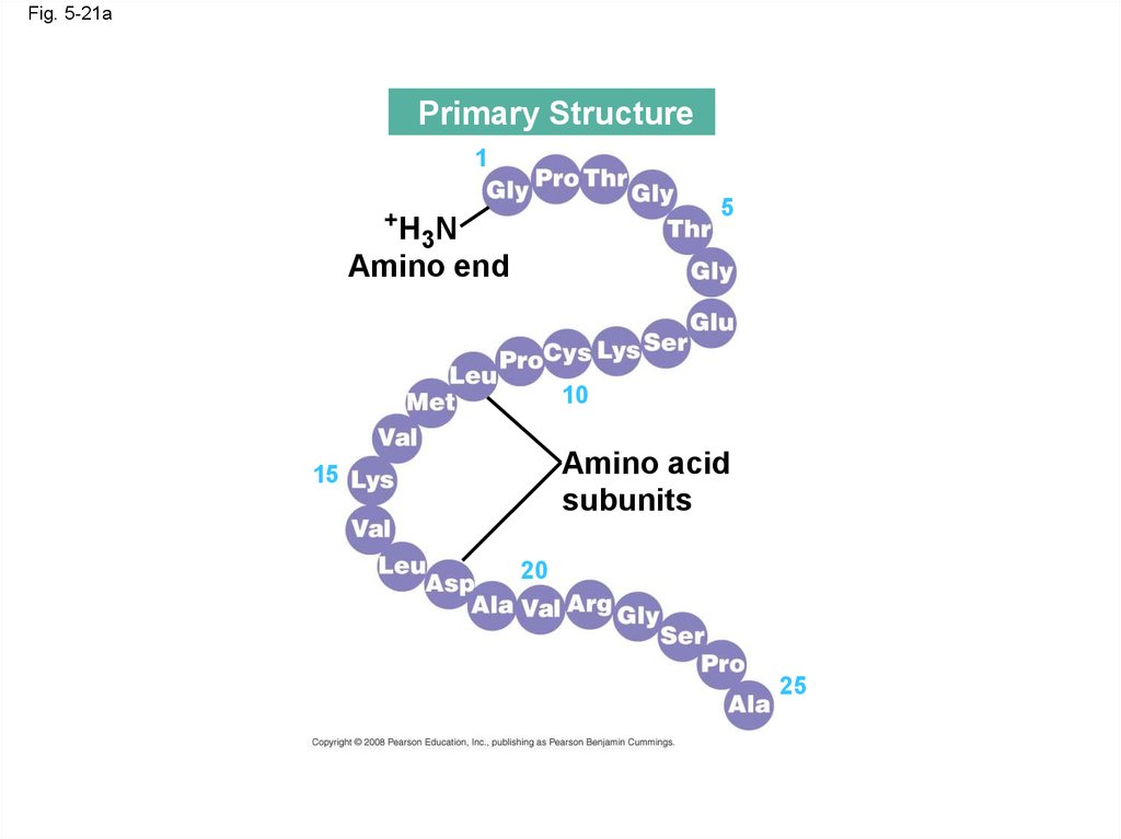
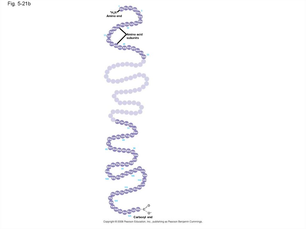
75. Four Levels of Protein Structure
• The primary structure of a protein is its uniquesequence of amino acids
• Secondary structure, found in most proteins,
consists of coils and folds in the polypeptide
chain
• Tertiary structure is determined by interactions
among various side chains (R groups)
• Quaternary structure results when a protein
consists of multiple polypeptide chains
Animation: Protein Structure Introduction
Copyright © 2008 Pearson Education, Inc., publishing as Pearson Benjamin Cummings
76.
• Primary structure, the sequence of aminoacids in a protein, is like the order of letters in a
long word
• Primary structure is determined by inherited
genetic information
Animation: Primary Protein Structure
Copyright © 2008 Pearson Education, Inc., publishing as Pearson Benjamin Cummings
77.
Fig. 5-21Primary
Structure
Secondary
Structure
pleated sheet
+H N
3
Amino end
Examples of
amino acid
subunits
helix
Tertiary
Structure
Quaternary
Structure
78.
Fig. 5-21aPrimary Structure
1
+H
5
3N
Amino end
10
Amino acid
subunits
15
20
25
79.
Fig. 5-21b1
5
+H
3N
Amino end
10
Amino acid
subunits
15
20
25
75
80
90
85
95
105
100
110
115
120
125
Carboxyl end
80.
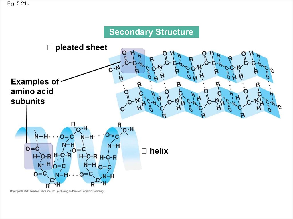
• The coils and folds of secondary structureresult from hydrogen bonds between repeating
constituents of the polypeptide backbone
• Typical secondary structures are a coil called
an helix and a folded structure called a
pleated sheet
Animation: Secondary Protein Structure
Copyright © 2008 Pearson Education, Inc., publishing as Pearson Benjamin Cummings
81.
Fig. 5-21cSecondary Structure
pleated sheet
Examples of
amino acid
subunits
helix
82.
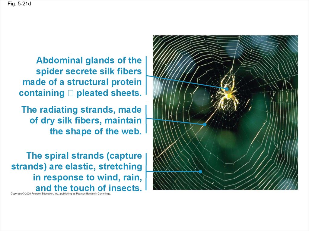
Fig. 5-21dAbdominal glands of the
spider secrete silk fibers
made of a structural protein
containing pleated sheets.
The radiating strands, made
of dry silk fibers, maintain
the shape of the web.
The spiral strands (capture
strands) are elastic, stretching
in response to wind, rain,
and the touch of insects.
83.
• Tertiary structure is determined byinteractions between R groups, rather than
interactions between backbone constituents
• These interactions between R groups include
hydrogen bonds, ionic bonds, hydrophobic
interactions, and van der Waals interactions
• Strong covalent bonds called disulfide
bridges may reinforce the protein’s structure
Animation: Tertiary Protein Structure
Copyright © 2008 Pearson Education, Inc., publishing as Pearson Benjamin Cummings
84.
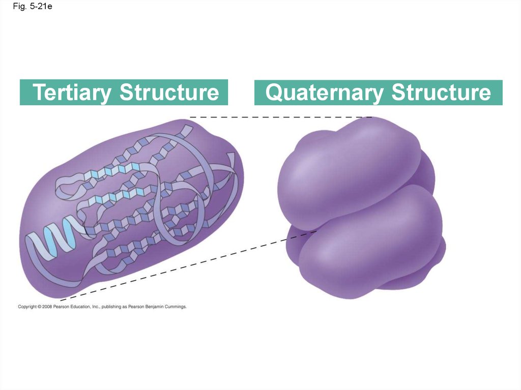
Fig. 5-21eTertiary Structure
Quaternary Structure
85.
Fig. 5-21fHydrophobic
interactions and
van der Waals
interactions
Polypeptide
backbone
Hydrogen
bond
Disulfide bridge
Ionic bond
86.
Fig. 5-21gPolypeptide
chain
Chains
Iron
Heme
Chains
Hemoglobin
Collagen
87.
• Quaternary structure results when two ormore polypeptide chains form one
macromolecule
• Collagen is a fibrous protein consisting of three
polypeptides coiled like a rope
• Hemoglobin is a globular protein consisting of
four polypeptides: two alpha and two beta
chains
Animation: Quaternary Protein Structure
Copyright © 2008 Pearson Education, Inc., publishing as Pearson Benjamin Cummings
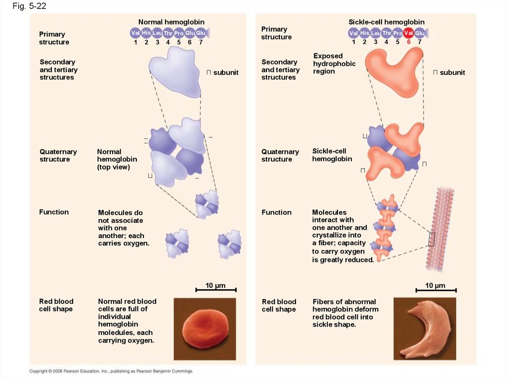
88. Sickle-Cell Disease: A Change in Primary Structure
• A slight change in primary structure can affecta protein’s structure and ability to function
• Sickle-cell disease, an inherited blood disorder,
results from a single amino acid substitution in
the protein hemoglobin
Copyright © 2008 Pearson Education, Inc., publishing as Pearson Benjamin Cummings
89.
Fig. 5-22Normal hemoglobin
Primary
structure
Sickle-cell hemoglobin
Primary
structure
Val His Leu Thr Pro Glu Glu
1
2
3
Secondary
and tertiary
structures
4
5
6
7
subunit
Secondary
and tertiary
structures
Val His Leu Thr Pro Val Glu
1
2
3
Exposed
hydrophobic
region
Quaternary
structure
Normal
hemoglobin
(top view)
Quaternary
structure
Sickle-cell
hemoglobin
Function
Molecules do
not associate
with one
another; each
carries oxygen.
Function
Molecules
interact with
one another and
crystallize into
a fiber; capacity
to carry oxygen
is greatly reduced.
10 µm
Red blood
cell shape
Normal red blood
cells are full of
individual
hemoglobin
moledules, each
carrying oxygen.
4
5
6
7
subunit
10 µm
Red blood
cell shape
Fibers of abnormal
hemoglobin deform
red blood cell into
sickle shape.
90.
Fig. 5-22aNormal hemoglobin
Primary
structure
Val His Leu Thr Pro Glu Glu
1
2
Secondary
and tertiary
structures
3
4
5
6
7
subunit
Quaternary
structure
Normal
hemoglobin
(top view)
Function
Molecules do
not associate
with one
another; each
carries oxygen.
91.
Fig. 5-22bSickle-cell hemoglobin
Primary
structure
Secondary
and tertiary
structures
Val His Leu Thr Pro Val Glu
1
2
3
Exposed
hydrophobic
region
Quaternary
structure
Sickle-cell
hemoglobin
Function
Molecules
interact with
one another and
crystallize into
a fiber; capacity
to carry oxygen
is greatly reduced.
4
5
6
7
subunit
92.
Fig. 5-22c10 µm
Normal red blood
cells are full of
individual
hemoglobin
molecules, each
carrying oxygen.
10 µm
Fibers of abnormal
hemoglobin deform
red blood cell into
sickle shape.
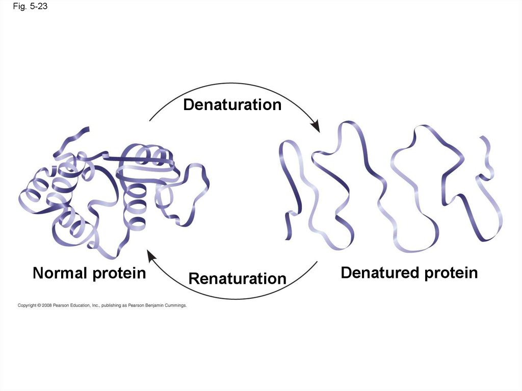
93. What Determines Protein Structure?
• In addition to primary structure, physical andchemical conditions can affect structure
• Alterations in pH, salt concentration,
temperature, or other environmental factors
can cause a protein to unravel
• This loss of a protein’s native structure is called
denaturation
• A denatured protein is biologically inactive
Copyright © 2008 Pearson Education, Inc., publishing as Pearson Benjamin Cummings
94.
Fig. 5-23Denaturation
Normal protein
Renaturation
Denatured protein
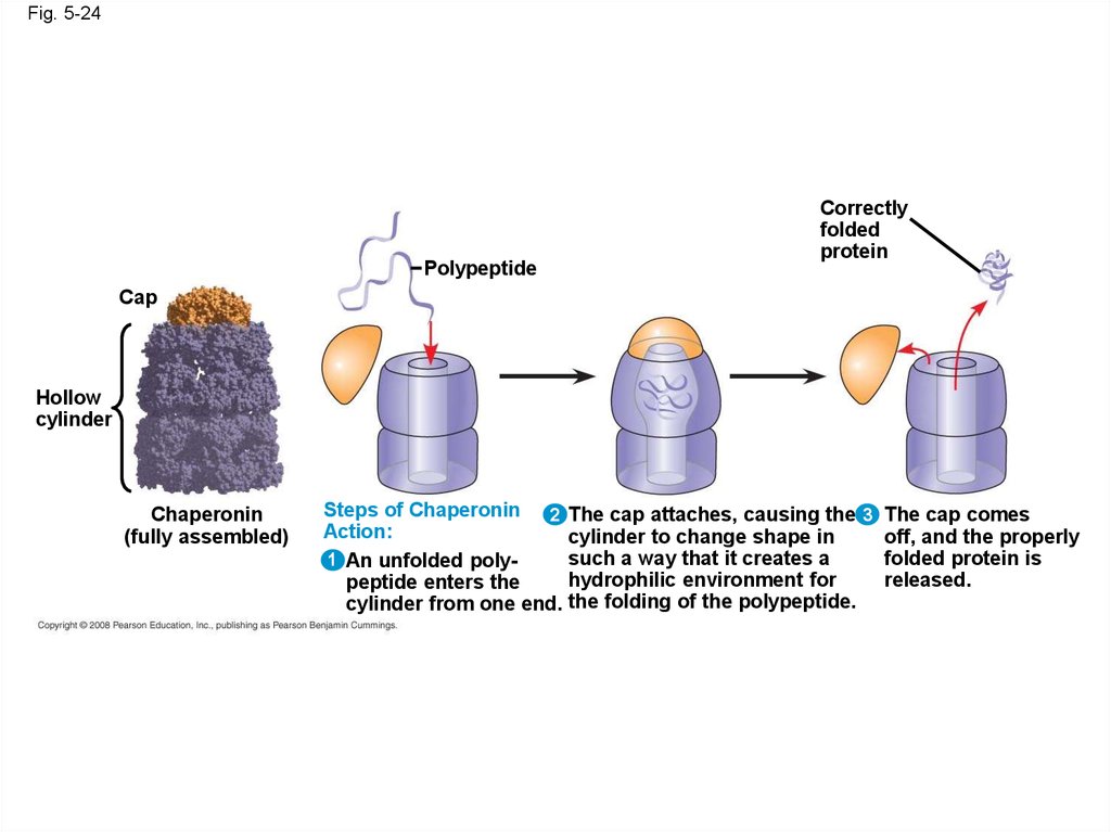
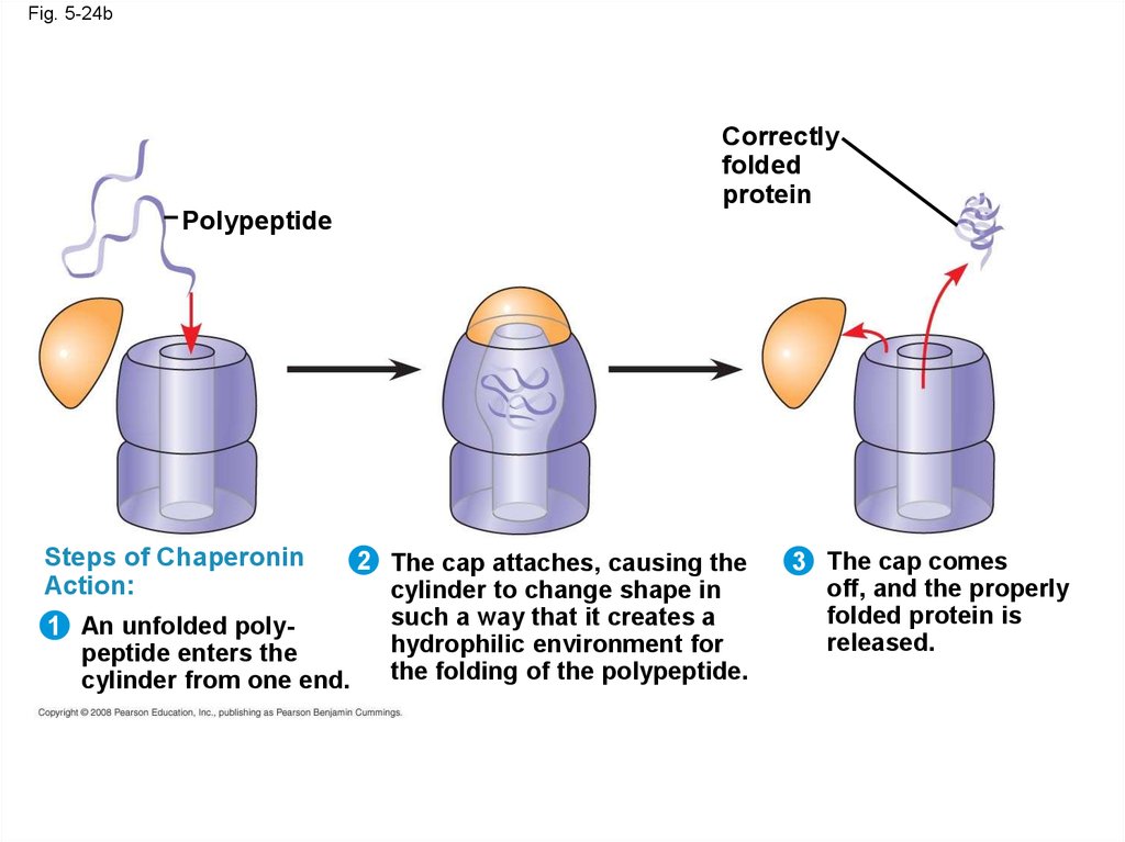
95. Protein Folding in the Cell
• It is hard to predict a protein’s structure from itsprimary structure
• Most proteins probably go through several
states on their way to a stable structure
• Chaperonins are protein molecules that assist
the proper folding of other proteins
Copyright © 2008 Pearson Education, Inc., publishing as Pearson Benjamin Cummings
96.
Fig. 5-24Polypeptide
Correctly
folded
protein
Cap
Hollow
cylinder
Chaperonin
(fully assembled)
Steps of Chaperonin 2
Action:
1 An unfolded polypeptide enters the
cylinder from one end.
The cap attaches, causing the 3 The cap comes
cylinder to change shape in
off, and the properly
such a way that it creates a
folded protein is
hydrophilic environment for
released.
the folding of the polypeptide.
97.
Fig. 5-24aCap
Hollow
cylinder
Chaperonin
(fully assembled)
98.
Fig. 5-24bCorrectly
folded
protein
Polypeptide
Steps of Chaperonin
Action:
1 An unfolded polypeptide enters the
cylinder from one end.
2 The cap attaches, causing the
cylinder to change shape in
such a way that it creates a
hydrophilic environment for
the folding of the polypeptide.
3 The cap comes
off, and the properly
folded protein is
released.
99.
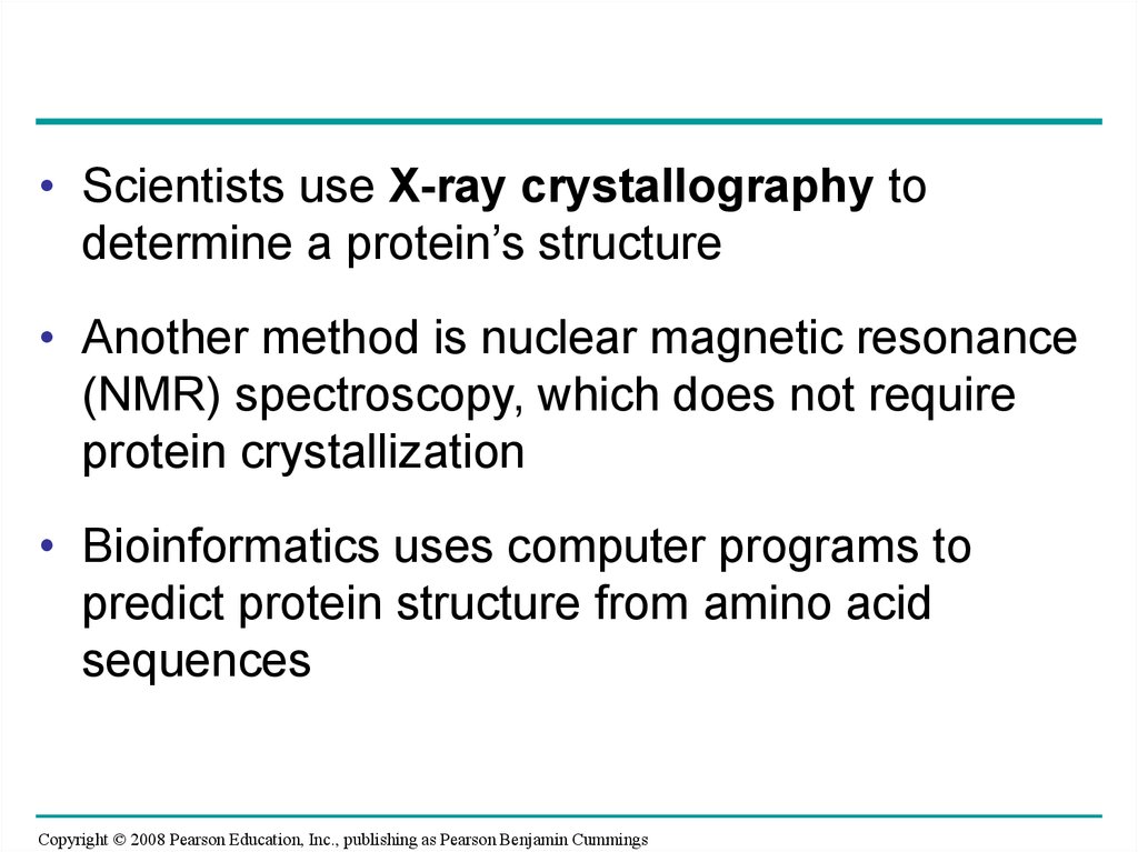
• Scientists use X-ray crystallography todetermine a protein’s structure
• Another method is nuclear magnetic resonance
(NMR) spectroscopy, which does not require
protein crystallization
• Bioinformatics uses computer programs to
predict protein structure from amino acid
sequences
Copyright © 2008 Pearson Education, Inc., publishing as Pearson Benjamin Cummings
100.
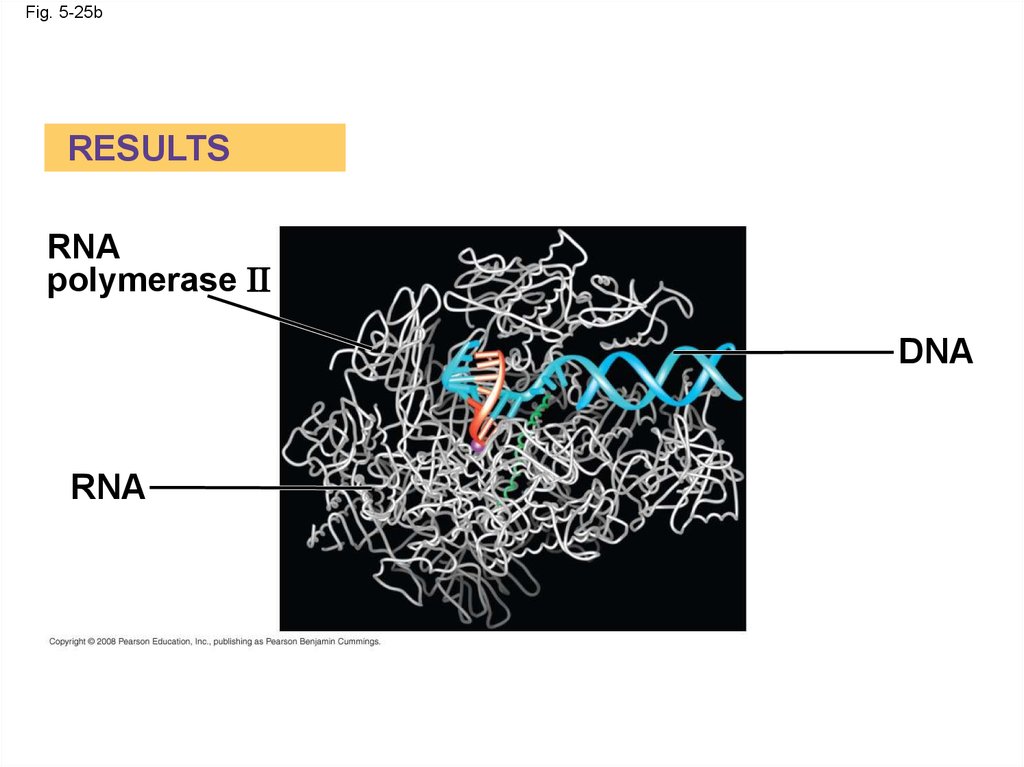
Fig. 5-25EXPERIMENT
Diffracted
X-rays
X-ray
source X-ray
beam
Crystal
Digital detector
X-ray diffraction
pattern
RESULTS
RNA
polymerase II
DNA
RNA
101.
Fig. 5-25aEXPERIMENT
Diffracted
X-rays
X-ray
source X-ray
beam
Crystal
Digital detector
X-ray diffraction
pattern
102.
Fig. 5-25bRESULTS
RNA
polymerase II
DNA
RNA
103. Concept 5.5: Nucleic acids store and transmit hereditary information
• The amino acid sequence of a polypeptide isprogrammed by a unit of inheritance called a
gene
• Genes are made of DNA, a nucleic acid
Copyright © 2008 Pearson Education, Inc., publishing as Pearson Benjamin Cummings
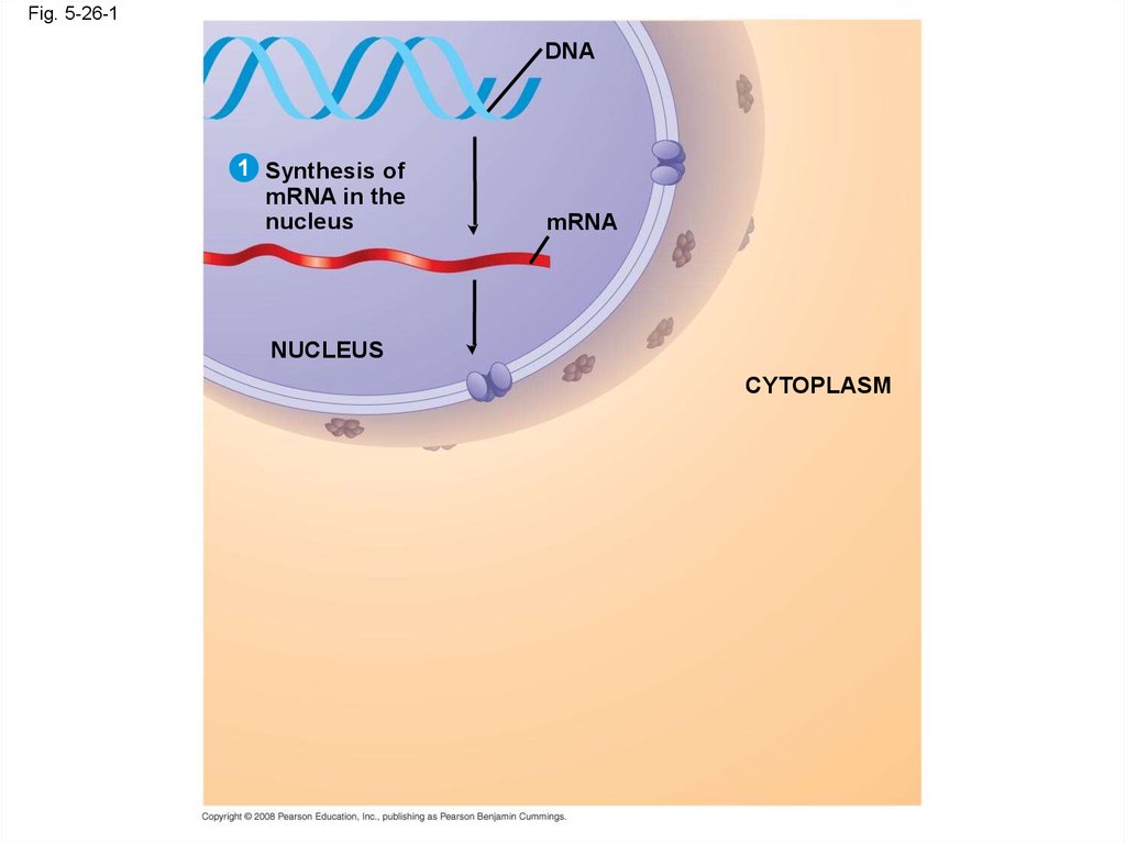
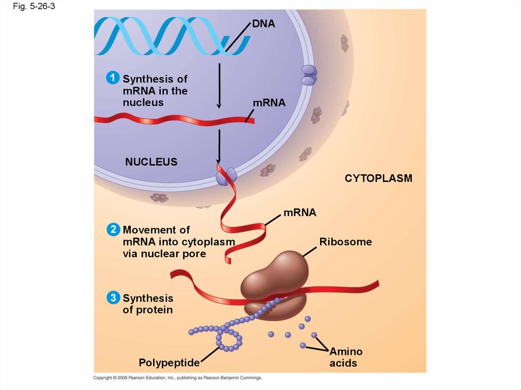
104. The Roles of Nucleic Acids
• There are two types of nucleic acids:– Deoxyribonucleic acid (DNA)
– Ribonucleic acid (RNA)
• DNA provides directions for its own replication
• DNA directs synthesis of messenger RNA
(mRNA) and, through mRNA, controls protein
synthesis
• Protein synthesis occurs in ribosomes
Copyright © 2008 Pearson Education, Inc., publishing as Pearson Benjamin Cummings
105.
Fig. 5-26-1DNA
1 Synthesis of
mRNA in the
nucleus
mRNA
NUCLEUS
CYTOPLASM
106.
Fig. 5-26-2DNA
1 Synthesis of
mRNA in the
nucleus
mRNA
NUCLEUS
CYTOPLASM
mRNA
2 Movement of
mRNA into cytoplasm
via nuclear pore
107.
Fig. 5-26-3DNA
1 Synthesis of
mRNA in the
nucleus
mRNA
NUCLEUS
CYTOPLASM
mRNA
2 Movement of
mRNA into cytoplasm
via nuclear pore
Ribosome
3 Synthesis
of protein
Polypeptide
Amino
acids
108. The Structure of Nucleic Acids
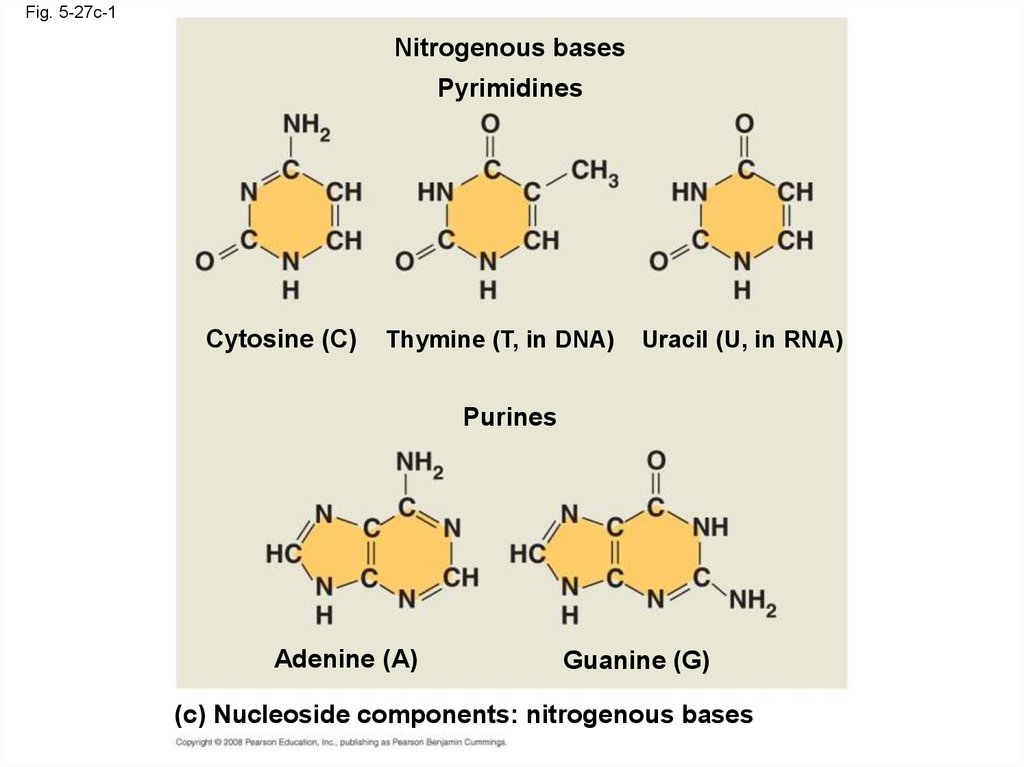
• Nucleic acids are polymers calledpolynucleotides
• Each polynucleotide is made of monomers
called nucleotides
• Each nucleotide consists of a nitrogenous
base, a pentose sugar, and a phosphate group
• The portion of a nucleotide without the
phosphate group is called a nucleoside
Copyright © 2008 Pearson Education, Inc., publishing as Pearson Benjamin Cummings
109.
Fig. 5-275
end
Nitrogenous bases
Pyrimidines
5 C
3 C
Nucleoside
Nitrogenous
base
Cytosine (C)
Thymine (T, in DNA) Uracil (U, in RNA)
Purines
Phosphate
group
5 C
Sugar
(pentose)
Adenine (A)
Guanine (G)
(b) Nucleotide
3 C
3
Sugars
end
(a) Polynucleotide, or nucleic acid
Deoxyribose (in DNA)
Ribose (in RNA)
(c) Nucleoside components: sugars
110.
Fig. 5-27ab5' end
5'C
3'C
Nucleoside
Nitrogenous
base
5'C
Phosphate
group
5'C
3'C
(b) Nucleotide
3' end
(a) Polynucleotide, or nucleic acid
3'C
Sugar
(pentose)
111.
Fig. 5-27c-1Nitrogenous bases
Pyrimidines
Cytosine (C)
Thymine (T, in DNA)
Uracil (U, in RNA)
Purines
Adenine (A)
Guanine (G)
(c) Nucleoside components: nitrogenous bases
112.
Fig. 5-27c-2Sugars
Deoxyribose (in DNA)
Ribose (in RNA)
(c) Nucleoside components: sugars
113. Nucleotide Monomers
• Nucleoside = nitrogenous base + sugar• There are two families of nitrogenous bases:
– Pyrimidines (cytosine, thymine, and uracil)
have a single six-membered ring
– Purines (adenine and guanine) have a sixmembered ring fused to a five-membered ring
• In DNA, the sugar is deoxyribose; in RNA, the
sugar is ribose
• Nucleotide = nucleoside + phosphate group
Copyright © 2008 Pearson Education, Inc., publishing as Pearson Benjamin Cummings
114. Nucleotide Polymers
• Nucleotide polymers are linked together to builda polynucleotide
• Adjacent nucleotides are joined by covalent
bonds that form between the –OH group on the
3 carbon of one nucleotide and the phosphate
on the 5 carbon on the next
• These links create a backbone of sugarphosphate units with nitrogenous bases as
appendages
• The sequence of bases along a DNA or mRNA
polymer is unique for each gene
Copyright © 2008 Pearson Education, Inc., publishing as Pearson Benjamin Cummings
115. The DNA Double Helix
• A DNA molecule has two polynucleotides spiralingaround an imaginary axis, forming a double helix
• In the DNA double helix, the two backbones run in
opposite 5 → 3 directions from each other, an
arrangement referred to as antiparallel
• One DNA molecule includes many genes
• The nitrogenous bases in DNA pair up and form
hydrogen bonds: adenine (A) always with thymine
(T), and guanine (G) always with cytosine (C)
Copyright © 2008 Pearson Education, Inc., publishing as Pearson Benjamin Cummings
116.
Fig. 5-285' end
3' end
Sugar-phosphate
backbones
Base pair (joined by
hydrogen bonding)
Old strands
Nucleotide
about to be
added to a
new strand
3' end
5' end
New
strands
5' end
3' end
5' end
3' end
117. DNA and Proteins as Tape Measures of Evolution
• The linear sequences of nucleotides in DNAmolecules are passed from parents to offspring
• Two closely related species are more similar in
DNA than are more distantly related species
• Molecular biology can be used to assess
evolutionary kinship
Copyright © 2008 Pearson Education, Inc., publishing as Pearson Benjamin Cummings
118. The Theme of Emergent Properties in the Chemistry of Life: A Review
• Higher levels of organization result in theemergence of new properties
• Organization is the key to the chemistry of life
Copyright © 2008 Pearson Education, Inc., publishing as Pearson Benjamin Cummings
119.
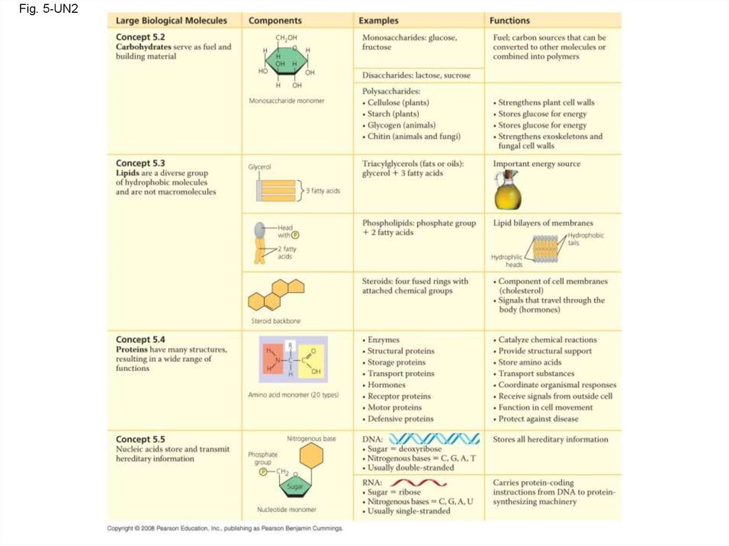
Fig. 5-UN2120.
Fig. 5-UN2a121.
Fig. 5-UN2b122.
Fig. 5-UN3123.
Fig. 5-UN4124.
Fig. 5-UN5125.
Fig. 5-UN6126.
Fig. 5-UN7127.
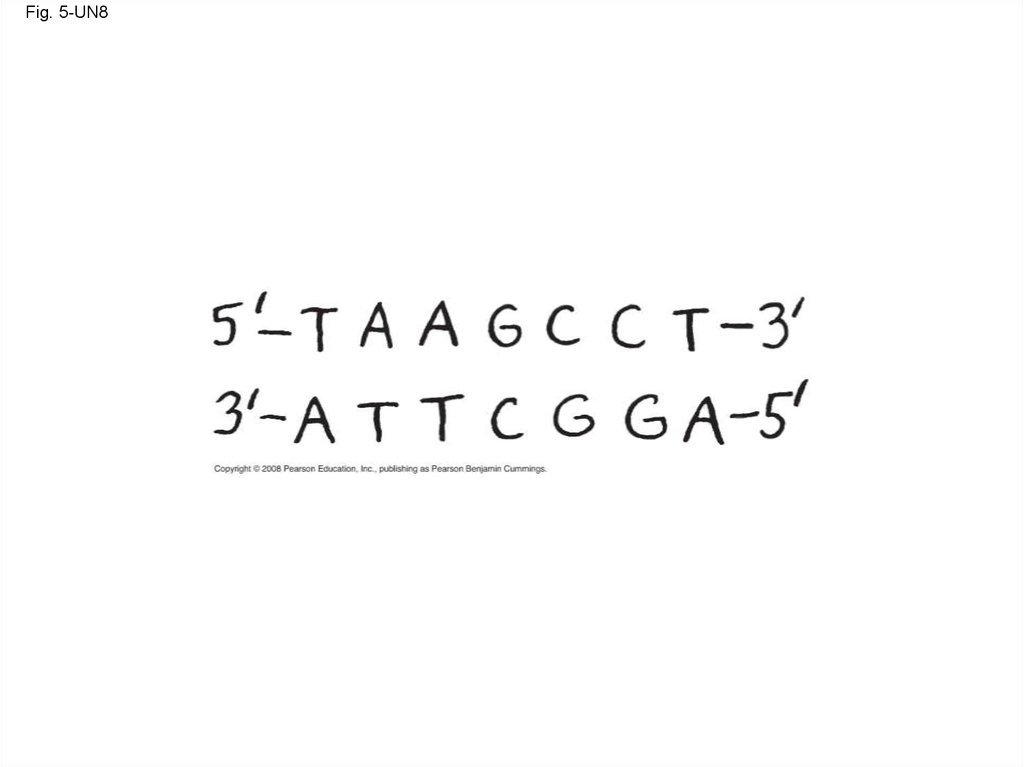
Fig. 5-UN8128.
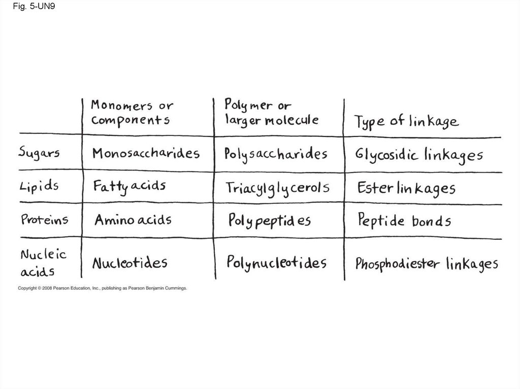
Fig. 5-UN9129.
Fig. 5-UN10130. You should now be able to:
1. List and describe the four major classes ofmolecules
2. Describe the formation of a glycosidic linkage
and distinguish between monosaccharides,
disaccharides, and polysaccharides
3. Distinguish between saturated and
unsaturated fats and between cis and trans fat
molecules
4. Describe the four levels of protein structure
Copyright © 2008 Pearson Education, Inc., publishing as Pearson Benjamin Cummings
131. You should now be able to:
5. Distinguish between the following pairs:pyrimidine and purine, nucleotide and
nucleoside, ribose and deoxyribose, the 5
end and 3 end of a nucleotide
Copyright © 2008 Pearson Education, Inc., publishing as Pearson Benjamin Cummings