


 Nodules (Follicles)")


 Биология
Похожие презентации:
")


")


Immune system
1.
IMMUNE SYSTEM2.
3.
The major organsof the immune system are:
Central:
• Bone marrow
• Thymus
Peripheral:
• Spleen
• Lymph nodes
• Tonsils
4.
5.
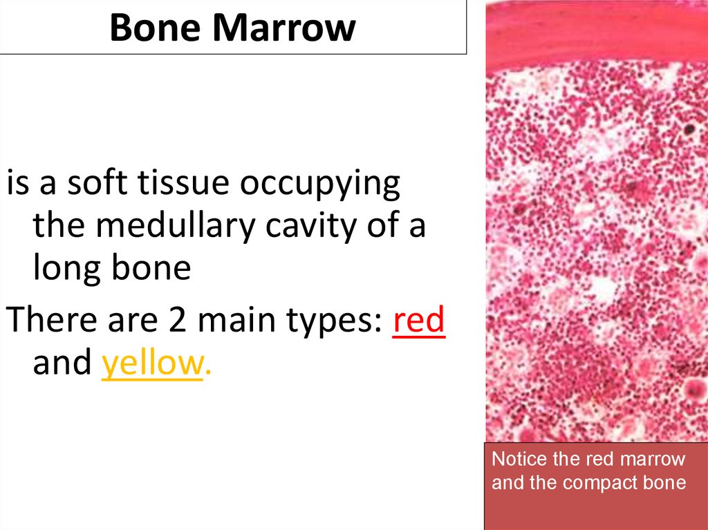
Bone Marrowis a soft tissue occupying
the medullary cavity of a
long bone
There are 2 main types: red
and yellow.
Notice the red marrow
and the compact bone
6.
Red bone marrow is blood cell forming tissueand
it is composed of stroma (reticular tissue) and
hematopoietic cords.
7. Bone Marrow
Hematopoietic cords consists of blood cells of alltypes and at all stages of differentiation
Erythroblastic islands are clusters of developing
erythrocytes surrounding macrophages and receiving
iron from them.
Sinusoids (capillaries) have openings in their walls
through which maturing blood cells and platelets enter
the circulation.
8.
ThymusFunctions:
1. Production of T- lymphocyte.
2. Production of hormone - thymosin
Consists of epithelial reticular cells (Stroma) and
lymphocytes
A thin capsule send septa (trabecula) dividing Thymus
into incomplete lobules.
Lobules consists of cortex + medulla
9.
Thymus14
10.
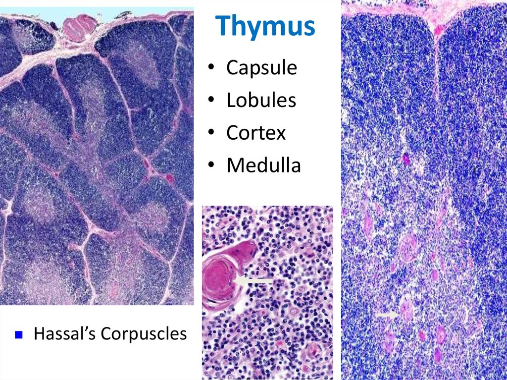
ThymusHassal’s Corpuscles
Capsule
Lobules
Cortex
Medulla
11.
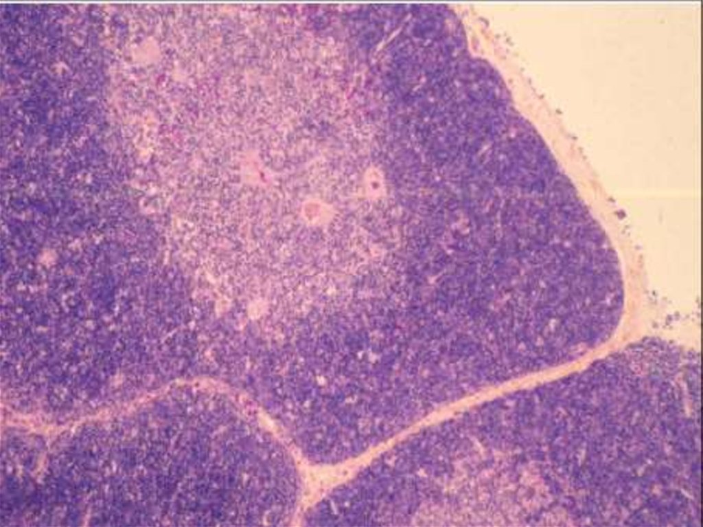
Cortex--- dark-staining periphery of each lobule. Smalllymphocytes predominate
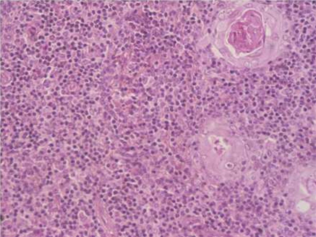
Medulla is the light core of each lobules.
It has more epithelial reticular cells and fewer lymphocytes
than in the cortex.
The spheric Hassall’s corpuscles are composed of concentric
layers of flattened epithelial reticular cells.
12.
13.
14. Thymus
Peripheral part of I. S.15. Thymus
1. Lymphoid (= Lymph,Lymphatic) Nodules (Follicles)
16.
Lymphatic Nodule- have a darkstaining periphery, or
mantle zone, that
contains tightly
packed small
lymphocytes,
17. Figure 5-3 part 1 of 2
Lymphatic Noduleand a light-staining
core, or germinal
center, that contains
numerous
lymphoblasts lymphocytes
stimulated by antigens
to enlarge and
proliferate.
18.
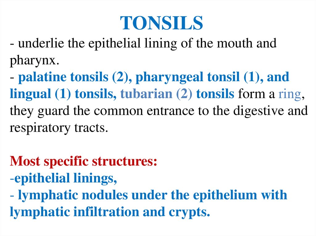
TONSILS- underlie the epithelial lining of the mouth and
pharynx.
- palatine tonsils (2), pharyngeal tonsil (1), and
lingual (1) tonsils, tubarian (2) tonsils form a ring,
they guard the common entrance to the digestive and
respiratory tracts.
Most specific structures:
-epithelial linings,
- lymphatic nodules under the epithelium with
lymphatic infiltration and crypts.
19.
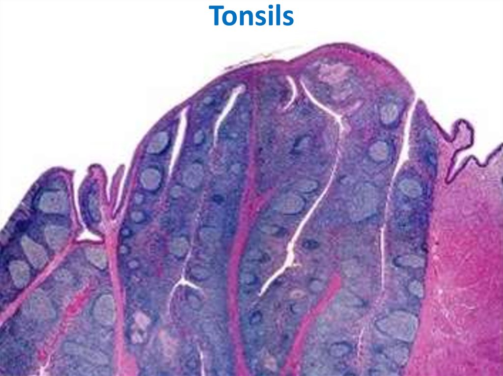
Tonsils20.
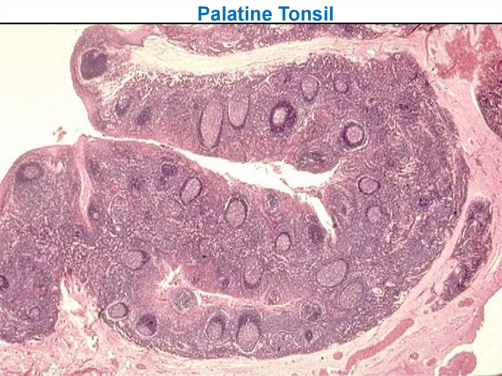
Palatine Tonsil21.
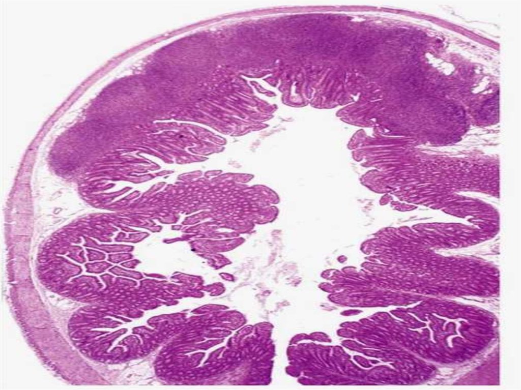
Peyer’s PatchesSmaller aggregates present under mucous membrane:
“Mucosa Associated Lymphoid Tissue” or MALT (in
Digestive sys)
22.
23. 1. Lymphoid (= Lymph, Lymphatic) Nodules (Follicles)
LYMPH NODESThese are
-the smallest but most numerous encapsulated lymphoid
organs.
Lie in groups along lymphatic vessels
Functions:
1. Filtration of lymph
2. Lymphocyte production (lymphopoiesis).
3. Immunoglobulin production.
24. Lymphatic Nodule
CM
25. Lymphatic Nodule
LYMPH NODES-- Inner space consists of reticular connective tissue
and has 3 zones:
1. cortex, adjacent to the convex surface,
2. - a central medulla lying near the depression
(hilum) in the concave surface,
and intermediate paracortical zone.
1. Cortex consists of layer of typical lymphoid
nodules
26.
2. Paracortical zone.This is the T-dependent region, It contains mainly Tlymphocytes.
3. Medulla.
is composed of cords of lymphoid tissue (medullary
cords) separated by medullary sinuses.
The cords contain many plasma cells that have
migrated from the cortex.
27. Tonsils
Lymphatic vessels inside LN are Sinuses.Types: subcapsular,
peritrabecular,
medullary
28.
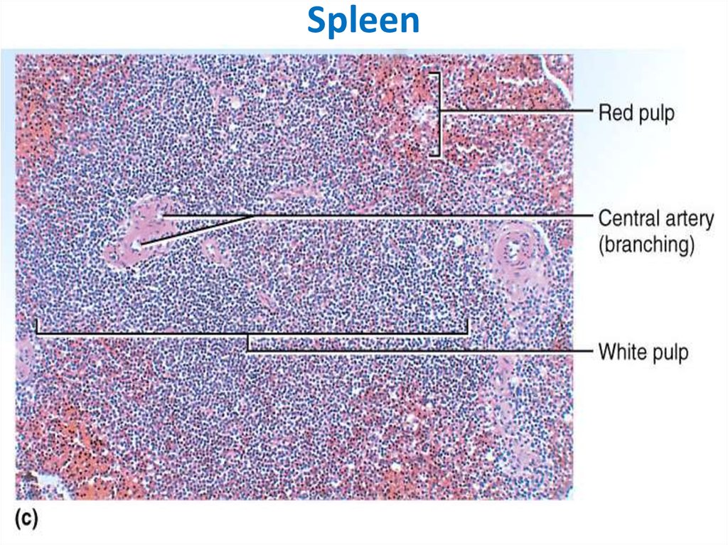
SPLEEN --- Is the largest of the lymphoid organsFunctions:
1. Filtration of blood.
2. Lymphocyte production (lymphopoiesis).
3. Destruction of worn red blood cells
4. Extramedullary hematopoiesis (in embryonic
period)
29. Peyer’s Patches
Inner space -- Splenic pulp -- is composed of:1. reticular tissue consisting of reticular cells and
reticular fibers,
2. as well as blood vessels -- usual and sinusoid
capillaries.
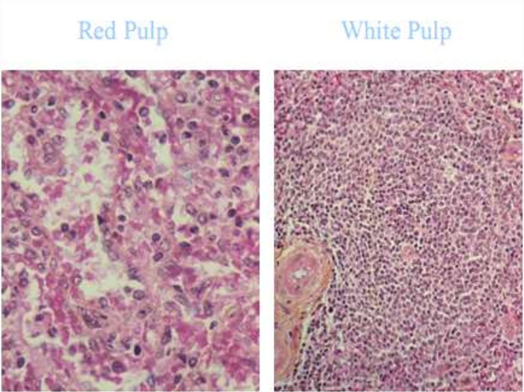
Splenic pulp = White pulp + Red pulp
30.
White pulp- consists of lymphocytes;
-- surround small arteries;
--- has 2 major components:
1. Periarterial lymphatic sheaths (PALS) - W.P.
immediately surrounding each small artery
(called “central artery”). These contain mainly T
lymphocytes and constitute the T-dependent
regions of the spleen.
2. Peripheral white pulp (PWP) -- includes a
typical lymphoid nodules (usually with a germinal
center). These contain mainly B lymphocytes and
constitute the B-dependent regions of the spleen.
31. Lymph Node
Red pulp -- collects blood andmakes up most of the spleen
and also has 2 major components:
- the red pulp cords and
-- the splenic sinusoids that lie between them.
32.
Spleen45