


 Биология
Похожие презентации:


Problems of genetic engineering in the creation of transgenic animals
1.
Problems of genetic engineering in the creation oftransgenic animals
1.The methods of introduction of foreign DNA into
the host cells;
2.Status and prospects of obtaining and using
transgenic animals;
3.Problems in creating of transgenic animals
2.
A transgenic animal is one that carries a foreign gene that has beendeliberately inserted into its genome.
Transgenic technology developed and refined on laboratory mice. Since the
early 1980's hundreds of genes were introduced to different strains of mice.
The introduction of foreign DNA into mice was carried out by different
methods:
- by using retroviral vectors to infect embryo cells in the early stages of its
development before implantation into a female recipient,
- by microinjection DNA into the sperm nucleus (male pronucleus) of
fertilized egg,
- by introduction of genetically modified embryonic stem cells in preimplanted
embryo
in
its
early
stages
of
development.
3.
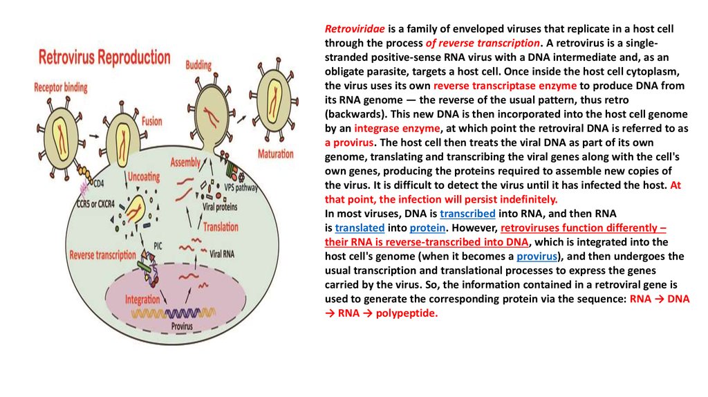
Retroviridae is a family of enveloped viruses that replicate in a host cellthrough the process of reverse transcription. A retrovirus is a singlestranded positive-sense RNA virus with a DNA intermediate and, as an
obligate parasite, targets a host cell. Once inside the host cell cytoplasm,
the virus uses its own reverse transcriptase enzyme to produce DNA from
its RNA genome — the reverse of the usual pattern, thus retro
(backwards). This new DNA is then incorporated into the host cell genome
by an integrase enzyme, at which point the retroviral DNA is referred to as
a provirus. The host cell then treats the viral DNA as part of its own
genome, translating and transcribing the viral genes along with the cell's
own genes, producing the proteins required to assemble new copies of
the virus. It is difficult to detect the virus until it has infected the host. At
that point, the infection will persist indefinitely.
In most viruses, DNA is transcribed into RNA, and then RNA
is translated into protein. However, retroviruses function differently –
their RNA is reverse-transcribed into DNA, which is integrated into the
host cell's genome (when it becomes a provirus), and then undergoes the
usual transcription and translational processes to express the genes
carried by the virus. So, the information contained in a retroviral gene is
used to generate the corresponding protein via the sequence: RNA → DNA
→ RNA → polypeptide.
4.
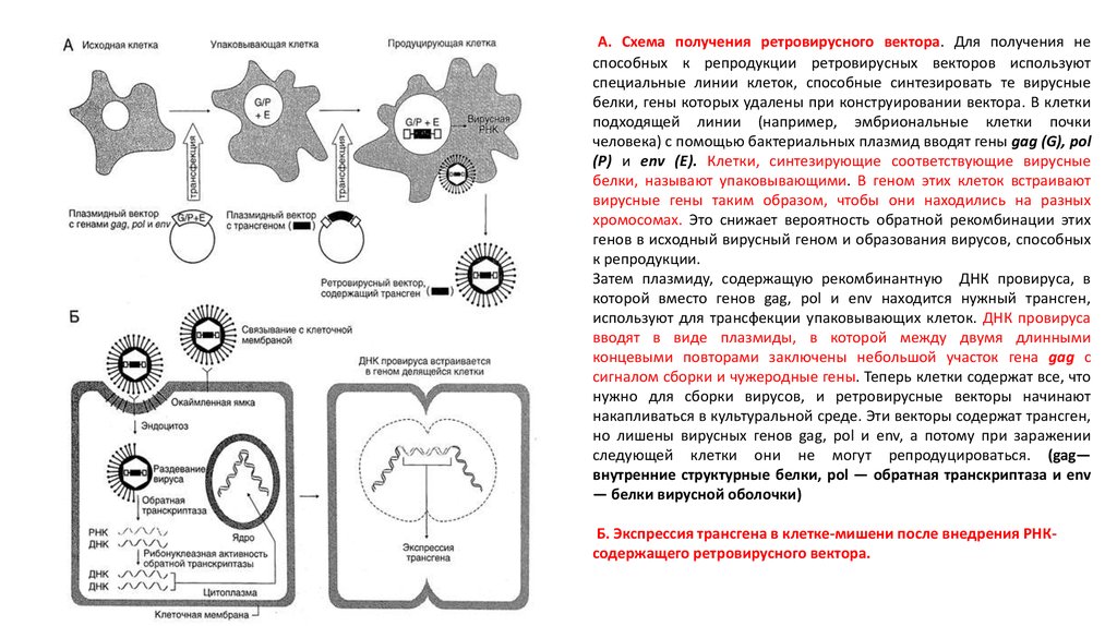
А. Схема получения ретровирусного вектора. Для получения неспособных к репродукции ретровирусных векторов используют
специальные линии клеток, способные синтезировать те вирусные
белки, гены которых удалены при конструировании вектора. В клетки
подходящей линии (например, эмбриональные клетки почки
человека) с помощью бактериальных плазмид вводят гены gag (G), pol
(Р) и env (Е). Клетки, синтезирующие соответствующие вирусные
белки, называют упаковывающими. В геном этих клеток встраивают
вирусные гены таким образом, чтобы они находились на разных
хромосомах. Это снижает вероятность обратной рекомбинации этих
генов в исходный вирусный геном и образования вирусов, способных
к репродукции.
Затем плазмиду, содержащую рекомбинантную ДНК провируса, в
которой вместо генов gag, pol и env находится нужный трансген,
используют для трансфекции упаковывающих клеток. ДНК провируса
вводят в виде плазмиды, в которой между двумя длинными
концевыми повторами заключены небольшой участок гена gag с
сигналом сборки и чужеродные гены. Теперь клетки содержат все, что
нужно для сборки вирусов, и ретровирусные векторы начинают
накапливаться в культуральной среде. Эти векторы содержат трансген,
но лишены вирусных генов gag, pol и env, а потому при заражении
следующей клетки они не могут репродуцироваться. (gag—
внутренние структурные белки, pol — обратная транскриптаза и env
— белки вирусной оболочки)
Б. Экспрессия трансгена в клетке-мишени после внедрения РНКсодержащего ретровирусного вектора.
5.
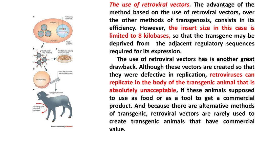
The use of retroviral vectors. The advantage of themethod based on the use of retroviral vectors, over
the other methods of transgenosis, consists in its
efficiency. However, the insert size in this case is
limited to 8 kilobases, so that the transgene may be
deprived from the adjacent regulatory sequences
required for its expression.
The use of retroviral vectors has is another great
drawback. Although these vectors are created so that
they were defective in replication, retroviruses can
replicate in the body of the transgenic animal that is
absolutely unacceptable, if these animals supposed
to use as food or as a tool to get a commercial
product. And because there are alternative methods
of transgenic, retroviral vectors are rarely used to
create transgenic animals that have commercial
value.
6.
Microinjection of DNA. Currently for thecreation
of
transgenic
mice
microinjection of DNA are most
commonly used. It consists in the
following.
The work begins with the stimulation of
hyper ovulation in female donors to
increase the number of eggs in which
foreign DNA will be injected. At first
Pregnant Mare Serum are injected to
females
and after about 48 hours
Human Chorionic Gonadotropin is
administrated. As a result of hyper
ovulation about 35 eggs are formed
instead of usual 5-10. Then females with
hyper ovulation is crossing with males
after which they were sacrificed,
fertilized eggs are washed out from the
oviduct, and immediately DNA is injected
into fertilized eggs.
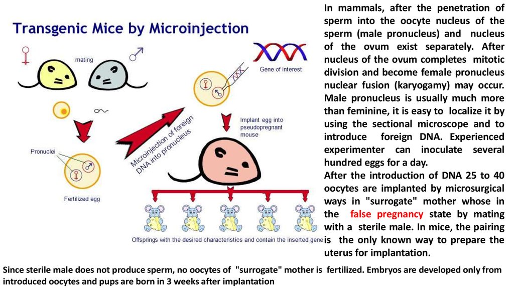
7.
In mammals, after the penetration ofsperm into the oocyte nucleus of the
sperm (male pronucleus) and nucleus
of the ovum exist separately. After
nucleus of the ovum completes mitotic
division and become female pronucleus
nuclear fusion (karyogamy) may occur.
Male pronucleus is usually much more
than feminine, it is easy to localize it by
using the sectional microscope and to
introduce foreign DNA. Experienced
experimenter can inoculate several
hundred eggs for a day.
After the introduction of DNA 25 to 40
oocytes are implanted by microsurgical
ways in "surrogate" mother whose in
the false pregnancy state by mating
with a sterile male. In mice, the pairing
is the only known way to prepare the
uterus for implantation.
Since sterile male does not produce sperm, no oocytes of "surrogate" mother is fertilized. Embryos are developed only from
introduced oocytes and pups are born in 3 weeks after implantation
8.
Sterilization (vasoectomy) of malesVasectomy (lat. Vas - a vessel, duct
+ ectomy) - a surgical procedure in
which the ligation or deleting part
of the vas deferens of male is
carried out (lat. Ductus deferens).
This operation leads to sterility
(inability to have offspring) while
preserving sexual function. After
vasectomy the male saved sexual
behavior: libido, erection,
ejaculation. But the obstruction of
the vas deferens results in the
absence of sperm in the ejaculate
(azoospermia).
9.
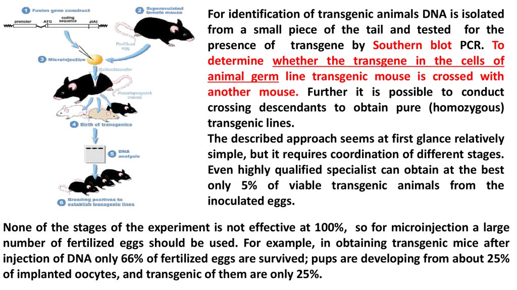
For identification of transgenic animals DNA is isolatedfrom a small piece of the tail and tested for the
presence of transgene by Southern blot PCR. To
determine whether the transgene in the cells of
animal germ line transgenic mouse is crossed with
another mouse. Further it is possible to conduct
crossing descendants to obtain pure (homozygous)
transgenic lines.
The described approach seems at first glance relatively
simple, but it requires coordination of different stages.
Even highly qualified specialist can obtain at the best
only 5% of viable transgenic animals from the
inoculated eggs.
None of the stages of the experiment is not effective at 100%, so for microinjection a large
number of fertilized eggs should be used. For example, in obtaining transgenic mice after
injection of DNA only 66% of fertilized eggs are survived; pups are developing from about 25%
of implanted oocytes, and transgenic of them are only 25%.
10.
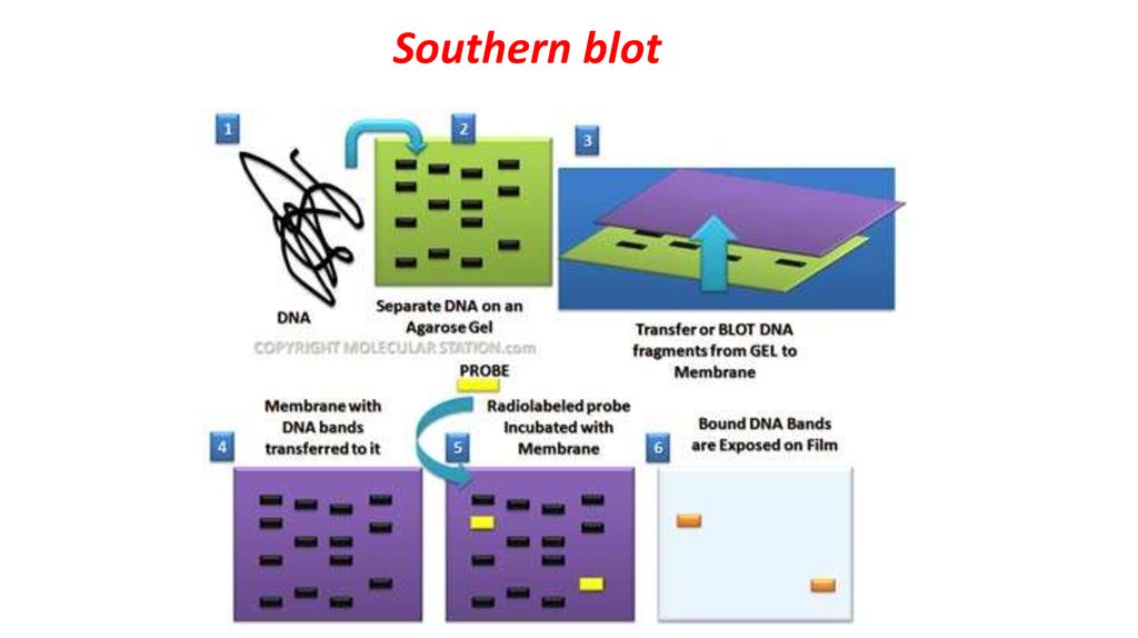
Southern blot11.
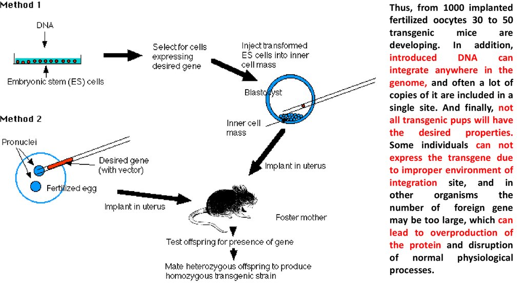
Thus, from 1000 implantedfertilized oocytes 30 to 50
transgenic
mice
are
developing. In addition,
introduced
DNA
can
integrate anywhere in the
genome, and often a lot of
copies of it are included in a
single site. And finally, not
all transgenic pups will have
the desired properties.
Some individuals can not
express the transgene due
to improper environment of
integration site, and in
other
organisms
the
number of foreign gene
may be too large, which can
lead to overproduction of
the protein and disruption
of normal physiological
processes.
12.
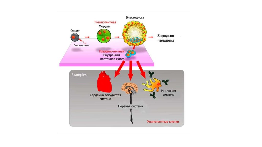
Using of the modified embryonicstem cells. Cells from mouse
embryos at the blastocyst stage,
can
proliferate
in
culture
maintaining the ability to
differentiate into all types of cells,
including the cells of the germ
line when they are administered
to another embryo at the
blastocyst stage. Such cells are
called pluripotent embryonic
stem cells (ES). ES-cells in culture
easily modified by genetic
engineering
without
compromising their pluripotency
For example, at one nonessential
gene site of
their genome
functional transgene can be
integrated.
Then it is possible to select the modified cells, cultivate them and use for the production of transgenic
animals. This prevents accidental insertion characteristic by microinjection and retroviral vector systems.
13.
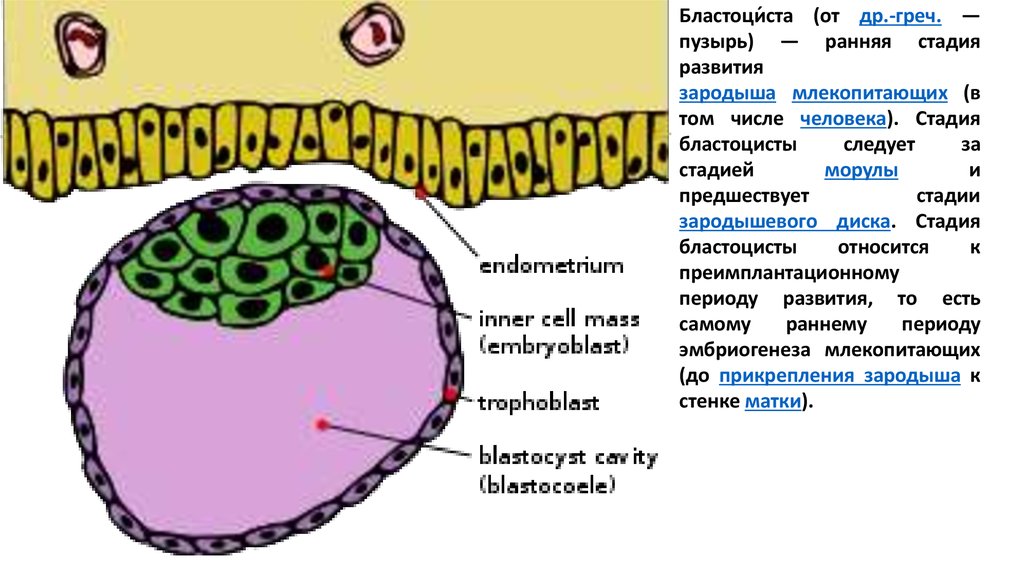
Бластоци́ста (от др.-греч. —пузырь) — ранняя стадия
развития
зародыша млекопитающих (в
том числе человека). Стадия
бластоцисты
следует
за
стадией
морулы
и
предшествует
стадии
зародышевого диска. Стадия
бластоцисты
относится
к
преимплантационному
периоду развития, то есть
самому
раннему
периоду
эмбриогенеза млекопитающих
(до прикрепления зародыша к
стенке матки).
14.
15.
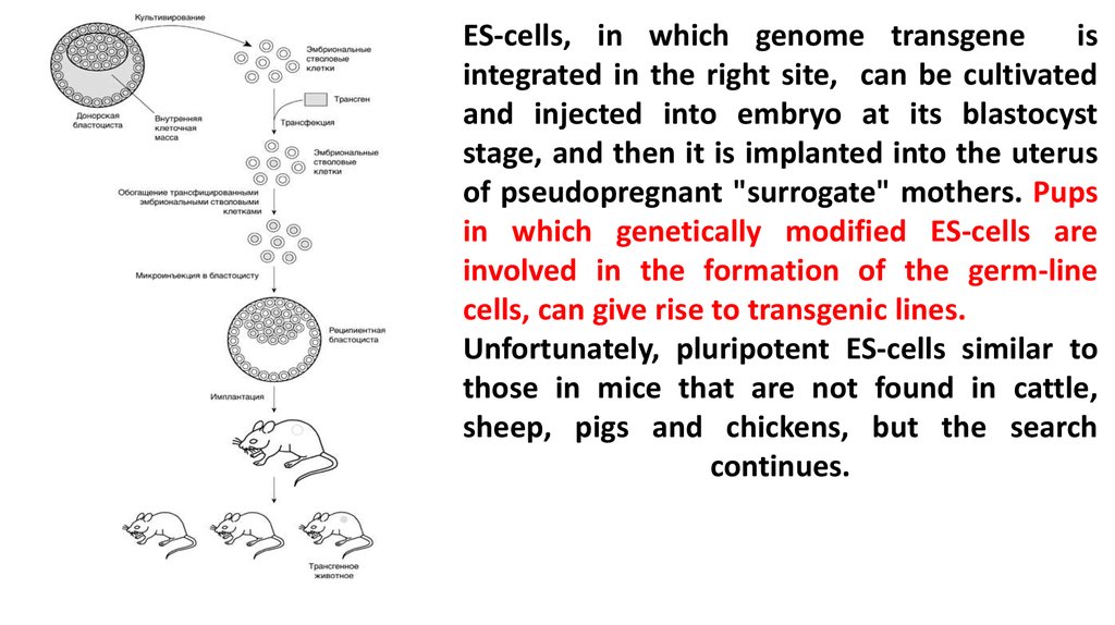
ES-cells, in which genome transgeneis
integrated in the right site, can be cultivated
and injected into embryo at its blastocyst
stage, and then it is implanted into the uterus
of pseudopregnant "surrogate" mothers. Pups
in which genetically modified ES-cells are
involved in the formation of the germ-line
cells, can give rise to transgenic lines.
Unfortunately, pluripotent ES-cells similar to
those in mice that are not found in cattle,
sheep, pigs and chickens, but the search
continues.
16.
To create transgenic cows a modifiedscheme of mice transgenosis by
microinjection of DNA are used. The
procedure includes the following
stages:
a) collection of oocytes of cows
slaughtered at the slaughterhouse;
b) oocytes maturation in vitro;
c) fertilization of oocytes with bovine
sperm in vitro;
d) centrifugation of fertilized eggs to
concentrate of yolks, which interferes
with visualization of the male
pronucleus in normal oocyte by
sectional microscopy;
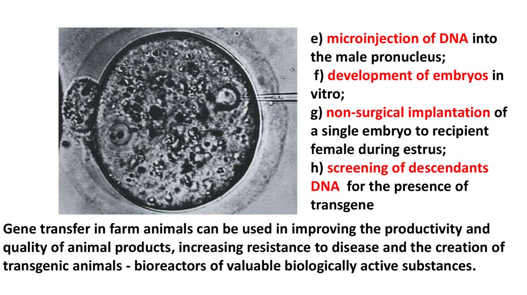
17.
e) microinjection of DNA intothe male pronucleus;
f) development of embryos in
vitro;
g) non-surgical implantation of
a single embryo to recipient
female during estrus;
h) screening of descendants
DNA for the presence of
transgene
Gene transfer in farm animals can be used in improving the productivity and
quality of animal products, increasing resistance to disease and the creation of
transgenic animals - bioreactors of valuable biologically active substances.
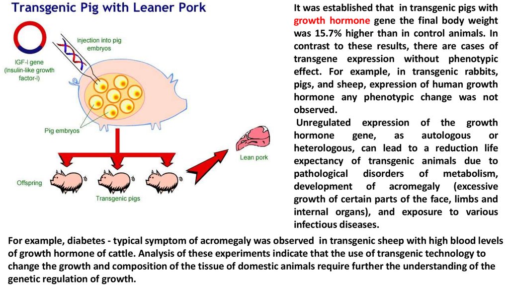
18.
It was established that in transgenic pigs withgrowth hormone gene the final body weight
was 15.7% higher than in control animals. In
contrast to these results, there are cases of
transgene expression without phenotypic
effect. For example, in transgenic rabbits,
pigs, and sheep, expression of human growth
hormone any phenotypic change was not
observed.
Unregulated expression of the growth
hormone
gene,
as
autologous
or
heterologous, can lead to a reduction life
expectancy of transgenic animals due to
pathological disorders of metabolism,
development of acromegaly (excessive
growth of certain parts of the face, limbs and
internal organs), and exposure to various
infectious diseases.
For example, diabetes - typical symptom of acromegaly was observed in transgenic sheep with high blood levels
of growth hormone of cattle. Analysis of these experiments indicate that the use of transgenic technology to
change the growth and composition of the tissue of domestic animals require further the understanding of the
genetic regulation of growth.
19.
20.
Creation of transgenic animals opens realprospects for improving the quality or
composition of animal products. For example, it
is possible to reduce lactose in milk by creating
transgenic cows and sheep, which have specific
for mammary gland promoter, linked to the
gene lactase. Thus in cow (sheep) milk lactose
can be cleaved into glucose and galactose. Such
milk could be used in nutrition of newborn
children suffering from hereditary lactose
intolerance. For these children during infancy
milk should be given only after processing by
enzyme. In addition, milk would be useful in a
variety of gastrointestinal human diseases
associated with decreased activity of lactase
(beta-galactosidase).
(Promoter - region of DNA to which RNA polymerase binds to start the synthesis of mRNA)
21.
The presence in the milk of various microflora caused problemsassociated with the storage, processing, consumption of milk and
animal health. In this regard, the genes which are responsible for
the production of antibodies against specific pathogens are great
interest. An important task is getting milk and dairy products
containing thermostable enzyme lysozyme are constructed.
During pasteurization of milk this enzyme, which has strong
antibacterial property does not lose its activity, which will
significantly increase shelf life of milk and milkproducts. The
possibility of the introduction of genes encoding antibodies with
protective effects against agents of cows mastitis are considered.
22.
Institute of Cytology and Genetics of RAS(Novosibirsk) and the Institute of Molecular
Genetics, RAS (Moscow) established genetic
construction pGoatcasGMCSF, which
containe regulatory region of goat’s gene
alpha-S1-casein that carry the human gene of
granulocyte-macrophage colony-stimulating
factor -GM-CSF.
Ген регулятор - Ген, кодирующий белок репрессор,
взаимодействующий с геном оператором и таким
образом регулирующий транскрипцию “своего”
транскриптона;
23.
By injection of recombinantDNA into zygotes pronuclei 4
transgenic mice were obtained.
PCR
shows
the
tissue
specificity of expression of
human GM-CSF only in the
mammary gland of lactating
females. Because mentioned
construction is tissue-specific, it
falls under the regulation of
physiological
signals
of
pregnancy and lactation.
24.
The possibility of including in organism’s cells the genes responsible for synthesis of proteins of greatimportance in human and veterinary medicine, formed the basis of the strategy of transgenic animals as
bioreactors. To date most of these proteins are extracted from the tissues and biological fluids of man. For
example, a clotting factor, interferon, alpha-1-antitrypsin, and other proteins are prepared from blood,
growth hormone - from the pituitary gland. They are produced in small quantities because of the high cost
and difficulty of extraction of human tissues. In addition, they may be contaminated with pathogens such as
Hepatitis, AIDS, etc.
Transgenic animals used for the production of valuable biological products have several advantages over
microorganisms-producers, as well as cellular systems. In simple recombinant systems, of microorganisms
glycosylation, B-hydroxylation or carboxylation of mammalian proteins in most cases it is impossible or
possible, but with insufficient accuracy. This changes the structure of proteins, which reflect on their
biological activity. Along with this, in drugs which are used by humans for therapeutic agents admixture of
bacterial proteins is undesirable. The main disadvantage of genetically engineered cell culture is the low yield
of protein. Industrial reactors used for the cultivation of producer cells, are expensive, both in terms of their
value, and in respect of their service. Creation of transgenic animals also requires more resources and
moreover it is not easy, but once bred line of such animals can produce a large number of proteins with low
cost, which will pay back all the expenses for a short time.
Production of biologically active human proteins from transgenic agricultural animals guarantee their
environmental cleanliness, which practically comes to exploitation of animals-producers.
25.
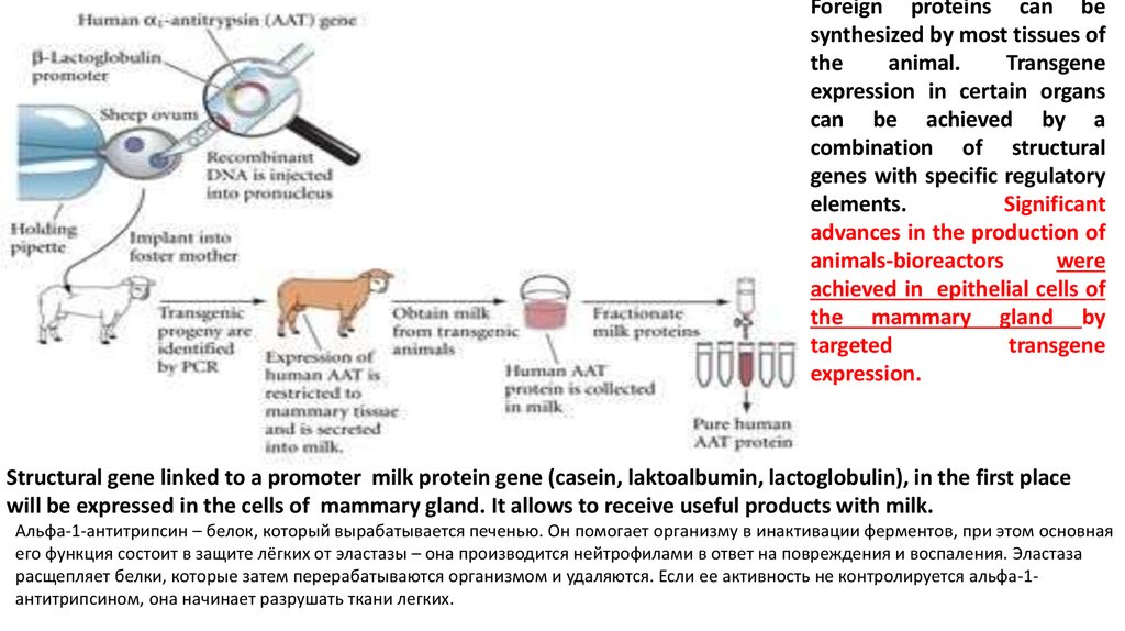
Foreign proteins can besynthesized by most tissues of
the
animal.
Transgene
expression in certain organs
can be achieved by a
combination of structural
genes with specific regulatory
elements.
Significant
advances in the production of
animals-bioreactors
were
achieved in epithelial cells of
the mammary gland by
targeted
transgene
expression.
Structural gene linked to a promoter milk protein gene (casein, laktoalbumin, lactoglobulin), in the first place
will be expressed in the cells of mammary gland. It allows to receive useful products with milk.
Альфа-1-антитрипсин – белок, который вырабатывается печенью. Он помогает организму в инактивации ферментов, при этом основная
его функция состоит в защите лёгких от эластазы – она производится нейтрофилами в ответ на повреждения и воспаления. Эластаза
расщепляет белки, которые затем перерабатываются организмом и удаляются. Если ее активность не контролируется альфа-1антитрипсином, она начинает разрушать ткани легких.
26.
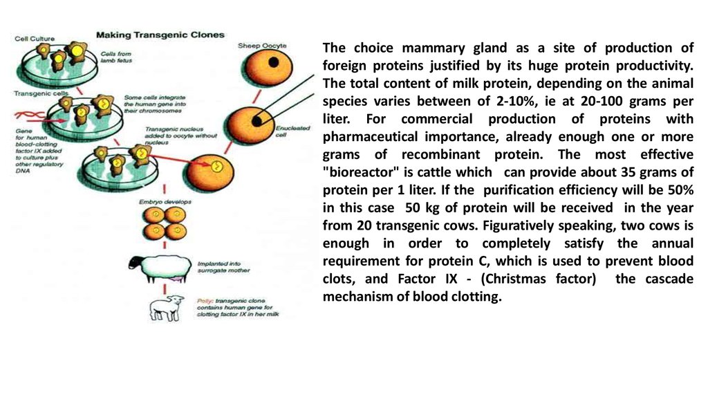
The choice mammary gland as a site of production offoreign proteins justified by its huge protein productivity.
The total content of milk protein, depending on the animal
species varies between of 2-10%, ie at 20-100 grams per
liter. For commercial production of proteins with
pharmaceutical importance, already enough one or more
grams of recombinant protein. The most effective
"bioreactor" is cattle which can provide about 35 grams of
protein per 1 liter. If the purification efficiency will be 50%
in this case 50 kg of protein will be received in the year
from 20 transgenic cows. Figuratively speaking, two cows is
enough in order to completely satisfy the annual
requirement for protein C, which is used to prevent blood
clots, and Factor IX - (Christmas factor) the cascade
mechanism of blood clotting.
27.
To date, a number of recombinant proteins is known, such as human protein C,antihemophilic factor 1X, alpha-1-antitrypsin, tissue plasma activator, lactoferrin, human
serum albumin, interleukin-2, urokinase, chymosin, etc., obtained from the milk of transgenic
animals . Works on the production of these proteins, with the exception of alpha-1antitrypsin, interleukin-2 and chymosin, are at the level of laboratory research and have not
reached a stage which would be of commercial interest.
Recently, much attention is paid to the use of animal organs for transplantation to man.
The main problem of interspecies transplantation is hyperacute rejection. Hyperacute
rejection involves the binding of antibodies of the host to carbohydrate antigenic
determinant on the surface of cells of transplanted organ. Antibodies cause acute
inflammatory response (activation of the complement cascade) that is why mass death of
cells bearing antibodies is occured and rapid loss of the transplanted organ is observed. In
natural conditions inflammatory response is blocked by special proteins. These proteins complement inhibitors are species specific. It has been suggested that if the donor animals
carried one or more genes of the human protein that inhibits the complement, the
transplanted organ would have been protected from the primary inflammatory response.
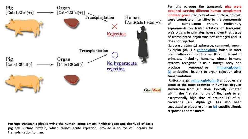
28.
For this purpose the transgenic pigs wereobtained carrying different human complement
inhibitor genes. The cells of one of these animals
were completely insensitive to the components
of
complement
system.
Preliminary
experiments on transplantation of transgenic
pig’s organs to primates have shown that tissue
of transplanted organ was not damaged and it
does not rejected.
Galactose-alpha-1,3-galactose, commonly known
as alpha gal, is a carbohydrate found in most
mammalian cell membranes. It is not found in
primates, including humans, whose immune
systems recognize it as a foreign body and
produce
xenoreactive
immunoglobulin
M antibodies, leading to organ rejection after
transplantation.
Anti-alpha gal immunoglobulin G antibodies are
some of the most common in humans. Regular
stimulation from gut flora, typically initiated
within the first six months of life, leads to an
exceptionally high titre of around 1% of all
circulating IgG. Alpha gal has also been
suggested to play a role in an IgE-specific allergic
response to some meats.
Perhaps transgenic pigs carrying the human complement inhibitor gene and deprived of basic
pig cell surface protein, which causes acute rejection, provide a source of organs for
transplantation to man.
29.
The first work on getting transgenic animals - producers of interleukin-2 turned out encouraging.Interleukin-2 being a soluble factor of T-helper lymphocytes involved in cell proliferation and differentiation
of T-cell killer, plays an important role in ensuring the required level of immunity. Using a gene construct
consisting of rabbit beta-casein DNA and structural human interleukin-2 gene, rabbits were obtained
secreting with milk active form of the protein.
Thus, integration of one or more genes in mammalian embryos is achieved and their expression as well
as the transmission to the offspring is proved. However, the difficulties and uncertainties should be
emphasized with which still related technique for producing transgenic animals. Mechanism of integration
of the gene in mammalian cells is still poorly understood. A)This integration occurs randomly and not
connected with a specific region of a chromosome. B) Another difficulty is due to the instability of the cells
in which gene (s) is introduced: it may be lost or modified as a result becomes inactive. C) Finally, the
activity of genes is determined not only by sequences of nucleotides that provide gene transcription with
the formation of mRNA, but as well as other sequences of nucleotides, which are often far from their own
gene. These sequences are administered with a gene to achieve full expression of the it.
The results achieved in the field of genetic engineering on getting transgenic mammals allow to deepen our
knowledge about gene expression that in the future facilitate gene transfer and identification of factors that
contribute to a more complete expression of the genetic information stored in transgene.
30.
Featured Article on lecturesJ. Livestock Science.-2013.- Vol. 153.-P. 1–9
Review article
Genetically modified farm animals and fish in agriculture:
F. Forabosco a,n, M.L.ohmus b, L.Rydhmer a, L.F.Sundstrom.
Department of Animal Breeding and Genetics,Swedish University of
Agricultural Sciences,Uppsala,Sweden
• Department of Chemistry, Environment and Feed Hygiene, Section of
Environment and Biosecurity, National Veterinary Institute, Swedish
• University of AgriculturalSciences,Uppsala,Sweden
• Department of Ecology and Genetics/Animal Ecology, Evolutionary Biology
Centre,Uppsala University, Uppsala,Sweden