


 slow the citric acid cycle (by drawing off oxaloacetate) and enhance the conversion of acetyl-CoA to acetoacetate. The released coenzyme A allows")


 Биология
Похожие презентации:


Обмін ліпідів
1. Обмін ліпідів
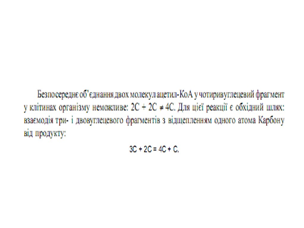
2.
A0217401.mov3. План лекції
1.Травлення ліпідів у кишковошлунковому тракті2.Розпад ліпідів у клітинах організму.
3.Окиснення жирних кислот.
4.Утворення кетонових тіл.
5.Біосинтез жирних кислот.
6.Біосинтез триацилгліцеролів.
4.
5. Запас енергетичних ресурсів у організмі людини
Energy is stored in the body in the
form of triglyceride and glycogen
within adipose tissue, liver, and
skeletal muscle. Triglyceride
present within adipose tissue is the
body’s major fuel reserve. A lean
adult has approximately 35 billion
adipocytes, and each adipocyte
contains about 0.4 to 0.6 µg of
triglyceride. An extremely obese
adult can have 4 times as many
adipocytes, each containing up to
twice as much lipid. Intramuscular
glycogen and triglyceride provide
an important source of fuel for
working muscles during exercise.
Triglycerides are a fivefold better
fuel per unit mass than glycogen,
because triglycerides are stored
compactly as an oil within
adipocytes and liberate 9.3 kcal/g
when oxidized, whereas glycogen
is stored intracellularly as a gel,
containing 2 g of water for every 1
g of glycogen, and liberate 4.1
kcal/g when oxidized.
6. Жовчні кислоти
7. Uptake of dietary lipid in the intestine of a vertebrate animal, and delivery of fatty acids to muscle and adipose tissues.
8. Молекулярна структура хіломікронів
Molecular structure of a
chylomicron. The surface is
covered with a layer of
phospholipids, with head groups
facing the aqueous phase. Tiiacylglycerols sequestered in the
interior make up more than 80% of
the mass. Several apoproteins that
protrude from the surface act as
signals in the uptake and
metabolism of chylomicron
contents. The diameter of
chylomicrons ranges from about
100 to about 500 nm.
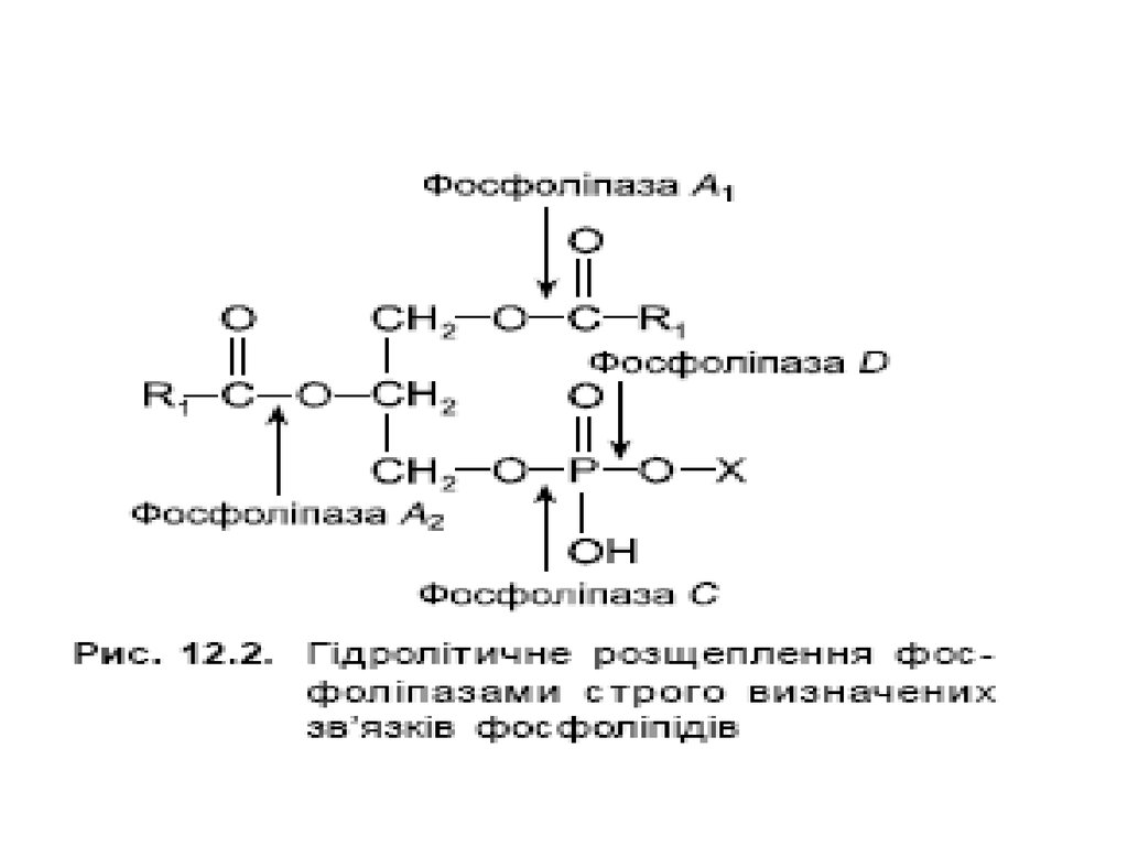
9. Гідроліз триацилгліцеролів у шлунково- кишковому тракті
Гідроліз триацилгліцеролів у шлунковокишковому тракті10.
11. Розпад ацилгліцеролів у клітинах
12. Вивільнення енергії з триацилгліцеролу
Mobilization of
triacylglycerols
stored in adipose
tissue. Low levels of
glucose in the blood
trigger the
mobilization of
triacylglycerols
through the action of
epinephrine and
glucagon on the
adipocyte adenylate
cyclase.
13. Шляхи включення гліцерину у гліколіз та глюконеогенез
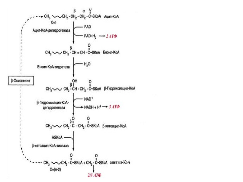
14. β-окиснення жирних кислот
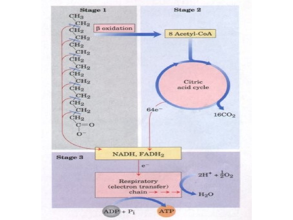
15. Stages of fatty acid oxidation.
Stage 1: A longchain fatty acid is
oxidized to yield acetyl residues in
the form of acetyl-CoA.
Stage 2: The acetyl residues are
oxidized to CO2 via the citric acid
cycle.
Stage 3: Electrons derived from the
oxidations of stages 1 and 2 are
passed to O2 via the mitochondrial
respiratory chain, providing the
energy for ATP synthesis by
oxidative phosphorylation.
16. β-окиснення жирних кислот
17. Активація жирних кислот
Fatty acid activation by the
formation of the fatty acyl-CoA
derivative occurs in two steps.
First, the carboxylate ion
displaces the outer two (β and
γ) phosphates of ATP to form
a fatty acyl-adenylate, the
mixed
anhydride
of
a
carboxylic
acid
and
a
phosphoric acid. The other
product is PPi, an excellent
leaving
group
that
is
immediately hydrolyzed to two
Pi, pulling the reaction in the
forward direction.
Coenzyme A carries out
nucleophilic attack on the
mixed anhydride, displacing
AMP and forming the thioester
fatty acyl-CoA. The overall
reaction is highly exergonic.
18. Проникнення жирних кислот через мембрану мітохондрії
19. Проникнення жирних кислот через мембрану мітохондрії
20. Транспорт жирних кислот через внутрішню мембрану мітохондрій
Ферментативні реакції
перенесення довголанцюгових
жирних кислот з цитозолю клітини
через внутрішню мембрану
мітохондрій за участі карнітину
зображено на рисунку.
Ацил-КоА вступає на шлях ßокиснення, а вільний карнітин
виходить з мітохондрій і в
цитозолі бере участь у
транспортуванні нової молекули
ацил-КоА.
21. Карнітин
22. Проникнення жирних кислот через мембрану мітохондрії
23. Стадія дегідрування
24. Стадія гідратації
25. Друга стадія дегідрування
26. Тіолазна реакція
27. β-окислення
The fatty acid oxidation (βoxidation) pathway.
(a) In each pass through this
sequence, one acetyl residue
(shaded in red) is removed in the
form of acetyl-CoA from the
carboxyl end of palmitate (C16),
which enters as palmitoyl-CoA.
(b) Six more passes through the
pathway yield
seven
more
molecules of acetyl-CoA, the
seventh arising from the last two
carbon atoms of the 16-carbon
chain. Eight molecules of acetylCoA are formed in all.
28. Енергетичний баланс β-окислення
Якщо жирна кислота містить n ?????? ???????, ?? ?? ??????? ?? ?????????????рюється (n : 2 ) ??????? ??????K?? ( ?????? ?????? ??????? ??? ????? ??????? ) ?? (n :
2 ) – 1 молекул ФАДН2 і НАДH(Н+), оскільки за останнього циклу
окиснення утворюються
дві молекули ацетилKоА, але по одній
молекулі ФАДH2 і НАДH (Н+).
Отже продуктами окиснення жирної кислоти з парним
числом атомів карбону є: ацетилКоА, ФАДH2 і НАДH(Н+). В
подальшому ацетилKоА вступає в ЦТК, а ФАДH2 і НАДH (Н+) –
безпосередньо в дихальний ланцюг.
За кожного циклу βокиснення утворюється : 1 молекула
ФАДH2 і 1 молекула НАДH(Н+). Останні у процесі окиснення в
дихальному ланцюзі та спряженого з ним фосфорилювання
дають: ФАДH2 (через KoQ) – 2 молекули АТФ, а НАДH(Н+) – 3
молекули АТФ,
тобто сумарно за один цикл утворюються 5 молекул АТФ.
29. Енергетичний баланс β-окислення
У випадку пальмітинової кислоти (С 16) відбувається 7 циклів β окиснення:
пальмітоїлKоА + 7ФАД + 7НАД+ + 7Н2O + 7HSKoA = 8ацетил
KоА + 7ФАДН2 + 7НАДН + 7Н+
Оскільки за окиснення жирної кислоти, яка містить n(16) атомів карбону,
відбувається (n : 2) – 1 = (16:2)1= 7 циклів β окиснення, це призводить до
утворення: 5 × 7 = 35 молекул АТФ.
У процесі βокиснення пальмітинової кислоти утворюються 8 молекул
ацетилКоА (n:2)=(16:2)=8, кожна з яких, згораючи в циклі трикарбонових
кислот, дає 12 молекул АТФ. Отже 12 × 8=96 молекул АТФ. Таким чином
усього за повного βокиснення пальмітинової кислоти утворюється:
35 + 96 = 131 молекула АТФ.
Одна молекула АТФ витрачається на активацію жирної кислоти, тому
баланс АТФ при повному окисненні пальмітинової кислоти складає 131 –
1= 130 молекул АТФ.
30.
31.
32. Окислення ненасичених жирних кислот
• The oxidation of amonounsaturated fatty
acyl-CoA, such as
oleoyl-CoA (Δ9),
requires an additional
enzyme, enoyl-CoA
isomerase. This
enzyme repositions the
double bond,
converting the cis
isomer to a trans
isomer, a normal
intermediate in β
oxidation.
33. Окислення ненасичених жирних кислот
• Oxidation ofpolyunsaturated fatty acids
requires a second auxiliary
enzyme in addition to
enoyl-CoA isomerase:
NADPH-dependent 2,4dienoyl-CoA reductase.
The combined action of
these two enzymes
converts a trans-Δ2,cisΔ4dienoyl-CoA
intermediate into the transΔ2-enoylCoA substrate
necessary for β oxidation.
34. Кетонові тіла
35. Утворення кетонових тіл
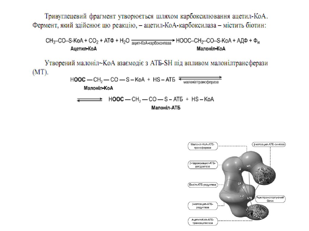
36. Синтез жирних кислот
37. Синтез жирних кислот
38. Синтаза вищих жирних кислот
39.
40.
41.
42.
43.
44.
45.
46.
47. Сумарне рівняння синтезу жирних кислот
Із пальмітинової кислоти синтезується стеаринова і інші вищі жирні кислоти шляхомприєднання ацетил-КоА ( в мітохондріях) чи малоніл–КоА ( е ендоплазматичному
ретикулумі)
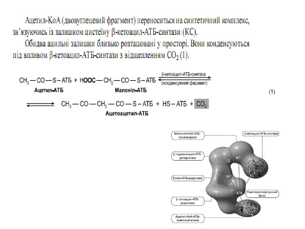
48. Синтез жирних кислот
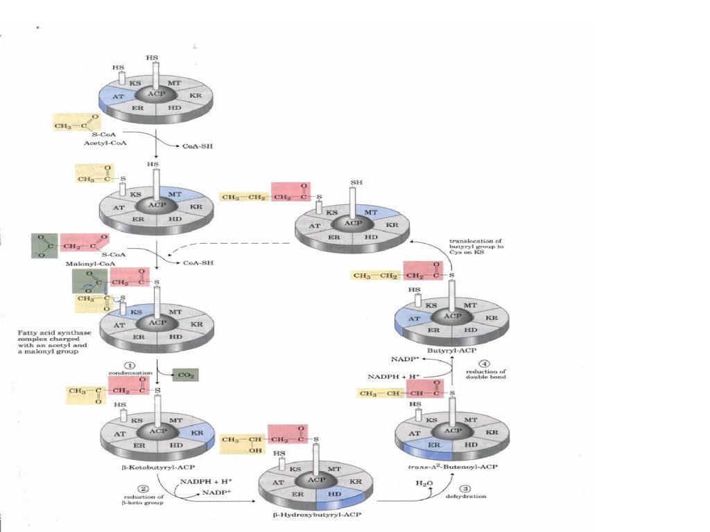
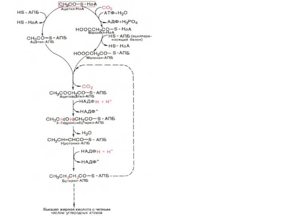
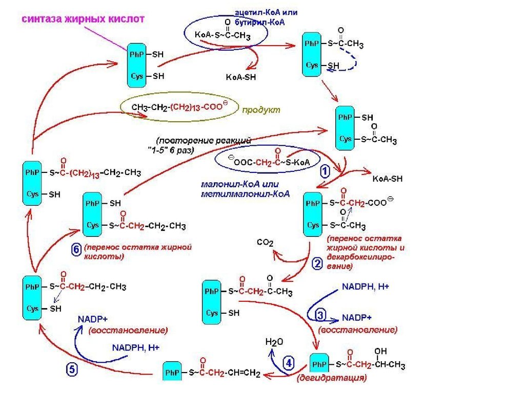
Утворений малоніл~KоА взаємодіє з АТБSHпід впливом малонілтрансацилази. Ацетил
KоА переноситься на синтетичний комплекс,
зв’язуючись із залишком цистеїну синтази.
Обидва
ацильні
залишки
близько
розташовані у просторі. Вони конденсуються
під впливом 3кетоацилАТБсинтази з
відщепленням
СО2.
При
конденсації
ацетильний залишок переноситься на
малонільний, витісняючі його карбоксильну
групу. АцетоацетилАТБ відновлюється за
кетогрупою до β оксипо
хідного під впливом 3кетоацилАТБ
редуктази. Донором гідрогену для цієї реакці
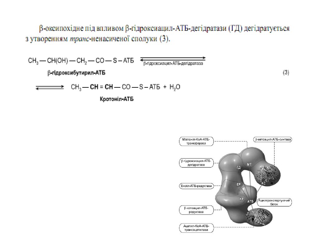
є НАДФH(Н+). βоксипохідне під впливом β
гідроксиацилАТБдегідратази дегідра
тується з утворенням трансненасиченої
сполуки , яка, у свою чергу, відновлюється за
рахунок НАДФH(Н+) під впливом ферменту
еноїлАТБредуктази. Таким чином з двох
окиснених
двовуглецевих
фрагментів
синтезується відновний чотиривуглецевий
бутирилАТБ. Він переноситься з АТБ на
залишок цистеїну ферменту.
49.
50.
51. Позамітохондріальна система біосинтезу жирних кислот
Позамітохондріальна система
біосинтезу жирних кислот
Як вже зазначалося, будівельним блоком для
синтезу жирних кислот в цитозолі клітини слугує
ацетил-KоА,
який
переважно
надходить
із
мітохондрій. Було з’ясовано, що цитрат стимулює
синтез жирних кислот в цитозолі клітини. Відомо
також, що утворений в мітохондріях у процесі
окиснювального декарбоксилювання пірувату та
окиснення жирних кислот ацетил-KоА не може
дифундувати
у
цитозоль
клітини,
оскільки
мітохондріальна мембрана непроникна для даного
субстрату.
Тому спочатку внутрішньомітохондріальний ацетилКоА взаємодіє з оксалоацетатом з утворенням
цитрату.
Реакція
каталізується
ферментом
цитратсинтазою. Утворений цитрат переноситься
через мембрану мітохондрій у цитозоль за
допомогою
спеціальної
трикарбоксилаттранспортуючої системи. В цитозолі цитрат реагує з
HS-KoA і АТФ, знову розщеплюючись на ацетил-KоА і
оксалоацетат. Дана реакція каталізується АТФцитратліазою. Вже у цитозолі оксалоацетат за
участю
цитозольної
малатдегідрогенази
відновлюється до малату. Останній за допомогою
дикарбоксилаттранспортувальної
системи
повертається в мітохондріальний матрикс, де
окиснюється до оксалоацетату, завершуючи тим
самим так званий човниковий цикл.
52. Утворення ненасичених жирних кислот
53. Біосинтез триацилгліцеролів
54. Біосинтез триацилгліцеролів
55. Біосинтез триацилгліцеролів
56. Біосинтез фосфогліцеридів
57. Біосинтез фосфогліцеридів
58. Біосинтез фосфогліцеридів
59. Біосинтез фосфогліцеридів
60. Біосинтез холестеролу
61.
62.
63. Ketone body formation and export from the liver. Conditions that increase gluconeogenesis (diabetes, fasting) slow the citric acid cycle (by drawing off oxaloacetate) and enhance the conversion of acetyl-CoA to acetoacetate. The released coenzyme A allows
continued β oxidation of fatty acids.64.
• The role of β oxidationin the conversion of
seed triacylglycerols
into glucose in
germinating seeds.
65. Утворення кетонових тіл
• Formation of ketone bodies fromacetyl-CoA. Under circumstances that
cause acetylCoA accumulation
(starvation or untreated diabetes, for
example), thiolase catalyzes the
condensation of two acetyl-CoA
molecules to acetoacetyl-CoA, the
parent of the three ketone bodies.
These reactions all occur within the
mitochondrial matrix. The six-carbon
compound β-hydroxy-βmethylglutarylCoA (HMG-CoA) is also an
intermediate of sterol biosynthesis,
but the enzyme that forms HMG-CoA
in that pathway is cytosolic. HMGCoA lyase is present in the
mitochondrial matrix but not in the
cytosol.
66.
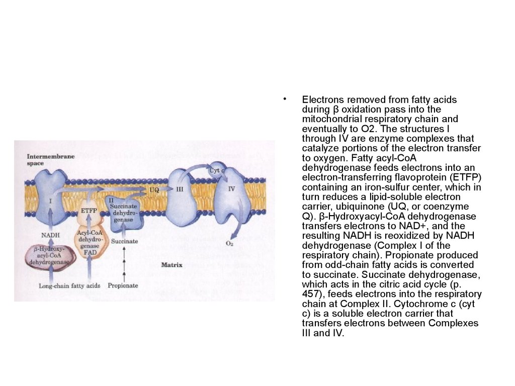
Electrons removed from fatty acids
during β oxidation pass into the
mitochondrial respiratory chain and
eventually to O2. The structures I
through IV are enzyme complexes that
catalyze portions of the electron transfer
to oxygen. Fatty acyl-CoA
dehydrogenase feeds electrons into an
electron-transferring flavoprotein (ETFP)
containing an iron-sulfur center, which in
turn reduces a lipid-soluble electron
carrier, ubiquinone (UQ, or coenzyme
Q). β-Hydroxyacyl-CoA dehydrogenase
transfers electrons to NAD+, and the
resulting NADH is reoxidized by NADH
dehydrogenase (Complex I of the
respiratory chain). Propionate produced
from odd-chain fatty acids is converted
to succinate. Succinate dehydrogenase,
which acts in the citric acid cycle (p.
457), feeds electrons into the respiratory
chain at Complex II. Cytochrome c (cyt
c) is a soluble electron carrier that
transfers electrons between Complexes
III and IV.
67.
68. Синтез жирних кислот
69.
• Acyl carrier protein (ACP).The prosthetic group is 4'phosphopantetheine, which is
covalently attached to the
hydroxyl group of a Ser
residue in ACP.
Phosphopantetheine contains
the B vitamin pantothenate,
also found in the coenzyme A
molecule. Its -SH group is the
site of entry of malonyl
groups during fatty acid
synthesis
70.
71.
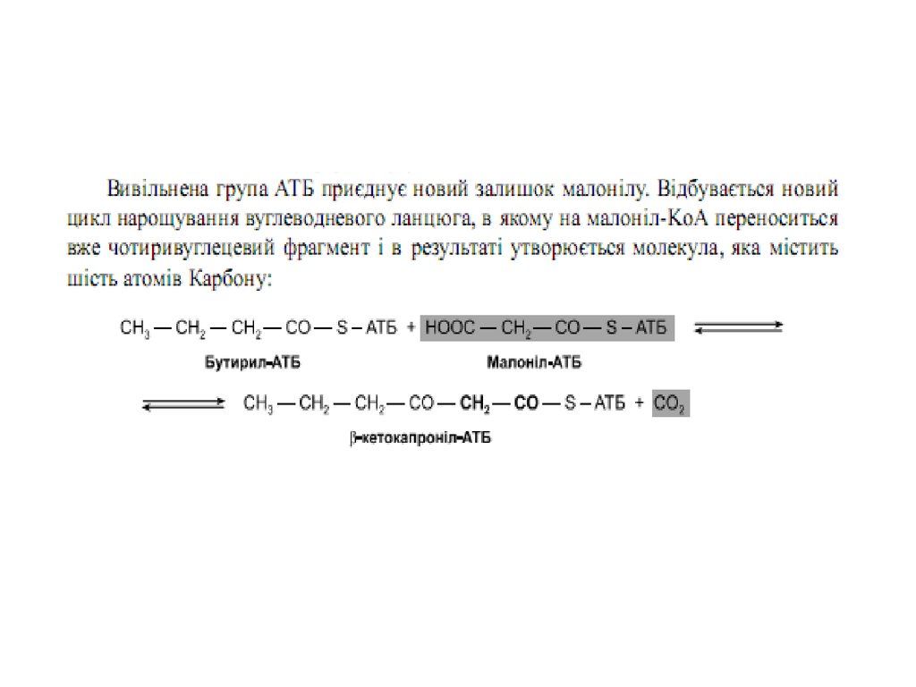
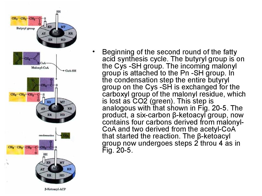
Beginning of the second round of the fatty
acid synthesis cycle. The butyryl group is on
the Cys -SH group. The incoming malonyl
group is attached to the Pn -SH group. In
the condensation step the entire butyryl
group on the Cys -SH is exchanged for the
carboxyl group of the malonyl residue, which
is lost as CO2 (green). This step is
analogous with that shown in Fig. 20-5. The
product, a six-carbon β-ketoacyl group, now
contains four carbons derived from malonylCoA and two derived from the acetyl-CoA
that started the reaction. The β-ketoacyl
group now undergoes steps 2 throu 4 as in
Fig. 20-5.
72.
The acetyl group
shuttle for transfer of
acetyl groups from
mitochondria to the
cytosol for fatty acid
synthesis. (The outer
mitochondrial
membrane is freely
permeable to all of
these compounds.)
Pyruvate derived
from amino acid
catabolism in the
matrix, or from
glucose by glycolysis
in the cytosol, is
converted to acetylCoA in the matrix.
Acetyl groups pass
out of the
mitochondrion as
citrate; in the cytosol
they are delivered as
acetylCoA for fatty
acid synthesis.
Malate returns to the
mitochondrial matrix,
where it is converted
to oxaloacetate. An
alternative fate for
cytosolic malate is
oxidation by malic
enzyme to generate
cytosolic NADPH; the
pyruvate produced
returns to the
mitochondrial matrix.
73.
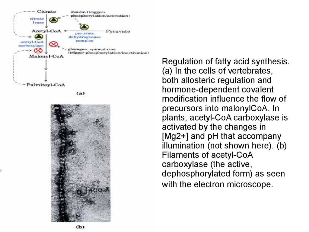
Regulation of fatty acid synthesis.
(a) In the cells of vertebrates,
both allosteric regulation and
hormone-dependent covalent
modification influence the flow of
precursors into malonylCoA. In
plants, acetyl-CoA carboxylase is
activated by the changes in
[Mg2+] and pH that accompany
illumination (not shown here). (b)
Filaments of acetyl-CoA
carboxylase (the active,
dephosphorylated form) as seen
with the electron microscope.
74.
Routes of synthesis of other fatty acids.
Palmitate is the precursor of stearate
and longer-chain saturated fatty acids,
as well as the monounsaturated acids,
palmitoleate and oleate. Mammals
cannot convert oleate into linoleate or αlinolenate (shaded red), which are
therefore required in the diet as
essential fatty acids. Conversion of
linoleate into other polyunsaturated fatty
acids and eicosanoids is outlined.
Unsaturated fatty acids are symbolized
by indicating the number of carbons and
the number and position of the double
bonds, as in Table 9-1.
75.
76.
77.
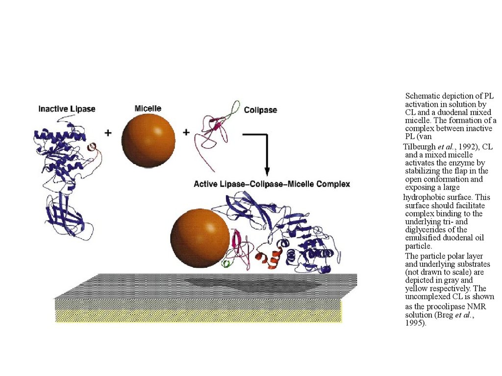
Schematic depiction of PLactivation in solution by
CL and a duodenal mixed
micelle. The formation of a
complex between inactive
PL (van
Tilbeurgh et al., 1992), CL
and a mixed micelle
activates the enzyme by
stabilizing the flap in the
open conformation and
exposing a large
hydrophobic surface. This
surface should facilitate
complex binding to the
underlying tri- and
diglycerides of the
emulsified duodenal oil
particle.
The particle polar layer
and underlying substrates
(not drawn to scale) are
depicted in gray and
yellow respectively. The
uncomplexed CL is shown
as the procolipase NMR
solution (Breg et al.,
1995).