


 NEURON")


 Биология
Похожие презентации:


Eye lecture
1. EYE
Assoc. prof/ Kharchenko S.V.Department of Histology and Embryology
Medical Academy named after S.I. Georgievsky
2. The sensory organs recieve information about the state of the external environment and the activity of the systems of the
THE SENSORY ORGANS RECIEVE INFORMATIONABOUT THE STATE OF THE EXTERNAL ENVIRONMENT
AND THE ACTIVITY OF THE SYSTEMS OF THE
ORGANISM ITSELF.
THE EYEBALL IS THE PERIPHERAL PART OF THE
VISUAL ANALYZER!
THE CENTRAL PART IS LOCATED IN THE OCCIPITAL
LOBE OF THE BRAIN!
BOTH SECTIONS OF THE OPTIC ANALYZER ARE
CONNECTED BY THE OPTIC NERVE!
3.
Eyeball (eye) - is the peripheral part of the visual analyzer.Through the organ of vision a person receives 80-85% of
information about the world around him. Vision is the most
important physiological process with the help of which an
idea is created about the size, shape and color of objects,
about their mutual location and distance. This information
allows a person to navigate the world around him.
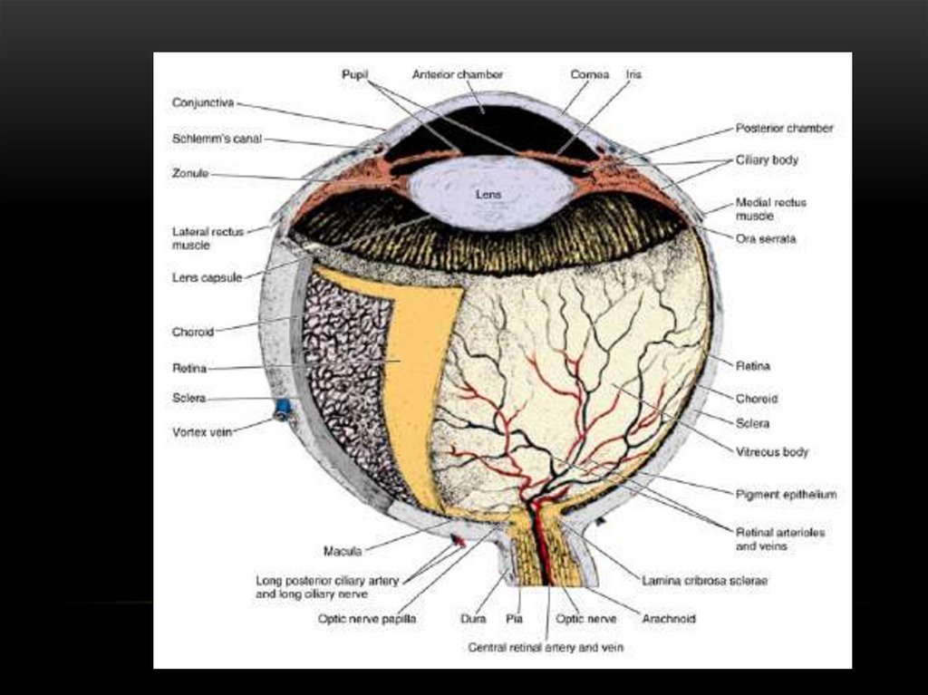
4.
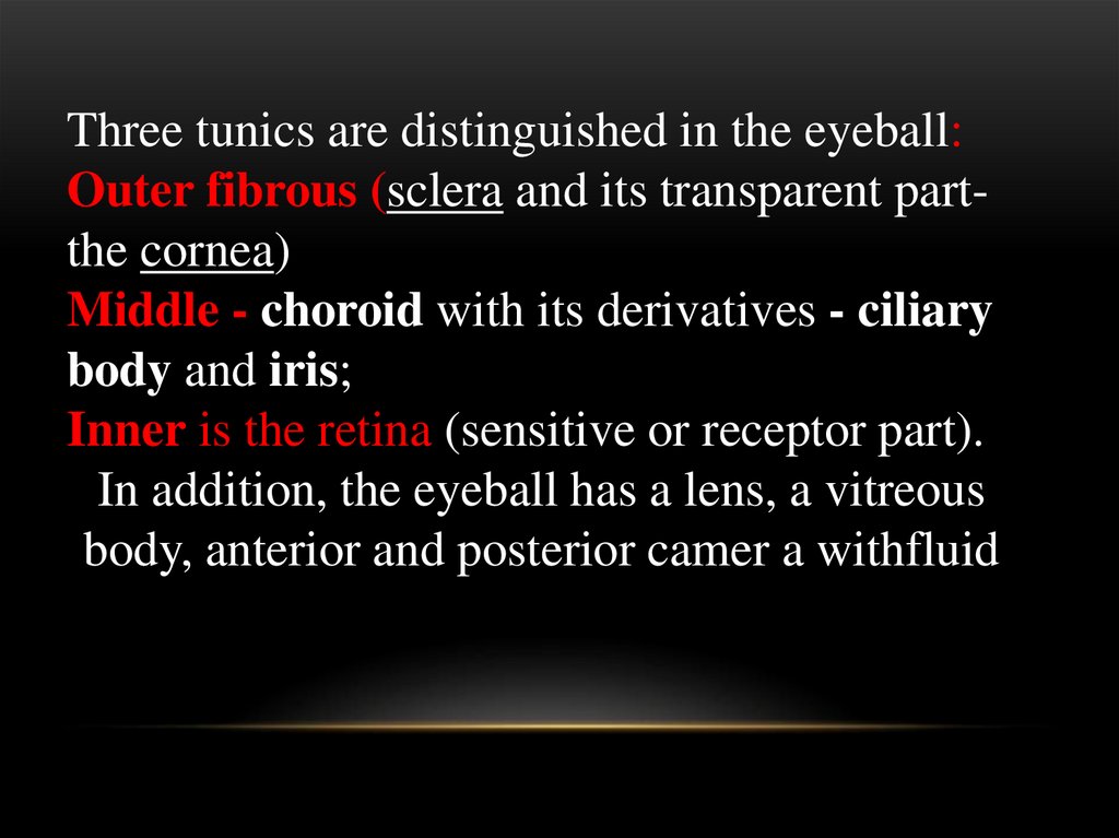
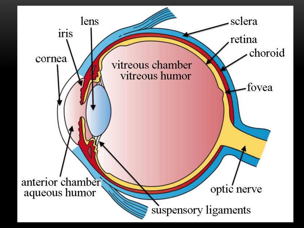
Three tunics are distinguished in the eyeball:Outer fibrous (sclera and its transparent partthe cornea)
Middle - choroid with its derivatives - ciliary
body and iris;
Inner is the retina (sensitive or receptor part).
In addition, the eyeball has a lens, a vitreous
body, anterior and posterior camer a withfluid
5.
6.
Functionally several apparatuses aredistinguished:
receptor (retina),
dioptric or light-refracting (cornea,
crystalline lens, vitreous body, fluid of the
anterior and posterior chambers of the
eye)
accommodative (iris, ciliary body)
Auxiliary (eyelids, lacrimal glands,
oculomotor
7. The organ of vision develops from 3 embryonic sources: ectoderm, neural tube and mesenchyme
THE ORGAN OF VISION DEVELOPS FROM 3EMBRYONIC SOURCES: ECTODERM, NEURAL
TUBE AND MESENCHYME
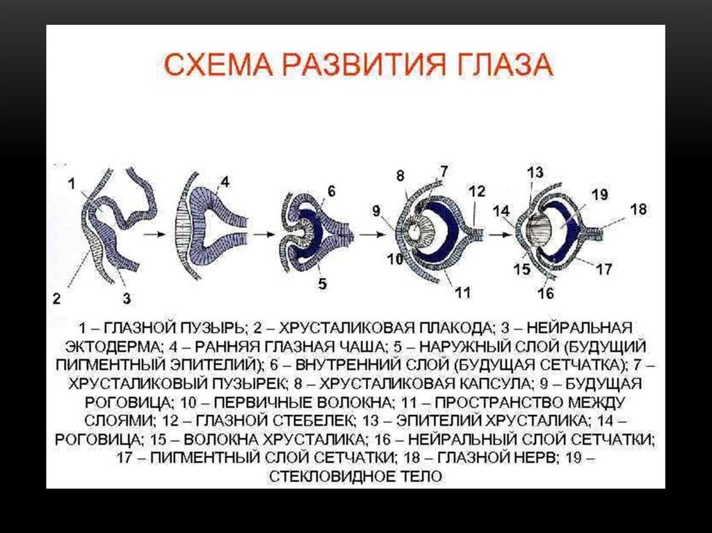
• DEVELOPMENT begins at the beginning of the 3rd week of embryonic
development in the form of eye fossae in the wall that is still not closed in the
neural tube, later 2 eye vesicles protrude from the wall of the diencephalon
from the zone of this fossa. The eye vesicles are connected to the
diencephalon using the eye stalk. The anterior wall of the vesicles is pushed
and the vesicles turn into double-walled eye cups.
8. DEVELOPMENT OF EYE
• At the same time, the ectoderm opposite the eye vesicles, while puffing,forms lens vesicles. Epithelial cells of the posterior hemisphere of the lens
vesicle lengthen and turn into long transparent structures - lens fibers. A
crystalline protein is synthesized in the lens fibers. Subsequently, in the lensfiber cells organoids disappear, the nuclei shrink and disappear. Thus, the
lens forms - a kind of elastic lens. From the ectoderm in front of the lens, the
anterior epithelium of the cornea is formed.
• The inner layert of the 2-wall ocular goblet differentiates into the retina, takes
part in the formation of the vitreous body, and the outer layer forms the
pigment layer of the retina.
• The material of the edge of the eye cup along with the mesenchyme is
involved in the formation of the iris. From the surrounding mesenchyme, the
choroid and sclera, the ciliary muscle, its own substance and the posterior
corneal epithelium are formed. Mesenchyma is also involved in the formation
of the vitreous, iris.
9.
10. STAGES OF EYE DEVELOPMENT
11.
12. TUNICA FIBROSA
• Consists of sclera - a dense opaque membrane and cornea - atransparent front
• The sclera is formed by a dense fibrous connective tissue,
consisting of collagen fibers bundels running parallel to the
surface of the organ and the fibroblasts and elastic fibers lying
between them.
• Contains blood vessels!
13. LIMBUS
• The sclera passes into the cornea in the limbregion, on the inner surface of which there is
a system of channels lined with the
endothelium leading to the venous sinus
(Schlemm canal) - the path of the outflow of
aqueous humor from the anterior chamber of
the eye
14. Cornea - has no blood vessels, is well innervated
CORNEA - HAS NO BLOOD VESSELS, IS WELLINNERVATED
Includes 5 layers:
1. anterior epithelium - stratified squamous non-keratinized
2. anterior limiting membrane (Bowman's membrane) consists of a network of collagen fibrils
3. substantia propiria or stroma - occupies 90% of the
thickness of the cornea and consists of connective tissue plates
(there are collagen fibrils, fibroblasts and the main substance,
glycoproteins (chondroitin and keratan sulfates) which ensure
the transparency of the cornea
4. posterior limiting membrane (Descemet's membrane)
5. posterior epithelium (endothelium) - single layer flat cells
15. CORNEA- transparent part of tunica fibrosa
CORNEA- TRANSPARENT PART OF TUNICA FIBROSA16.
17. HORoid
HOROIDIncludes:
choroid
ciliary body
iris
Actually, the choroid consists of Loose irregular connective tissue and
includes 4 layers:
- subvascular (at the border with the sclera),
- vascular (contains arteries and veins),
- choriocapillary (contains a network of capillaries)
- basal (includes the basal membrane of capillaries, the basement
membrane of the retinal pigment epithelium and the network of collagen
and elastic fibers)
18. CILIARY BODY
• It is formed by the ciliary muscle (smooth muscle)and ciliary processes that fix the lens
• It takes part in the accommodation of the eye,
changing the curvature of the lens
• Covered with a bilayer cubic ciliary epithelium that
produces watery moisture
19. IRIS
• the anterior part of the choroid, separates the anterior andposterior chambers of the eye, contains a hole of varying
diameter (pupil). It is formed by rhbc with blood vessels and
pigment cells.
• Consists of 5 layers:
-Anterior epithelium (single layer of squamose cells)
-Outer limiting membrane
-Vascular
-Inner limiting membrane
- Pigment epithelium - bilayed cubic pigment epithelium
• The iris contains 2 muscles of neural origin: m. sphinter
pupillae, m. dilatator pupillae.
20. Радужка и хрусталик
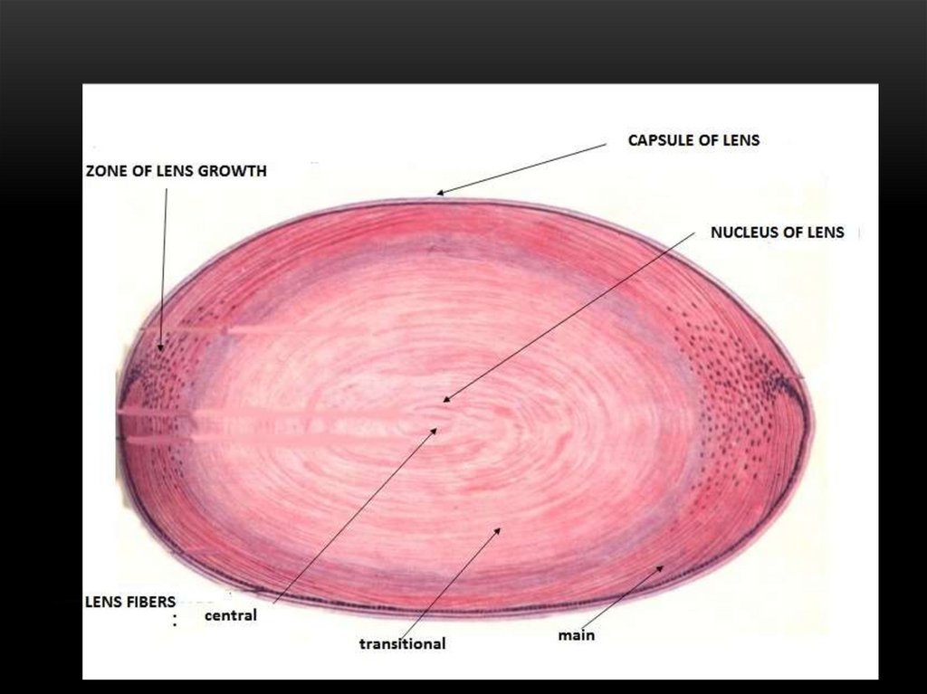
РАДУЖКА И ХРУСТАЛИК21. The lens - does not have blood vessels
THE LENS - DOES NOT HAVE BLOODVESSELS
Consists of:
1. Capsule - a thin transparent layer covering the lens from the
outside - is the basement membrane of its epithelium.
2. Lens epithelium - a layer of cuboidal cells lying under the
capsule on its anterior surface (they are a growth zone), in the
equatorial region the cells divide by mitosis, lengthen and
gradually turn into lens fibers
22. LENS FIBERS
• Elongated hexagonal epithelial cells lying parallel to the lenssurface in concentric layers and forming its matrix, which
consists of the cortex and nucleus. Contain transparent protein crystallin! Lens fibers of the cortex contain nuclei, the nucleus of
the lens consists of lens fibers without nuclei!
• With age, the elasticity of the lens decreases, which makes it
difficult to study closely located objects (presbyopia).
• In some older people, the lens loses its transparency (cataract)
23.
24.
25. VITREOUS BODY
• A transparent jelly-like mass filling the space between the lensand the retina.
• It consists of cells - hyalocytes, macrophages, lymphocytes and
intercellular substance, consisting of 99.9% water, collagen
fibrils and hyaluronic acid and a transparent protein vitrein!
• Provides the passage of light rays, maintaining the position of
the lens, participates in the metabolism of the retina, presses the
inner layers of the retina to the pigment epithelium
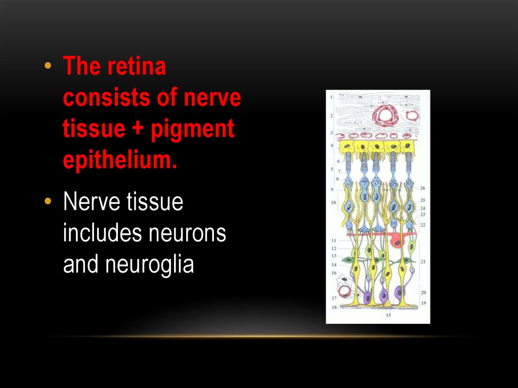
26. RETINA
• Photosensitive tunica of the eye.• It is divided into the visual part, lining the inside of the back most
of the eyeball and the front, the blind part, covering the ciliary
body and the back surface of the iris.
• There is a blind spot on the posterior surface of the retina (does
not contain photoreceptor cells) - the exit site of the optic nerve
and the central fossa is located more laterally along the axis of
the eye - the yellow spot is the concentration site of the
photoreceptor cells (best vision area)
27.
• The retinaconsists of nerve
tissue + pigment
epithelium.
• Nerve tissue
includes neurons
and neuroglia
28. NEURONS
They form a three-membered chain of radially located neuronsconnected to each other by synapses:
• Photoreceptor
• Bipolar (associative)
• Ganglionic (multipolar)
• In addition, horizontal and amacrine neurons are located in the
retina.
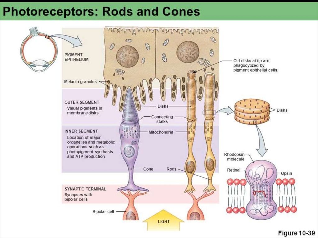
29. PHOTORECEPTORS
- are bipolar neurons - rods and cones.They consist of three parts: perikarion + modified dendrite + axon
Axons form synapses with bipolar and horizontal neurons
Modified dendrites consist of the outer and inner segments
connected by a cilia (connecting stalk)
The outer segments are surrounded by processes of the pigment
epithelium.
30. ROD
• - With narrow elongated outer segments• The outer segment of the process has a cylindrical shape and
contains a stack of 1000-1500 complited (closed) membrane
disks (flattened sacs).
• In the disc membranes is the visual pigment rhodopsin, which
includes protein and aldehyde vitamin A.
31. RODOPSIN
• Rodopsin decomposes under the influence of light with theappearance of an electrical signal, and regenerates in the dark.
• Disks are constantly updated due to their formation in the
proximal areas and displacement to the distal, where they are
phagocytosed by pigment epithelium.
• Vitamin A is necessary for the regeneration of discs, in its
absence they are destroyed (night blindness)
32. INNER SEGMENT
• It contains mitochondria, centriole, rER, sER, Golgi complex andprovides the outer segment with energy and substances
necessary for photoreception. The core is small, round.
• The axon ends with a spherical thickening (spherule) and forms
a synapse with a bipolar cell
• The sticks are located in the peripheral parts of the retina, are
responsible for black and white vision
33. CONES
The structure is similar to rodsBut
The outer segments of them are conical in shape, contain open
membrane disks (half-disk or semi-dikcs), formed by folds of
plasmolemma, in which the visual pigment iodopsin is located,
which decomposes under the action of red, green or blue light
34. CONS
• The inner segment has a drop of fat inside, surrounded bymitochondria (ellipsoid)
• Contains larger and lighter nuclei in the perikarion!
• Axon ends with an extension (leg) of a triangular shape
• Cones are located in the central parts of the retina. Provide
day and color vision.
• The absence of any type of cones causes color blindness
(color blindness)
35. RETINA
36.
37. BIPOLAR NEURONS
• Dendrites are associated with axons of photoreceptor cells, andtheir axons transmit impulses to the dendrites of ganglion and
amacrine cells.
38. MULTIPOLAR (GANGLIONIC) NEURON
• Large multipolar cells with an eccentrically located nucleus.Contain well-developed organelles. Their dendrites form bonds
with the axons of bipolar cells, and the axons, when they are
about to form the optic nerve.
39. HORIZONTAL NEURON
• Associative multipolar neurons, their dendrites and axon aresynaptically connected with the axons of the rods and cones, as
well as with dendrites of bipolar neurons
40. AMACRINE NEURONS
• Unipolar associative neurons whose dendrites formbonds with axons of bipolar cells and ganglionic
dendrites.
41. PIGMENT EPITHELIUM
• The outer layer of the retina is firmly connectedwith the choroid and is loosely connected with
the adjacent layers of the retina.
• This makes it possible to detach the retina
during pathological processes and leads to the
death of the photosensory layer.
42. Functions of the pigment epithelium
FUNCTIONS OF THE PIGMENTEPITHELIUM
• Accumulation and transport of vitamin A to
photoreceptors
• Phagocytosis and digestion of discs
• Providing nutrition to the outer layers of the retina
• Light absorption and prevention of excessive exposure
to photoreceptors
43. NEUROGLIA of retina
NEUROGLIA OF RETINA• It is represented by radial gliocytes (Muller cells), astrocytes
and microglia
• Astrocytes are located in the inner layers of the retina and pull
off capillaries with their processes - they form a blood-retinal
barrier
• Microglia are located in all layers of the retina and perform a
phagocytic function.
44. MULLER CELLS
• Large star shaped cells stretch across the entire thickness of theretina perpendicular to its layers. They occupy the space
between neurons and their processes.
• With their bases, they form the inner glial border membrane,
delimiting the retina from the vitreous, and the apical processes
form the outer glial border membrane.
• Lateral processes braid the bodies of neurons, perform
supportive and trophic functions
45. LAYERS OF RETINA
• 8 layers without limiting membraines• Pigment epithelium
• A layer of rods and cones
• Outer nuclear layer
• Outer plexiform layer
• Inner nuclear layer
• Inner plexiform layer
• A layer of ganglionic cells;
• A layer of nerve fibers.
46. LAYERS OF RETINA 10 layers
LAYERS OF RETINA 10 LAYERS1. pigment epithelium
2. layer of rods and cones - represented by photoreceptor dendrites
3. outer glial limiting membrane - formed by Muller cells
4. outer nuclear layer - contains photoreceptor perikarions
5. outer plexiform layer - the region of synapses between the
processes of photoreceptors and bipolar neurons
6. the inner nuclear layer - contains the bodies of bipolar, amacrine,
horizontal and muller cells
7. inner plexiform layer - the region of synapses between bipolar,
ganglionic and amacrine cells
8. ganglion layer - contains the bodies of ganglionic cells
9. layer of nerve fibers - consists of axons of ganglionic cells
10. the internal glial limiting membrane is formed by Muller cells