


 Математика
Похожие презентации:

Rehabilitation
1.
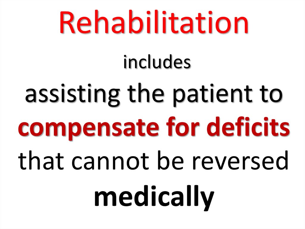
Rehabilitationincludes
assisting the patient to
compensate for deficits
that cannot be reversed
medically
2.
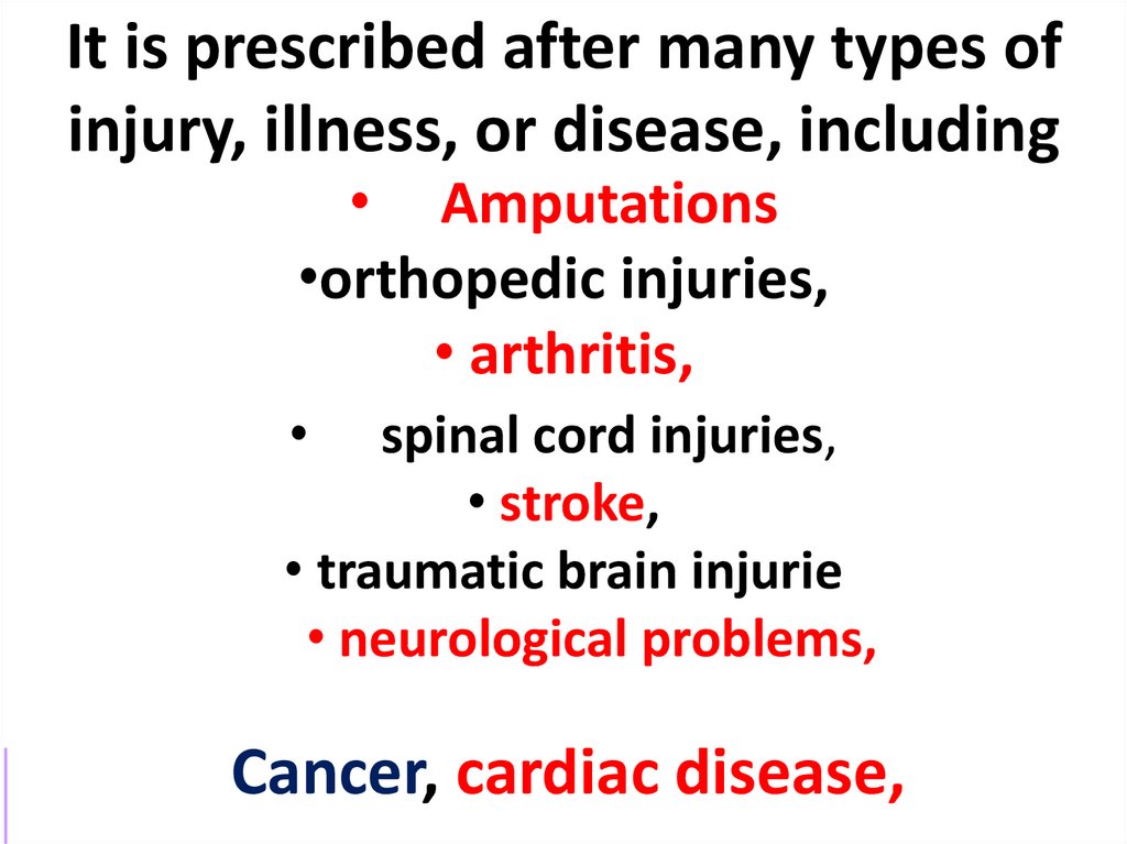
It is prescribed after many types ofinjury, illness, or disease, including
• Amputations
•orthopedic injuries,
• arthritis,
spinal cord injuries,
• stroke,
• traumatic brain injurie
• neurological problems,
Cancer, cardiac disease,
3.
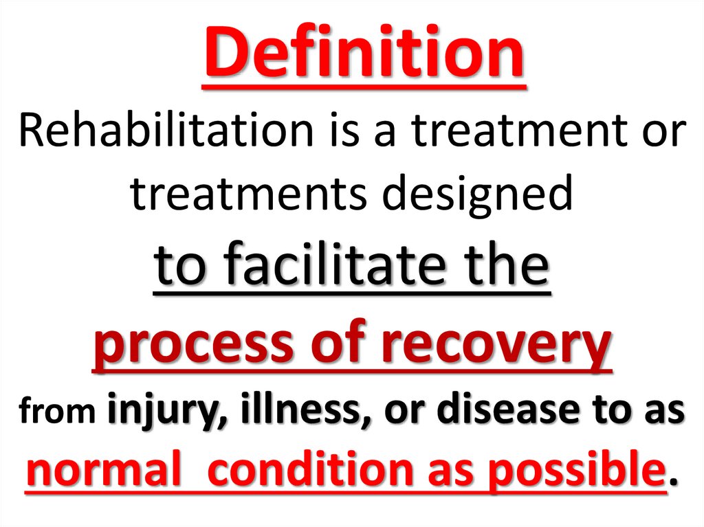
DefinitionRehabilitation is a treatment or
treatments designed
to facilitate the
process of recovery
from injury, illness, or disease to as
normal condition as possible.
4.
GOALSMinimize
deficits
Minimize functional
functional deficits
Prevent complications
complications
Use remaining function to maximum
5.
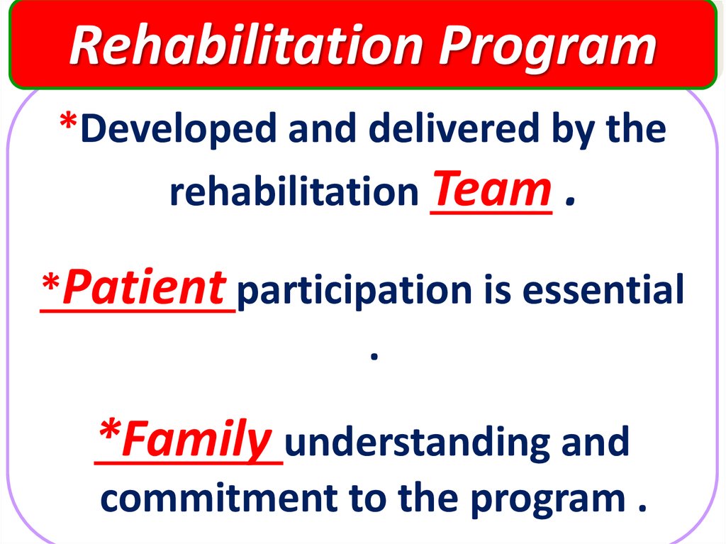
Rehabilitation Program*Developed and delivered by the
rehabilitation Team .
*Patient participation is essential
.
*Family understanding and
commitment to the program .
6.
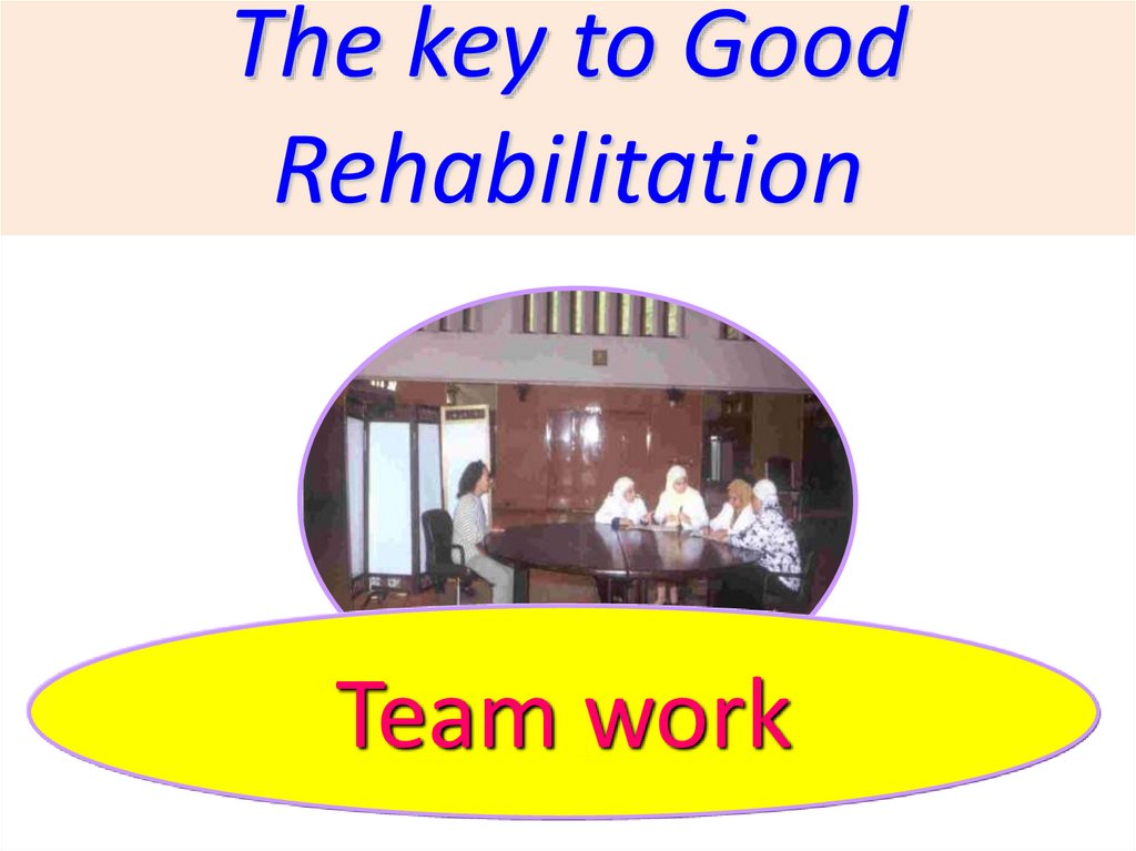
The key to GoodRehabilitation
Team work
7.
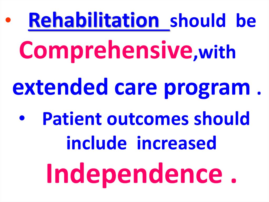
• Rehabilitation should beComprehensive,with
extended care program .
• Patient outcomes should
include increased
Independence .
8.
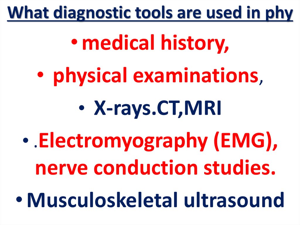
What diagnostic tools are used in phy• medical history,
• physical examinations,
• X-rays.CT,MRI
• .Electromyography (EMG),
nerve conduction studies.
• Musculoskeletal ultrasound
9.
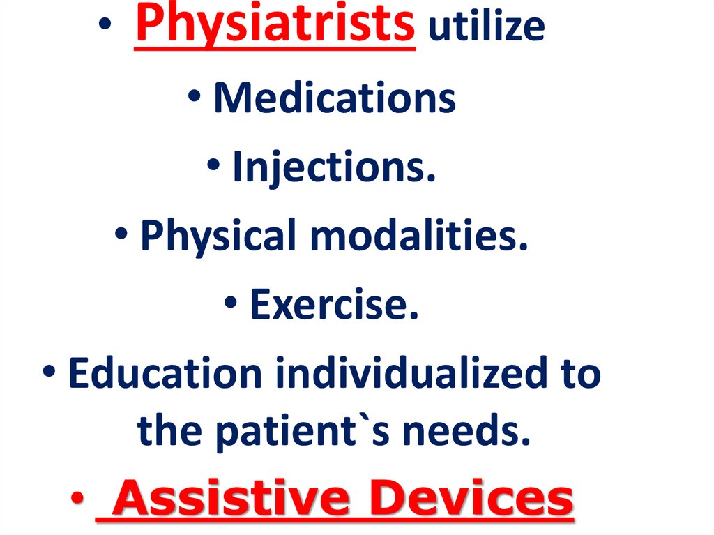
Physiatrists utilize
• Medications
• Injections.
• Physical modalities.
• Exercise.
• Education individualized to
the patient`s needs.
• Assistive Devices
10.
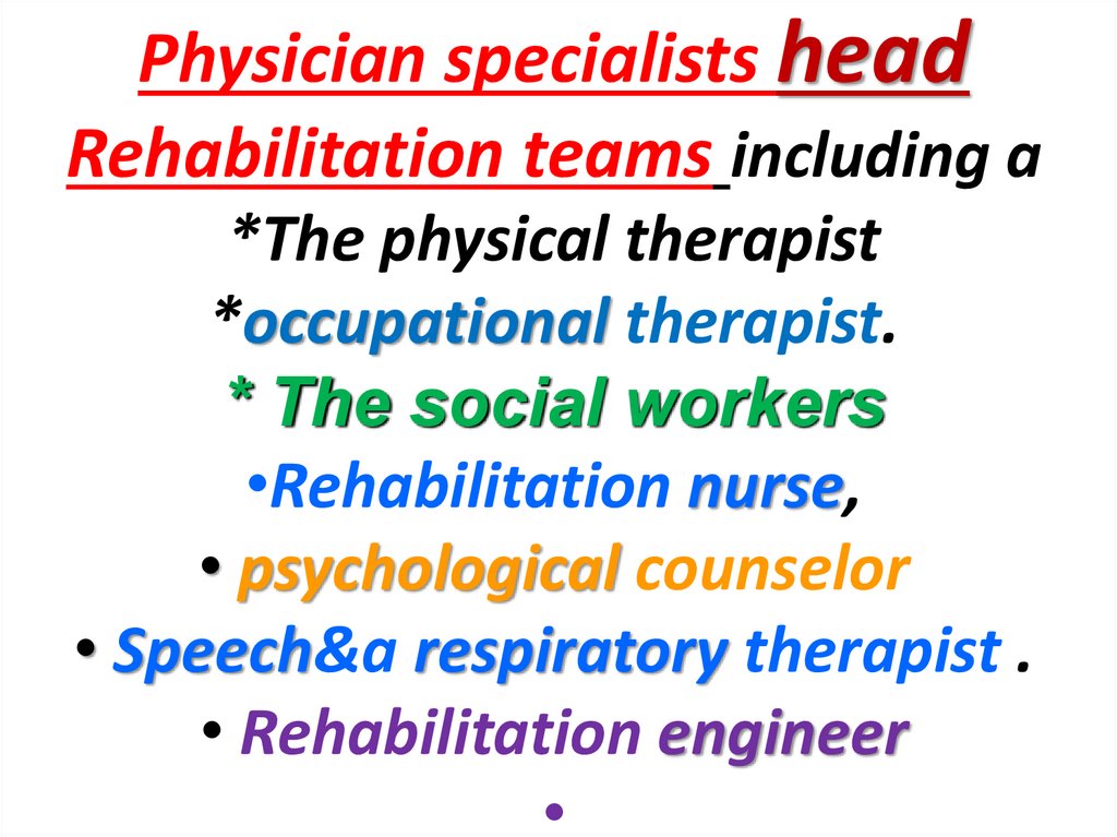
Physician specialists headRehabilitation teams including a
*The physical therapist
*occupational therapist.
* The social workers
•Rehabilitation nurse,
• psychological counselor
• Speech&a respiratory therapist .
• Rehabilitation engineer
11.
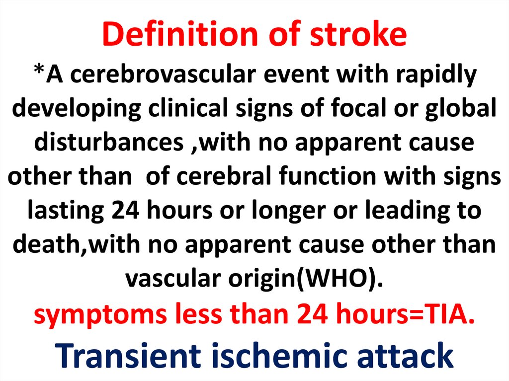
Definition of stroke*A cerebrovascular event with rapidly
developing clinical signs of focal or global
disturbances ,with no apparent cause
other than of cerebral function with signs
lasting 24 hours or longer or leading to
death,with no apparent cause other than
vascular origin(WHO).
symptoms less than 24 hours=TIA.
Transient ischemic attack
12.
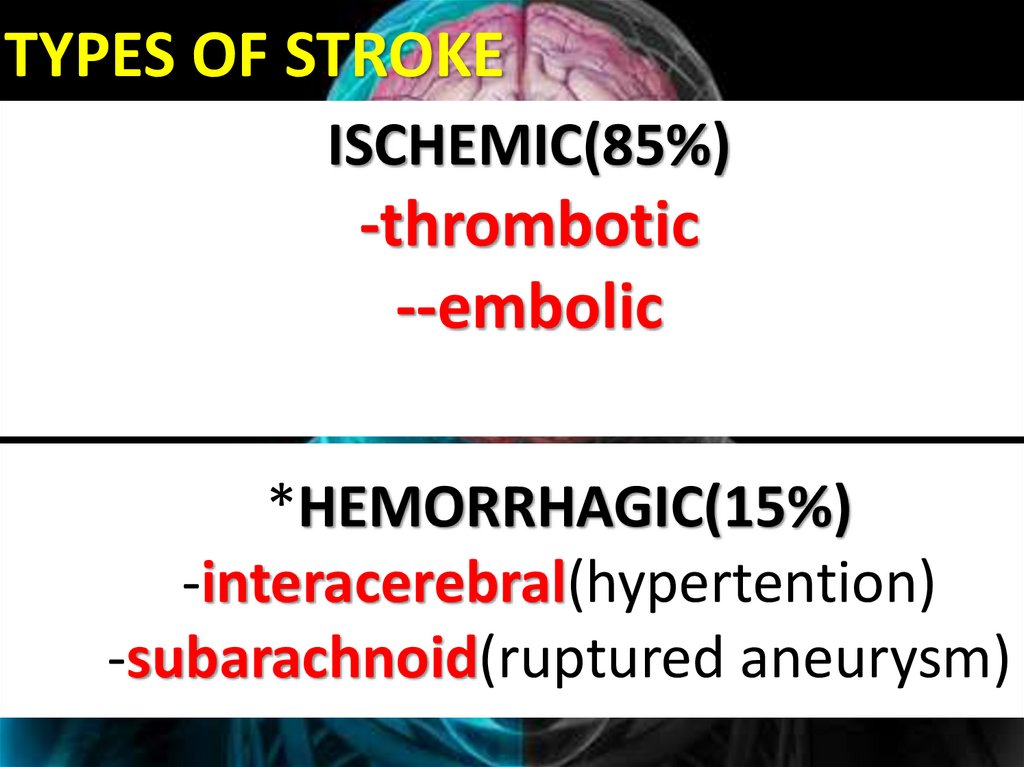
TYPES OF STROKEISCHEMIC(85%)
-thrombotic
--embolic
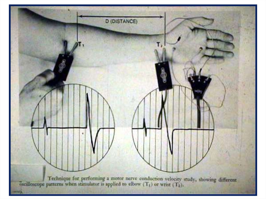
*HEMORRHAGIC(15%)
-interacerebral(hypertention)
-subarachnoid(ruptured aneurysm)
13.
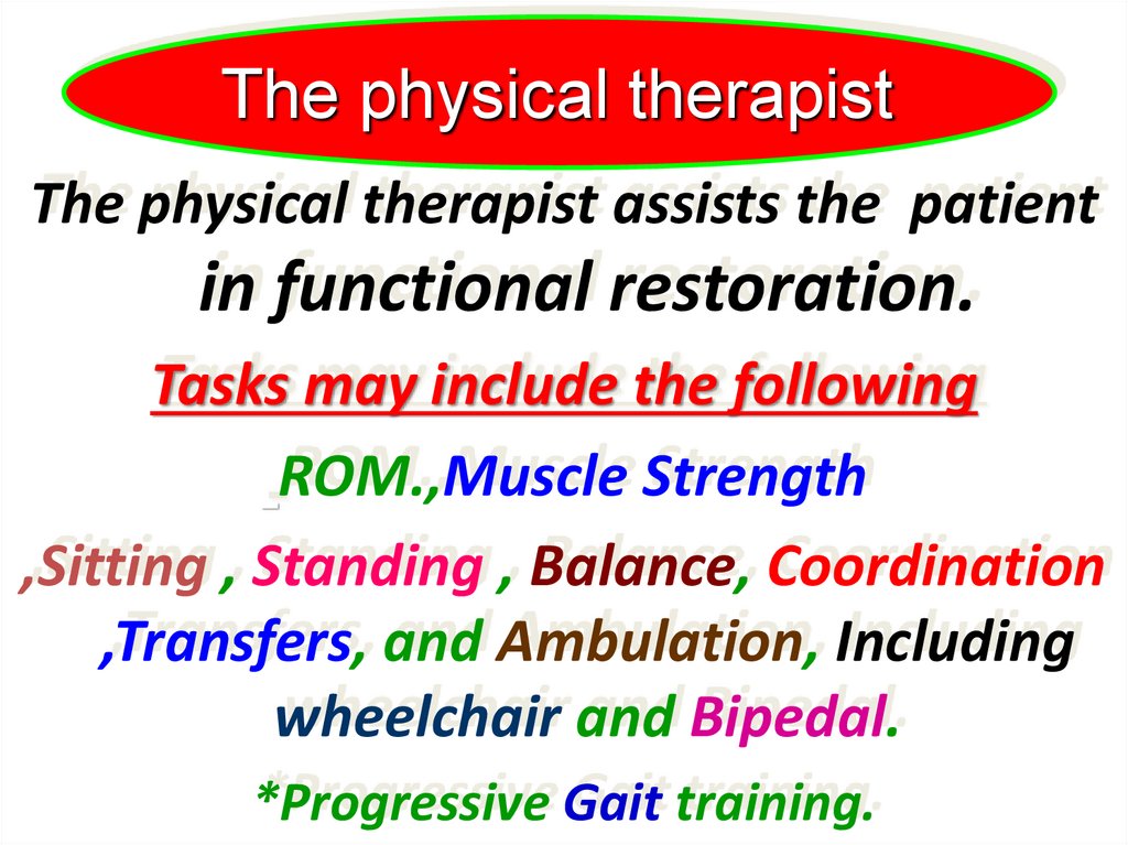
The physical therapistThe physical therapist assists the patient
in functional restoration.
Tasks may include the following
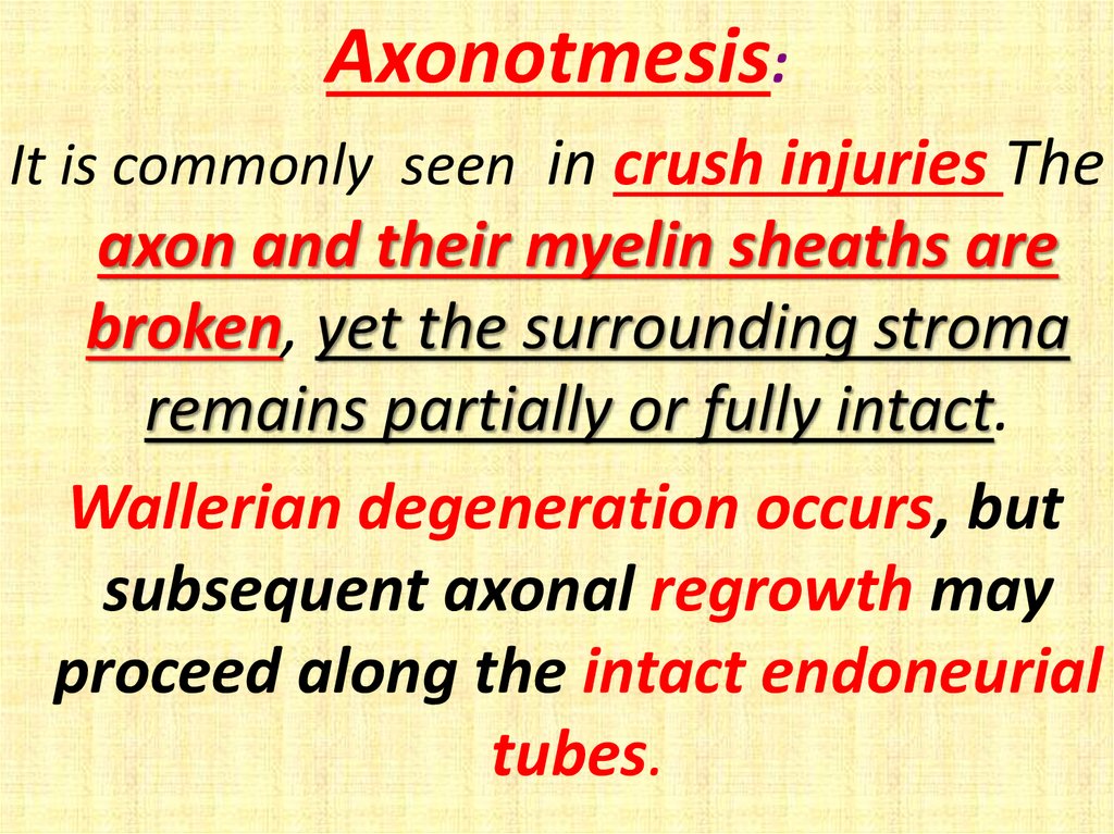
ROM.,Muscle Strength
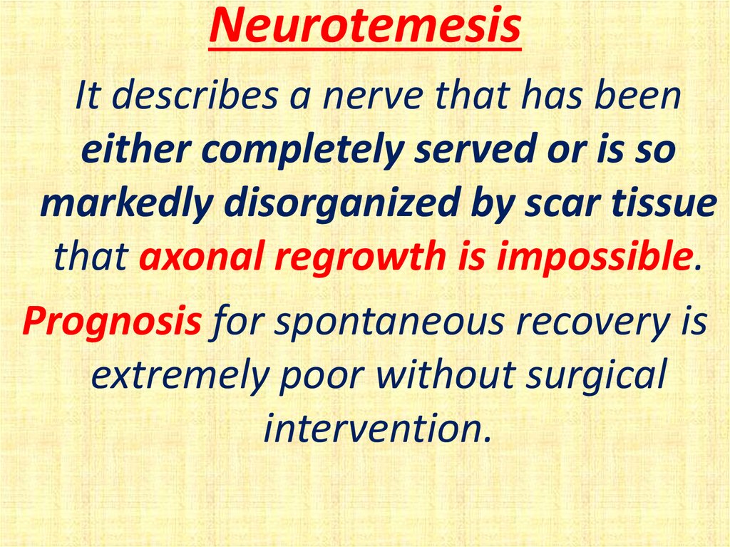
,Sitting , Standing , Balance, Coordination
,Transfers, and Ambulation, Including
wheelchair and Bipedal.
*Progressive Gait training.
14.
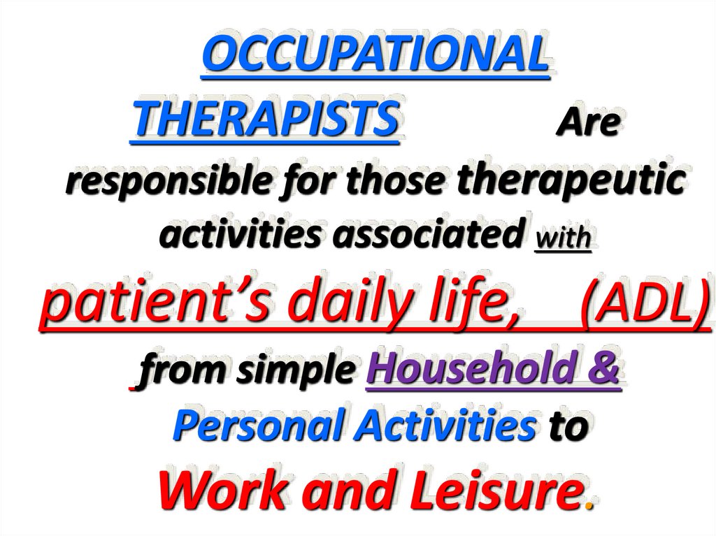
OCCUPATIONALTHERAPISTS
Are
responsible for those therapeutic
activities associated with
patient’s daily life, (ADL)
from simple Household &
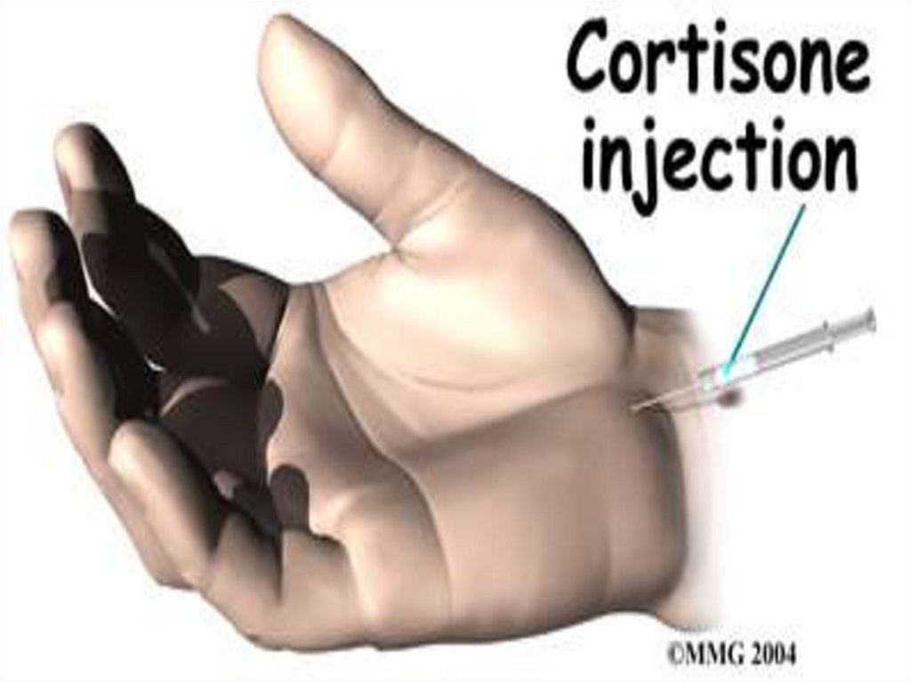
Personal Activities to
Work and Leisure.
15.
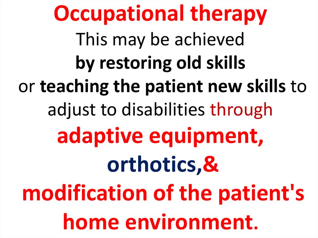
Occupational therapyThis may be achieved
by restoring old skills
or teaching the patient new skills to
adjust to disabilities through
adaptive equipment,
orthotics,&
modification of the patient's
home environment.
16.
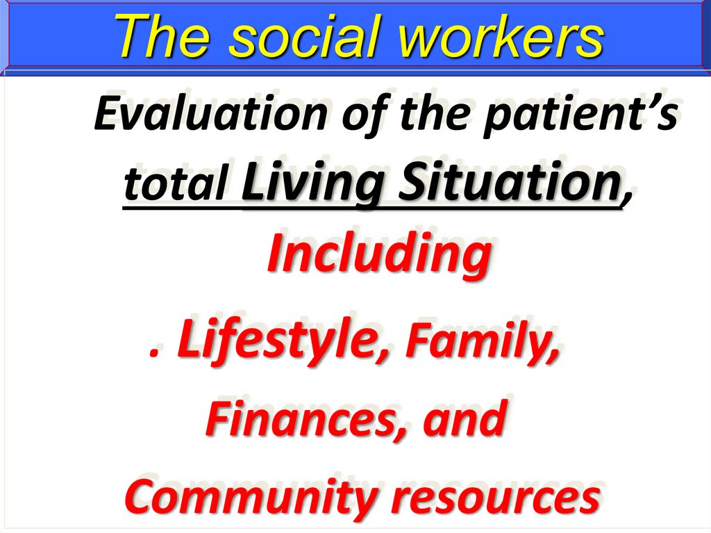
The social workersEvaluation of the patient’s
total Living Situation,
Including
. Lifestyle, Family,
Finances, and
Community resources
17.
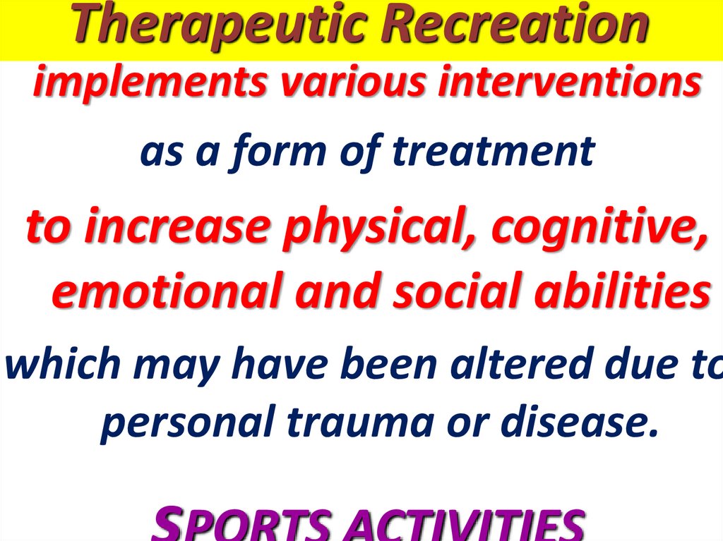
Therapeutic Recreationimplements various interventions
as a form of treatment
to increase physical, cognitive,
emotional and social abilities
which may have been altered due to
personal trauma or disease.
18.
What Are Assistive DevicesAssistive devices
can
help
Hand
a person function
better
Held
and be more
independent
Reacher
Assistive devices can
make daily tasks easier .
.
Flexible Sock Aid
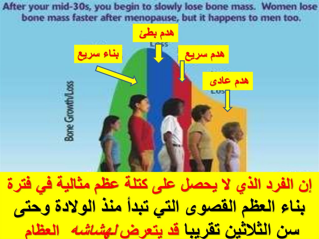
Grip Drink Holder
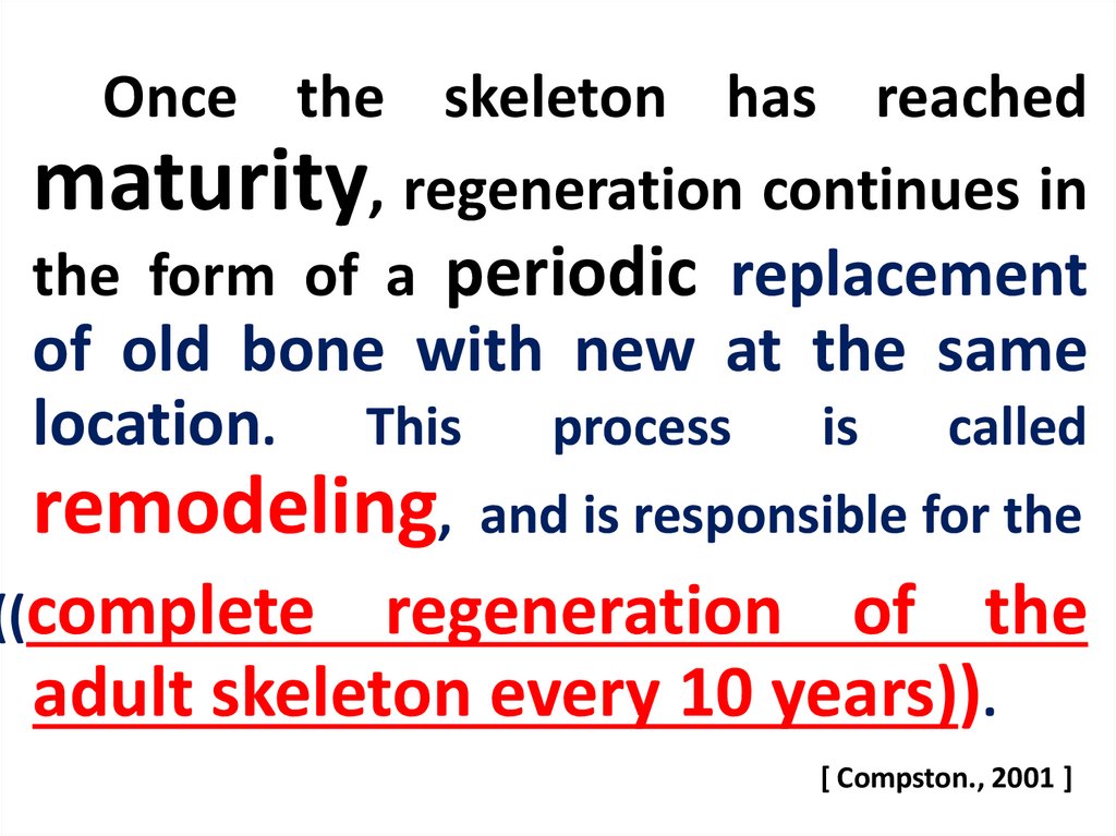
19.
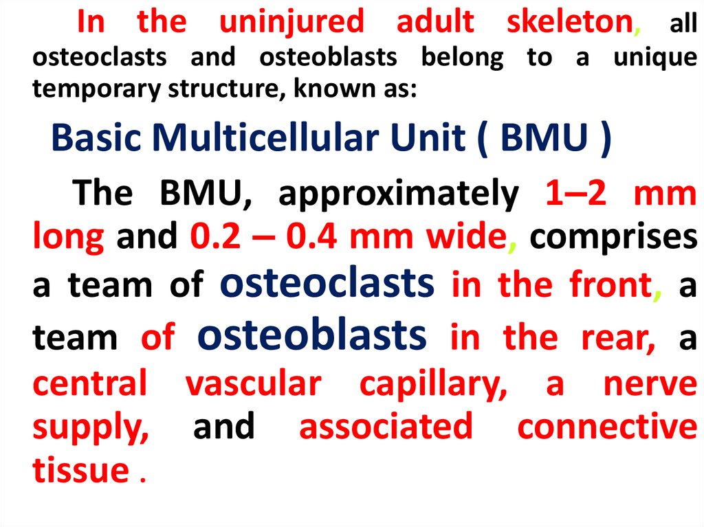
Types of Mobility Aidshelp with
walking or moving
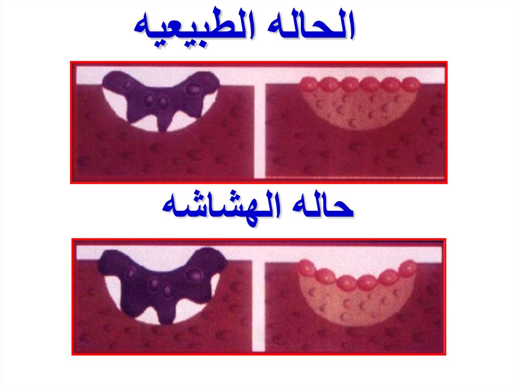
from place to place.
They can help
prevent falls
Walkers
improve independence .
Pediatric
20.
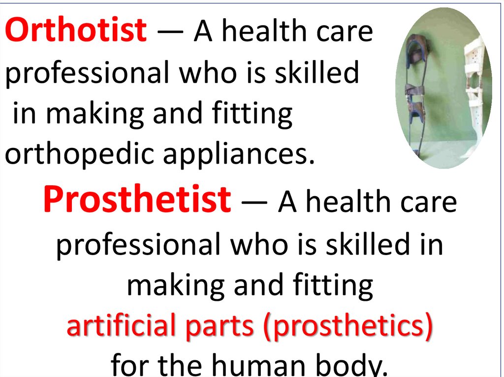
Orthotist — A health careprofessional who is skilled
in making and fitting
orthopedic appliances.
Prosthetist — A health care
professional who is skilled in
making and fitting
artificial parts (prosthetics)
for the human body.
21.
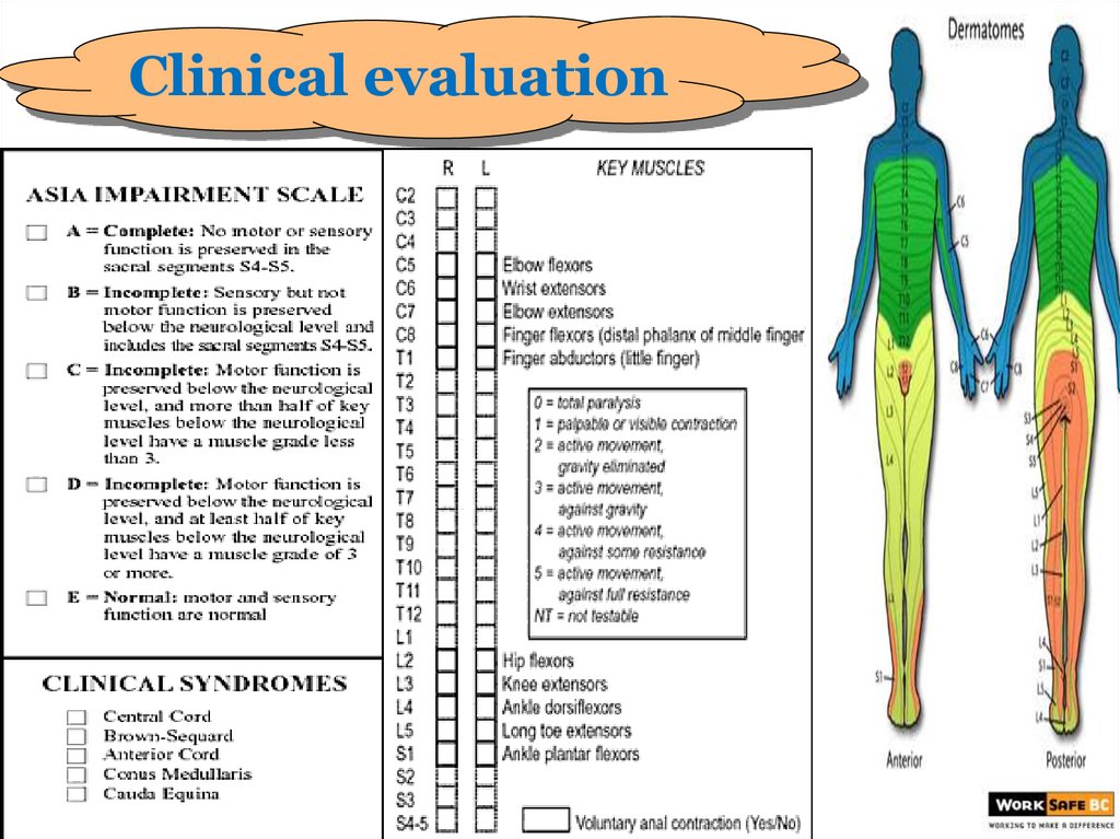
Clinical evaluation22.
23.
24.
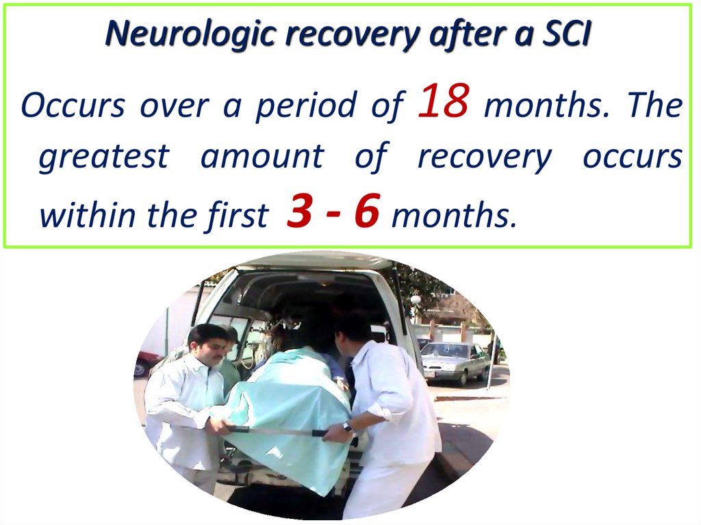
Neurologic recovery after a SCIOccurs over a period of 18 months. The
greatest amount of recovery occurs
within the first
3 - 6 months.
25.
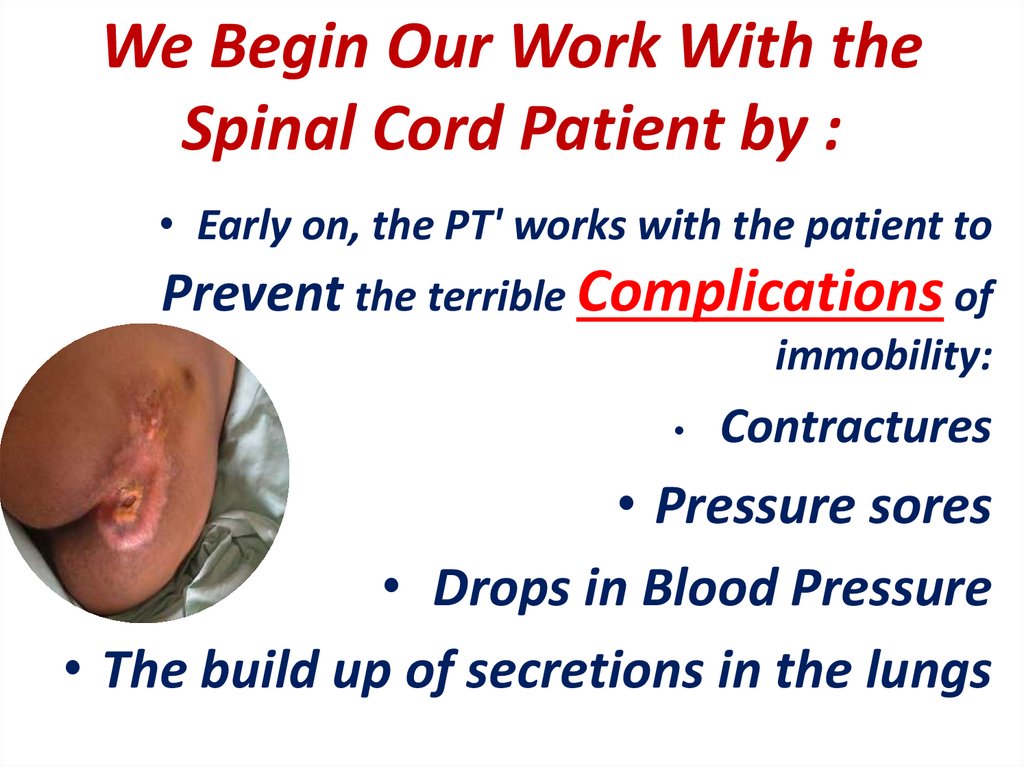
We Begin Our Work With theSpinal Cord Patient by :
• Early on, the PT' works with the patient to
Prevent the terrible Complications of
immobility:
Contractures
• Pressure sores
• Drops in Blood Pressure
• The build up of secretions in the lungs
26.
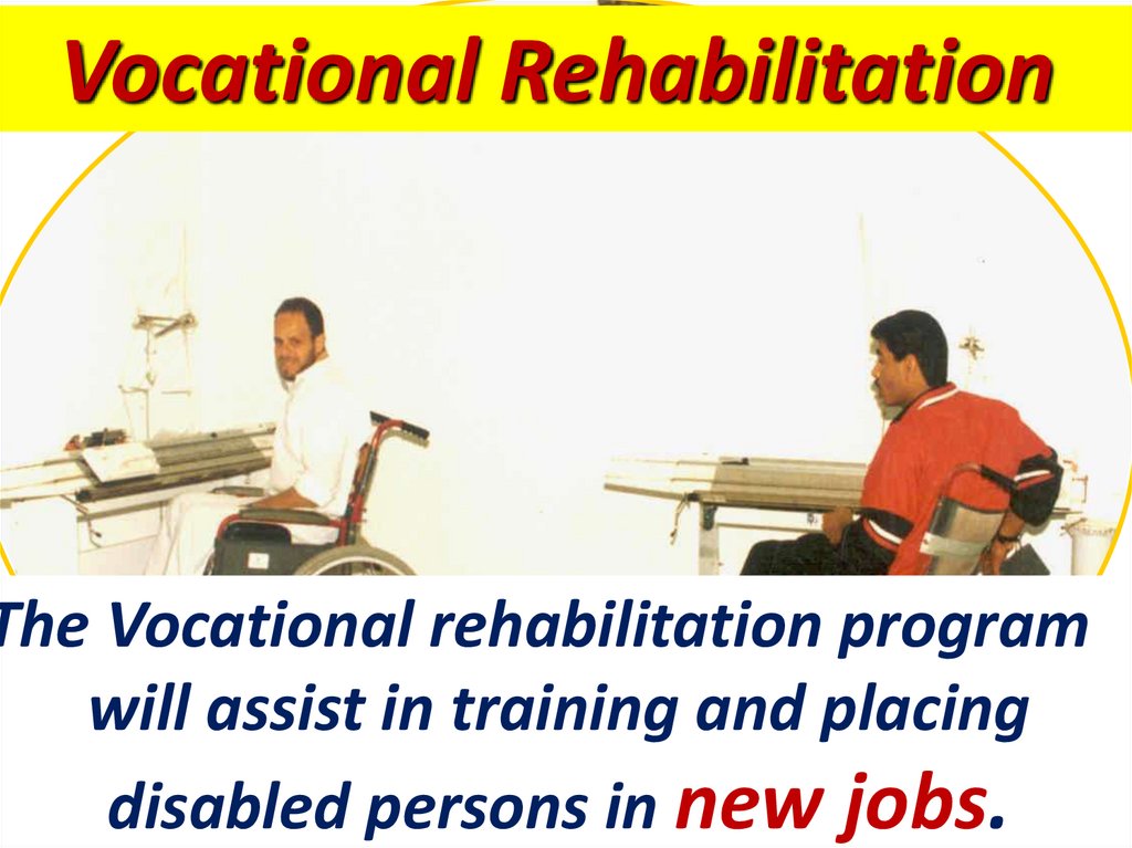
Vocational RehabilitationThe Vocational rehabilitation program
will assist in training and placing
disabled persons in new jobs.
27.
NECKPAIN
لواء
أد
رضا عوض
28.
AnatomyHead weighing 6:8 1b
7 cervical vertebrae
5 intervertebral discs
12 joints of Luschka
14 apophyseal joints.
System of ligaments
(ant. long, post. long ,lig. flavum , interspinous
and ligamentum nuchae)
Muscles
(14 paired anterior lateral & post)
29.
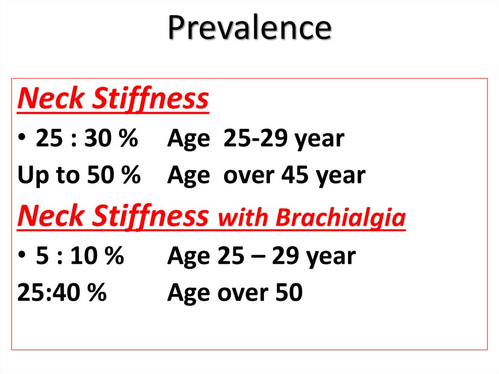
PrevalenceNeck Stiffness
• 25 : 30 % Age 25-29 year
Up to 50 % Age over 45 year
Neck Stiffness with Brachialgia
• 5 : 10 %
25:40 %
Age 25 – 29 year
Age over 50
30.
Causes31.
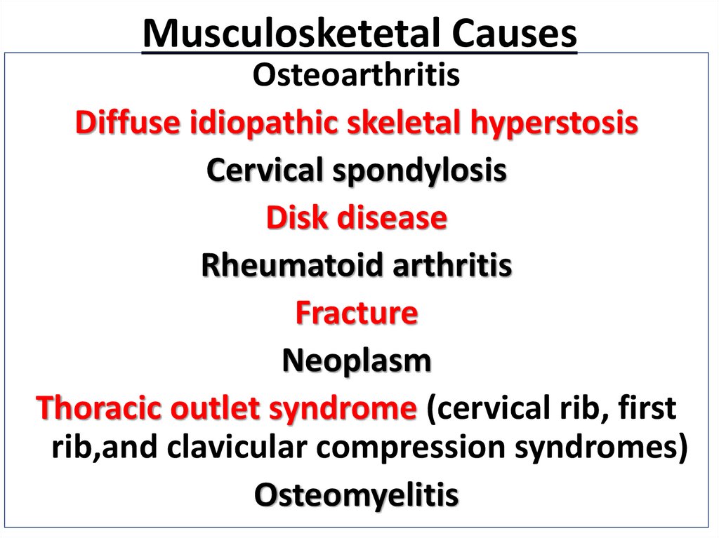
Musculosketetal CausesOsteoarthritis
Diffuse idiopathic skeletal hyperstosis
Cervical spondylosis
Disk disease
Rheumatoid arthritis
Fracture
Neoplasm
Thoracic outlet syndrome (cervical rib, first
rib,and clavicular compression syndromes)
Osteomyelitis
32.
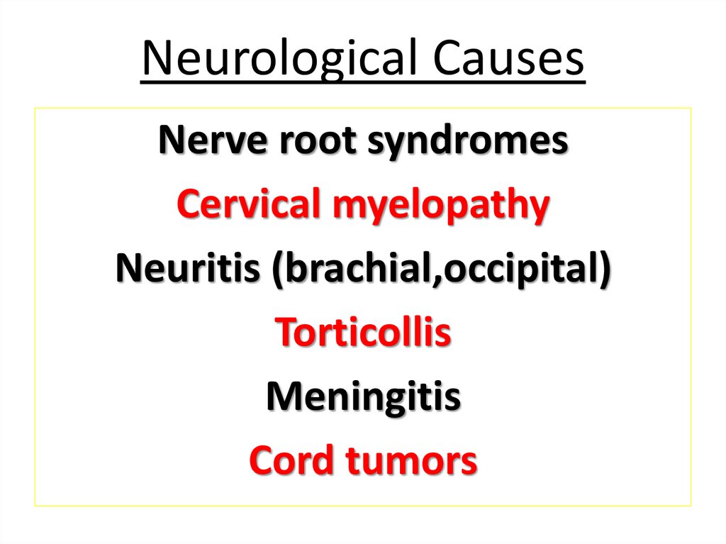
Neurological CausesNerve root syndromes
Cervical myelopathy
Neuritis (brachial,occipital)
Torticollis
Meningitis
Cord tumors
33.
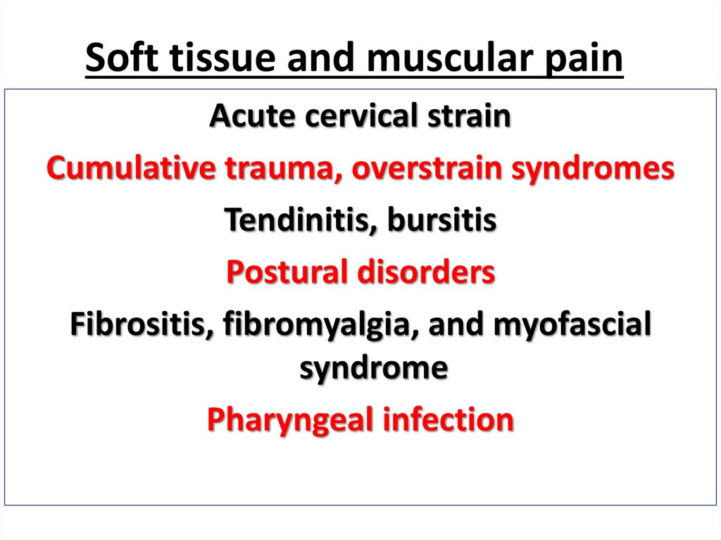
Soft tissue and muscular painAcute cervical strain
Cumulative trauma, overstrain syndromes
Tendinitis, bursitis
Postural disorders
Fibrositis, fibromyalgia, and myofascial
syndrome
Pharyngeal infection
34.
Referred PainHeart and coronary artery disease
Apex of lung: Pancoast’s tumor
Migraine
Muscle tension and myofascial pain
TMJ syndrome
Diaphragm, gallbladder, pancreas, hiatus
hernia
35.
36.
37.
38.
39.
40.
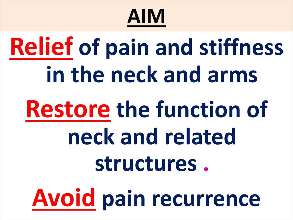
AIMRelief of pain and stiffness
in the neck and arms
Restore the function of
neck and related
structures .
Avoid pain recurrence
41.
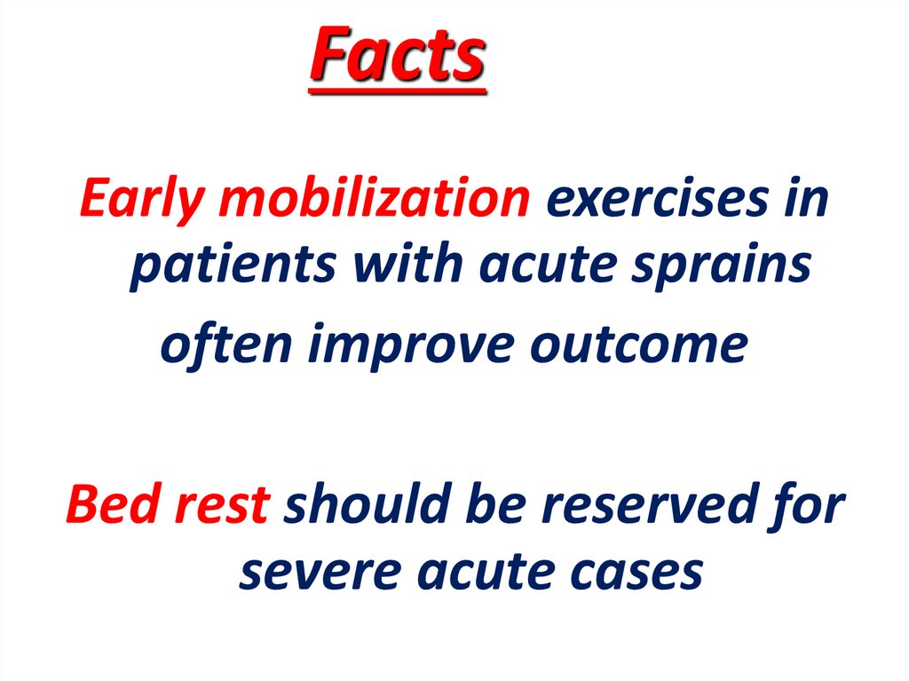
FactsEarly mobilization exercises in
patients with acute sprains
often improve outcome
Bed rest should be reserved for
severe acute cases
42.
توعيهالمريض
HAND
BOOK
43.
44.
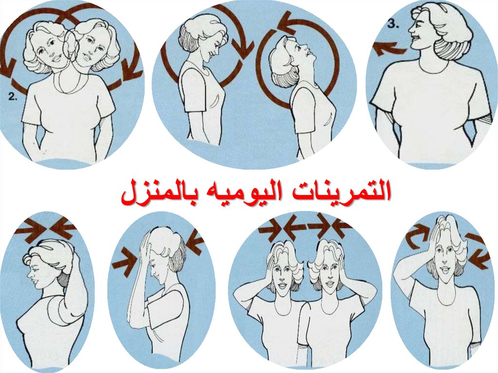
التمرينات اليوميه بالمنزل45.
Traction• Traction forces over 9kg cause
separation of 1- 1.5 mm at each
posterior vertebral level
• It is greatest with the neck in flexion
• 9-11 kg flattens the normal lordosis
• Rhythmic traction produces more
separation than sustained traction
46.
UP DATEMANAGEMENT OF
BACK PAIN
AT AGOUZA
SPECIALISED
SPINE CENTER
(ASSC)
PROF.
REDA AWAD
SUN RISE (SHARM.)
BY
DR /REDA AWAD
47.
LBP: Statistics• Second only to the common cold in
frequency among adult ailments
• Fifth most common reason for an office visit
• Source of LBP is “mechanical” in 90%
and the prognosis is good
• Acute: 50% are better in 1 week;
• 90% have resolved within 8 weeks
• Chronic: <5% of acute low back pain
progresses to chronic pain (6 month)
48.
•80% of all people experiencelow back pain at some time.
.. Up to 50% of working adults
have back pain each year.
• Lifetime recurrence rates of as
high as
85% have been documented
.
49.
The disc is made up of three basic structures:the nucleus pulposus,
the annulus fibrosus and
the vertebral end-plates,
50.
Disc innervation1981 Australian
clinical anatomist
and physician
Nikoli Bogduk
The outer 1/3 of
annulus receive
innervation with
small afferents.
51.
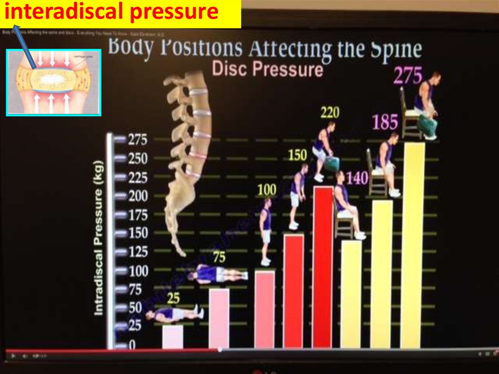
interadiscal pressure52.
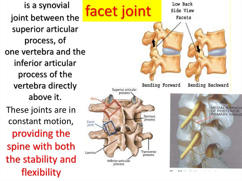
is a synovialjoint between the
superior articular
process, of
one vertebra and the
inferior articular
process of the
vertebra directly
above it.
These joints are in
constant motion,
facet joint
providing the
spine with both
the stability and
flexibility
53.
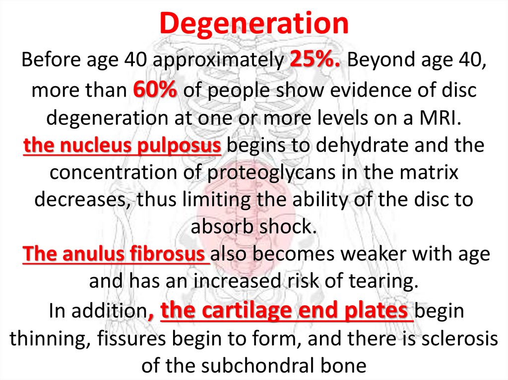
DegenerationBefore age 40 approximately 25%. Beyond age 40,
more than 60% of people show evidence of disc
degeneration at one or more levels on a MRI.
the nucleus pulposus begins to dehydrate and the
concentration of proteoglycans in the matrix
decreases, thus limiting the ability of the disc to
absorb shock.
The anulus fibrosus also becomes weaker with age
and has an increased risk of tearing.
In addition, the cartilage end plates begin
thinning, fissures begin to form, and there is sclerosis
of the subchondral bone
54.
As the disc dehydratesthe disc loose ability to support the
axial load of the body; this causes a
'weight bearing shift' from the nucleus,
outward, onto facet joints .
55.
56.
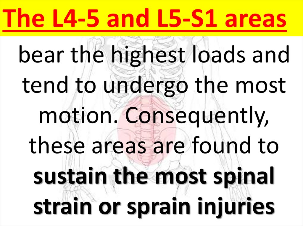
The L4-5 and L5-S1 areasbear the highest loads and
tend to undergo the most
motion. Consequently,
these areas are found to
sustain the most spinal
strain or sprain injuries
57.
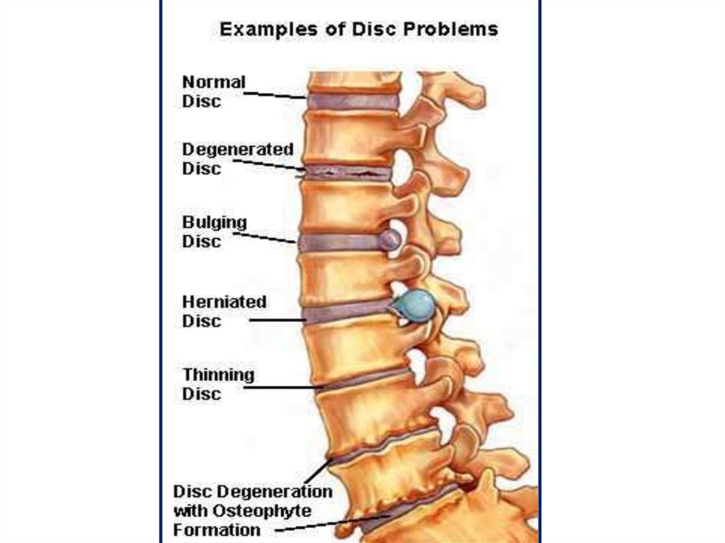
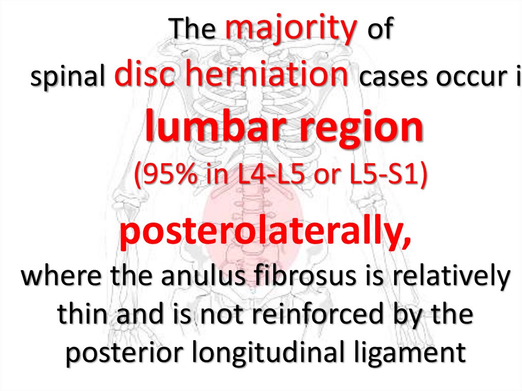
The majority ofspinal disc herniation cases occur in
lumbar region
(95% in L4-L5 or L5-S1)
posterolaterally,
where the anulus fibrosus is relatively
thin and is not reinforced by the
posterior longitudinal ligament
58.
59.
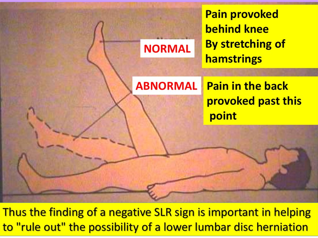
NORMALPain provoked
behind knee
By stretching of
hamstrings
ABNORMAL Pain in the back
provoked past this
point
Thus the finding of a negative SLR sign is important in helping
to "rule out" the possibility of a lower lumbar disc herniation
60.
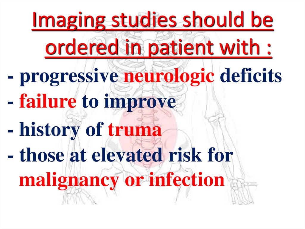
Imaging studies should beordered in patient with :
- progressive neurologic deficits
- failure to improve
- history of truma
- those at elevated risk for
malignancy or infection
61.
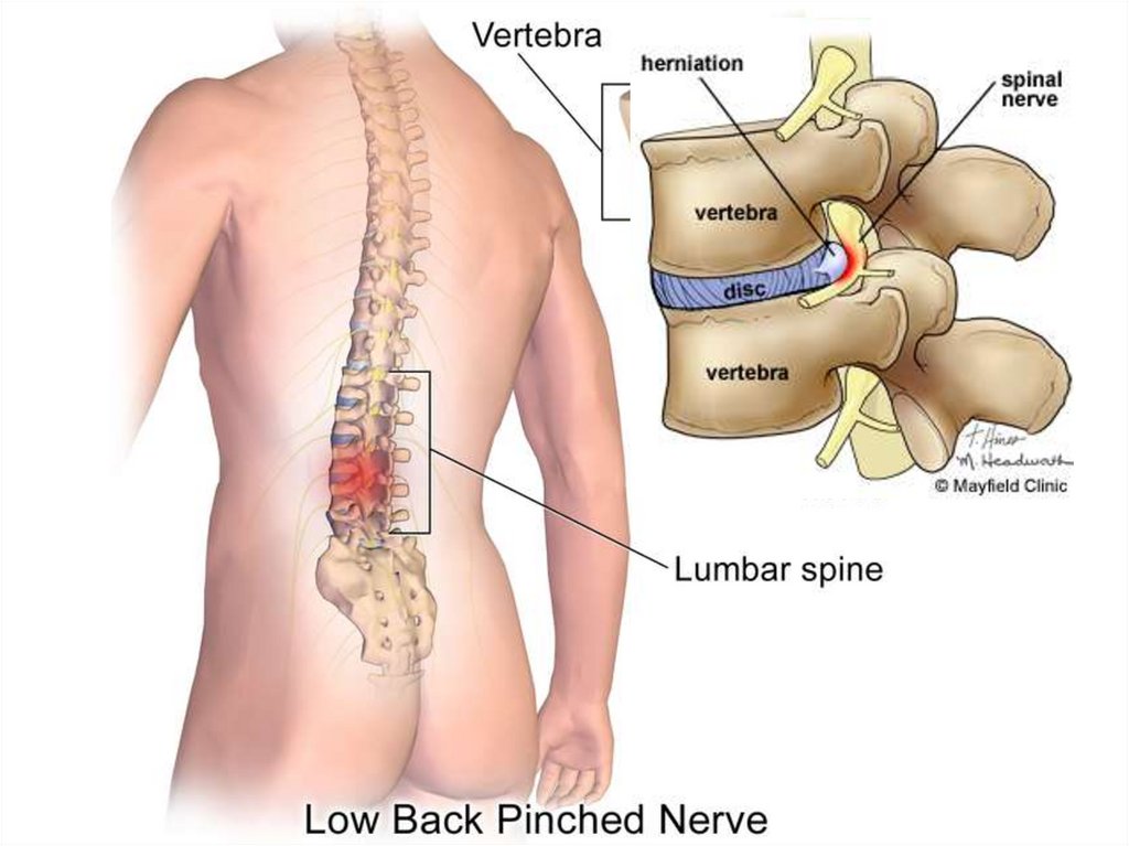
herniated disc atthe L5-S1
herniation (of the
disc between the
L4-L5
62.
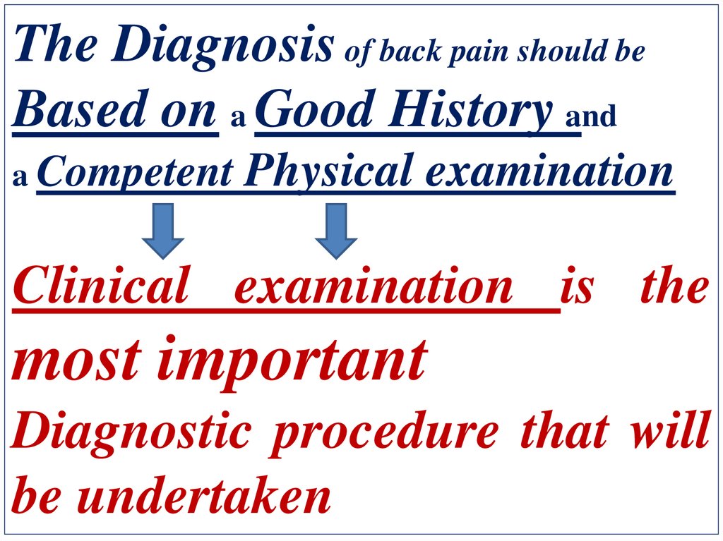
The Diagnosis of back pain should beBased on a Good History and
a Competent
Physical examination
Clinical examination is the
most important
Diagnostic procedure that will
be undertaken
63.
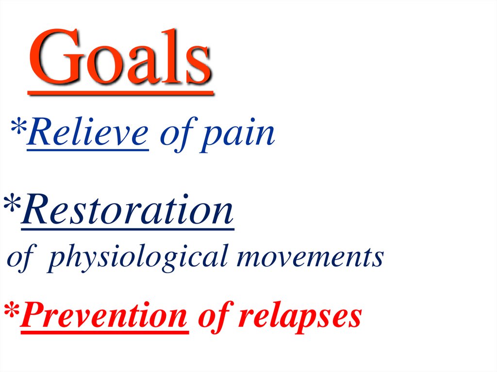
Goals*Relieve of pain
*Restoration
of physiological movements
*Prevention of relapses
64.
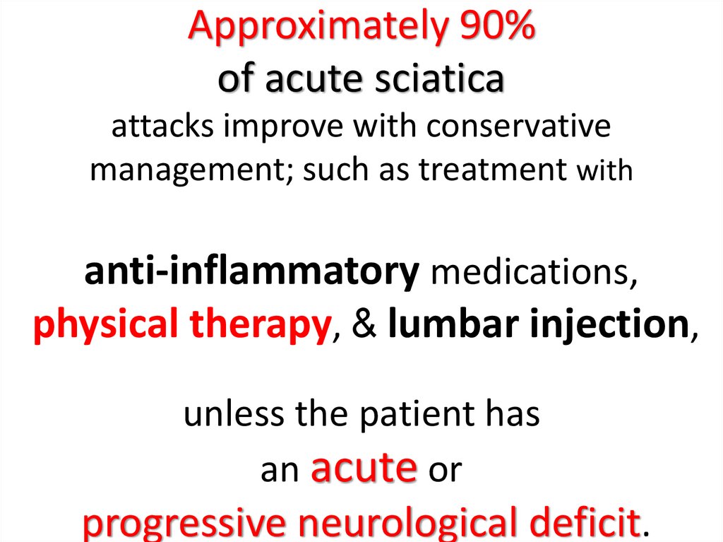
Approximately 90%of acute sciatica
attacks improve with conservative
management; such as treatment with
anti-inflammatory medications,
physical therapy, & lumbar injection,
unless the patient has
an acute or
progressive neurological deficit.
65.
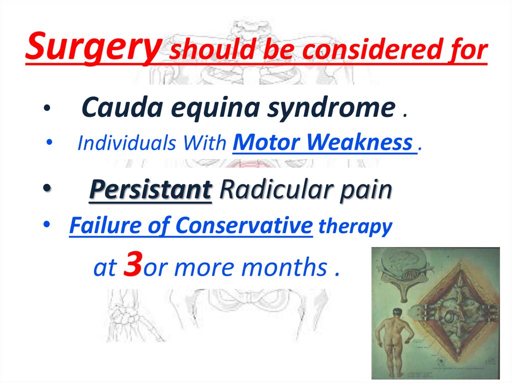
Surgery should be considered forCauda equina syndrome .
Individuals With Motor Weakness .
Persistant Radicular pain
• Failure of Conservative therapy
at 3or more months .
66.
Other Treatment•muscle relaxants or
• nonsteroidal anti-inflammatory
drugs to control muscle spasms.
•A lightweight lumbosacral corset may
also be used to help control muscle spasms.
Use of the corset should be discontinued as
soon as the spasms have resolved.
67.
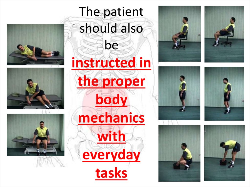
The patientshould also
be
instructed in
the proper
body
mechanics
with
everyday
tasks
68.
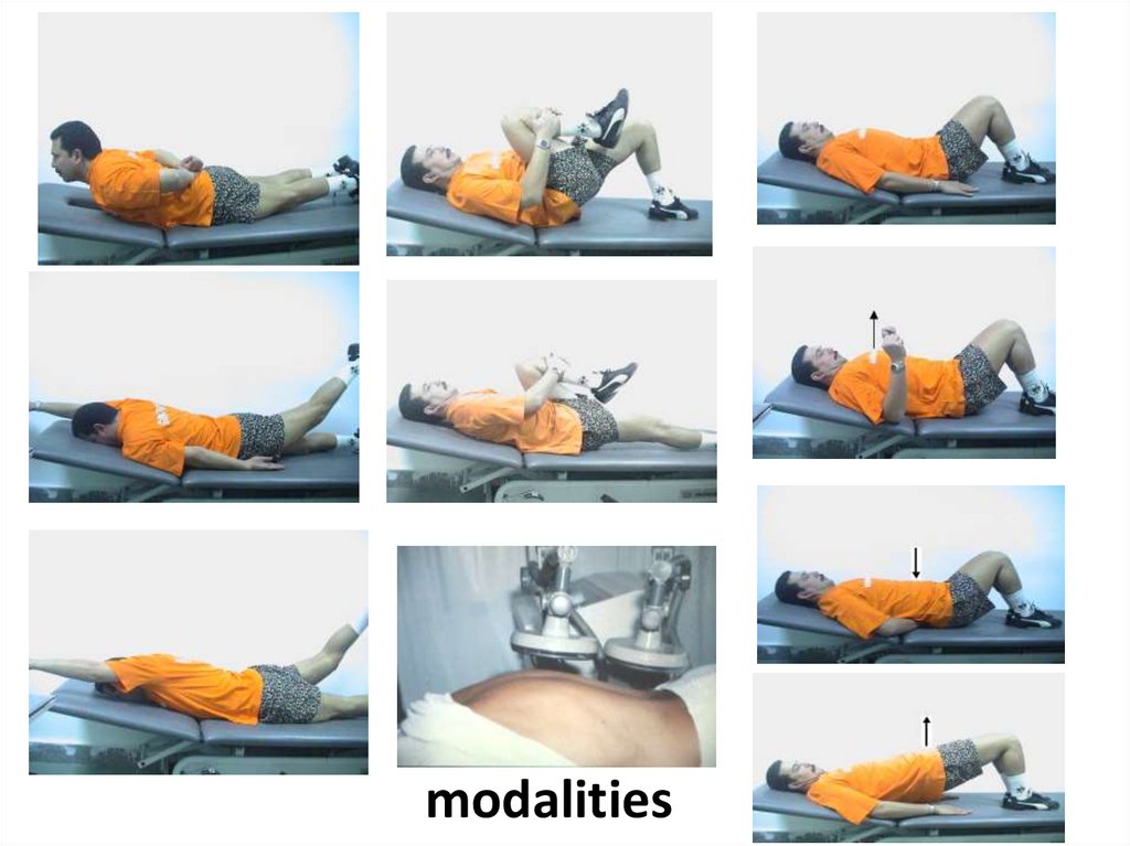
modalities69.
HANDBOOK
70.
71.
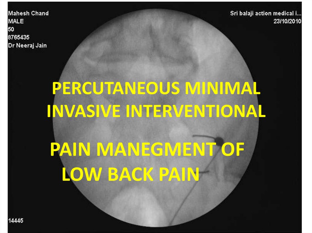
PERCUTANEOUS MINIMALINVASIVE INTERVENTIONAL
PAIN MANEGMENT OF
LOW BACK PAIN
72.
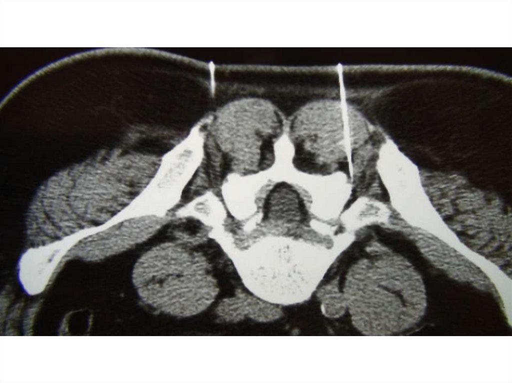
CTfluoroscopy
DISCECTOMY
73.
74.
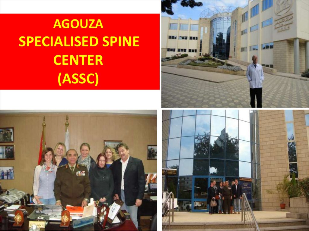
AGOUZASPECIALISED SPINE
CENTER
(ASSC)
75.
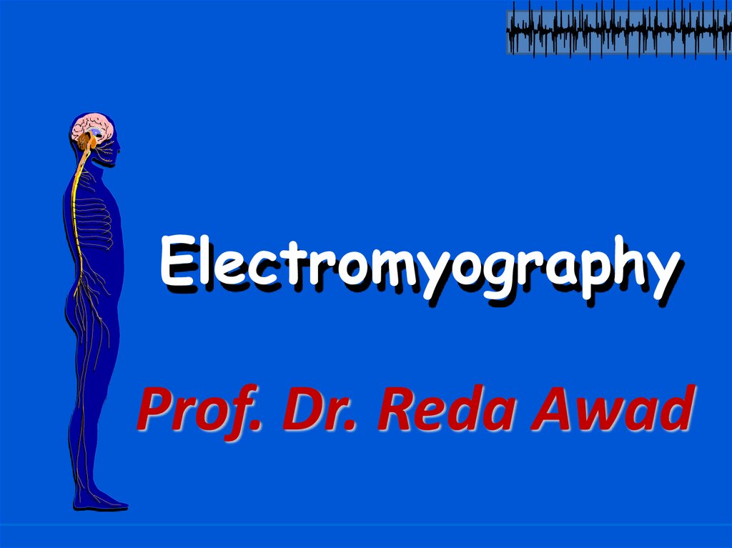
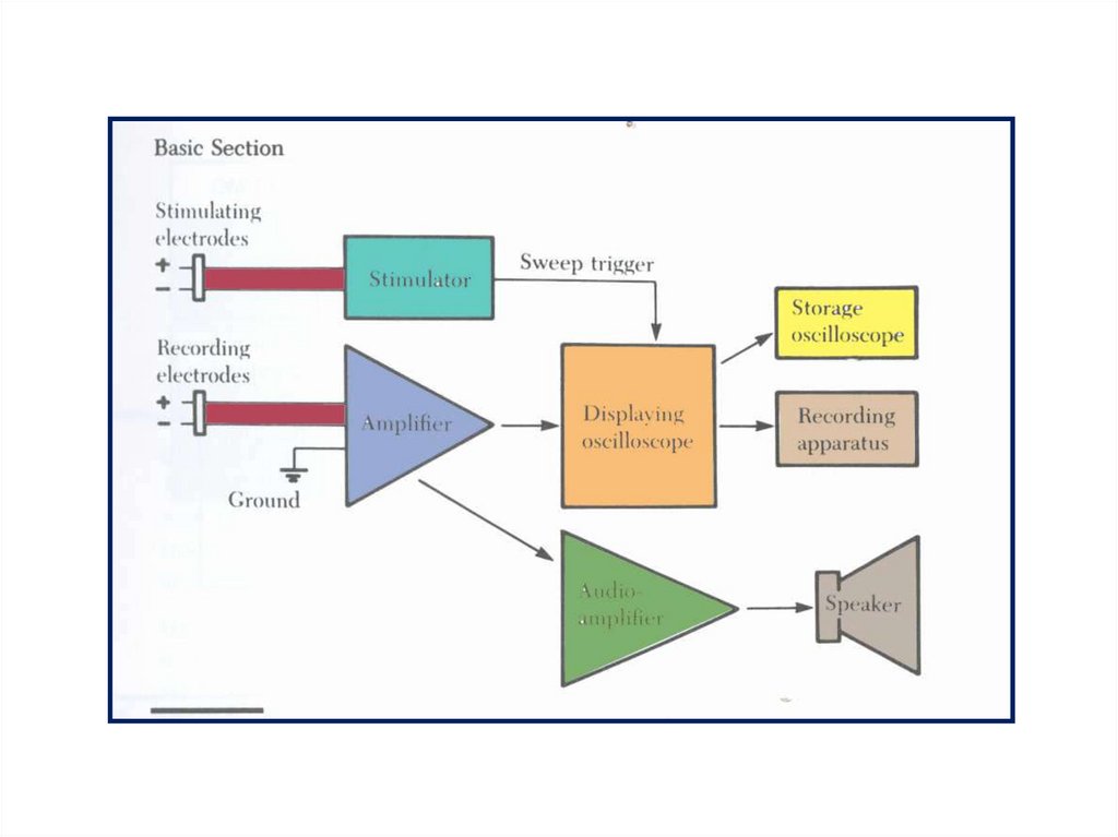
ElectromyographyProf. Dr. Reda Awad
76.
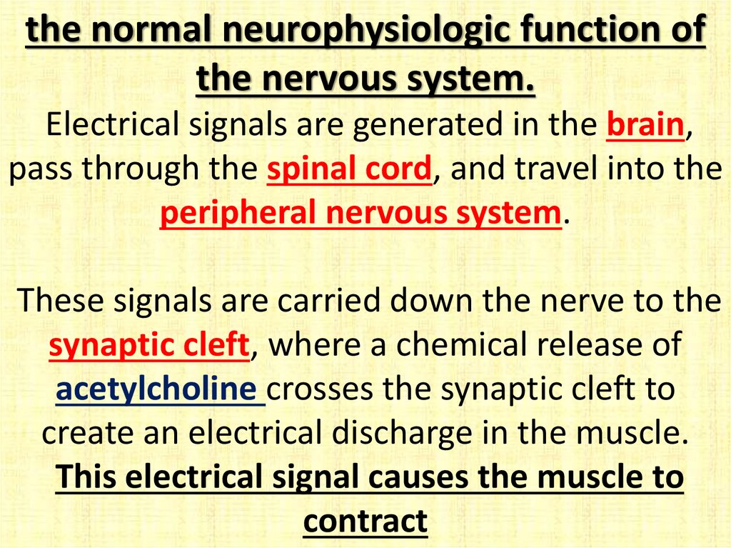
the normal neurophysiologic function ofthe nervous system.
Electrical signals are generated in the brain,
pass through the spinal cord, and travel into the
peripheral nervous system.
These signals are carried down the nerve to the
synaptic cleft, where a chemical release of
acetylcholine crosses the synaptic cleft to
create an electrical discharge in the muscle.
This electrical signal causes the muscle to
contract
77.
78.
79.
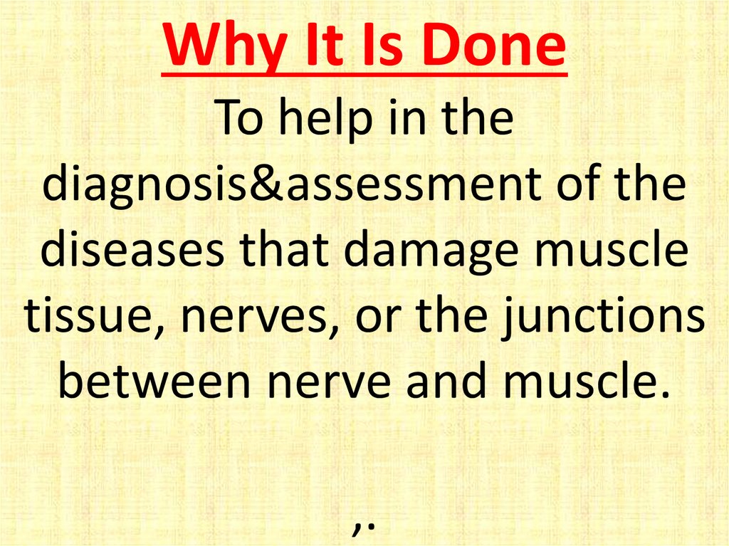
Why It Is DoneTo help in the
diagnosis&assessment of the
diseases that damage muscle
tissue, nerves, or the junctions
between nerve and muscle.
,.
80.
Electromyogram (EMG) and NerveConduction Studies
An electromyogram (EMG)
measures the electrical activity of
muscles at rest and during
contraction.
Nerve conduction studies
measure how well and how fast the
nerves can send electrical signals.
81.
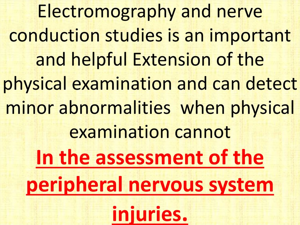
Electromography and nerveconduction studies is an important
and helpful Extension of the
physical examination and can detect
minor abnormalities when physical
examination cannot
In the assessment of the
peripheral nervous system
injuries.
82.
83.
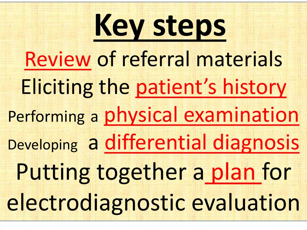
Key stepsReview of referral materials
Eliciting the patient’s history
Performing a physical examination
Developing a differential diagnosis
Putting together a plan for
electrodiagnostic evaluation
84.
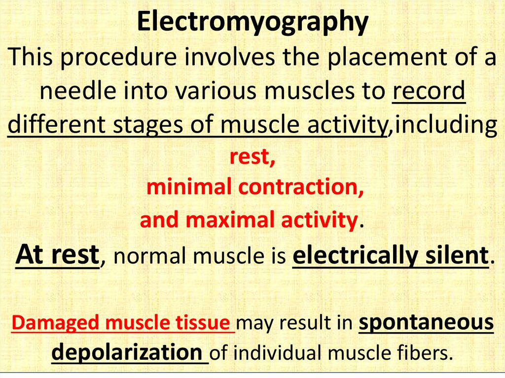
ElectromyographyThis procedure involves the placement of a
needle into various muscles to record
different stages of muscle activity,including
rest,
minimal contraction,
and maximal activity.
At rest, normal muscle is electrically silent.
Damaged muscle tissue may result in spontaneous
depolarization of individual muscle fibers.
85.
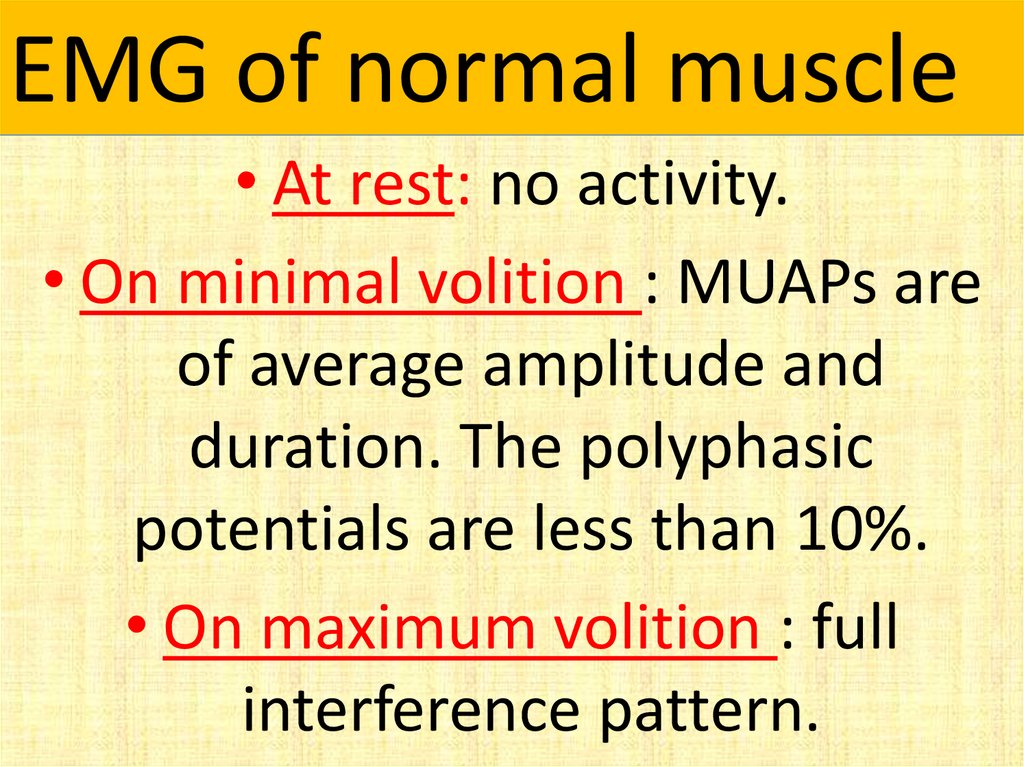
EMG of normal muscle• At rest: no activity.
• On minimal volition : MUAPs are
of average amplitude and
duration. The polyphasic
potentials are less than 10%.
• On maximum volition : full
interference pattern.
86.
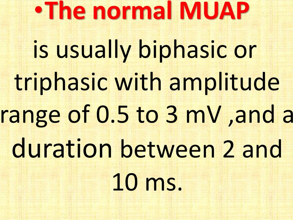
•The normal MUAPis usually biphasic or
triphasic with amplitude
range of 0.5 to 3 mV ,and a
duration between 2 and
10 ms.
87.
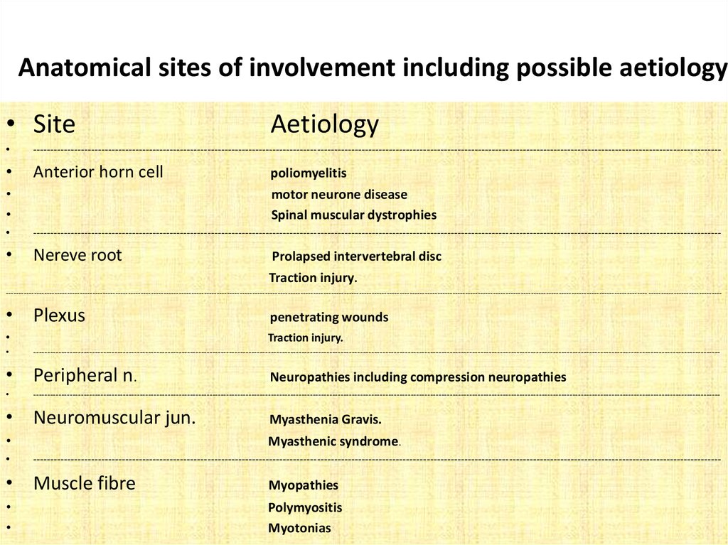
Anatomical sites of involvement including possible aetiology• Site
Aetiology
---------------------------------------------------------------------------------------------------------------------------------------------------------------------------------------------------------
Anterior horn cell
poliomyelitis
motor neurone disease
Spinal muscular dystrophies
---------------------------------------------------------------------------------------------------------------------------------------------------------------------------------------------------------
Nereve root
Prolapsed intervertebral disc
Traction injury.
------------------------------------------------------------------------------------------------------------------------------------------------------------------------------------------------------------------------------------------------------------- -----------------------------
• Plexus
penetrating wounds
Traction injury.
------------------------------------------------------------------------------------------------------------------------------------------------------------------------------------------------------------------------------------------------------
• Peripheral n.
Neuropathies including compression neuropathies
------------------------------------------------------------------------------------------------------------------------------------------------------------------------------------------------------------------------------------------------------
• Neuromuscular jun.
Myasthenia Gravis.
Myasthenic syndrome.
---------------------------------------------------------------------------------------------------------------------------------------------------------------------------------------------------------
• Muscle fibre
Myopathies
Polymyositis
Myotonias
88.
PeakLatency
Onset
Latency
Amplitudes
Negative
Deflection
Area
Peak to Peak
Stimulation
Artifact
Duration
Nerve
Conduction
Studies
Prof. Dr. Reda Awad
89.
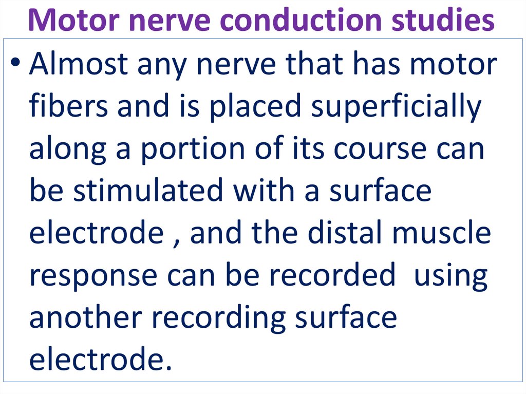
Motor nerve conduction studies• Almost any nerve that has motor
fibers and is placed superficially
along a portion of its course can
be stimulated with a surface
electrode , and the distal muscle
response can be recorded using
another recording surface
electrode.
90.
91.
TheTerm
Entrapment
describes the mechanical irritation
by which a specific peripheral
nerve becomes locally injured in a
vulnerable anatomic site
Familiarity with the Anatomy of
the peripheral Nerves is essential.
92.
Nerve compression CanOccur at any point where a
peripheral nerve passes
through
An opening In fibrous tissue
or through
An Osseo fibrous Canal.
93.
In addition to aneurologic examination,
the evaluation of every patient
with an entrapment neuropathy
should include
electromyography (EMG) motor
and sensory nerve conduction
velocity studies,
and appropriate radiographs
94.
EMG and nerve conductionvelocity measurements provide
localizing information often
necessary in the early diagnosis of
a compressive neuropathy and
reliably document the severity
and extent of nerve entrapment
95.
Classification of nerve injuries• Neurapraxia: It is a comparatively mild
injury with motor and sensory loss with
no evidence of Wallerian degeneration.
The nerve distally conducts normally.
Focal demyelination and/or ischemia
are thought to be the aetiology of the
conduction block.
• Recovery may occur within hours, days,
weeks, or up to a few months.
96.
Axonotmesis:It is commonly seen in crush injuries The
axon and their myelin sheaths are
broken, yet the surrounding stroma
remains partially or fully intact.
Wallerian degeneration occurs, but
subsequent axonal regrowth may
proceed along the intact endoneurial
tubes.
97.
NeurotemesisIt describes a nerve that has been
either completely served or is so
markedly disorganized by scar tissue
that axonal regrowth is impossible.
Prognosis for spontaneous recovery is
extremely poor without surgical
intervention.
98.
Characteristic features associated withVarious nerve compressions
Nerve
Clinical involvement
Median
Thumb and thenar eminence
Anterior
interosseous
Flexor pollicis longus, pronator Quadratus, flexor digitorum
Profundus to index and middle Fingers; normal sensation
Ulnar
Small finger and hypothenar Eminence
Musculocutaneous
Biceps
Radial
Wrist drop; sensory loss in dorsum Of thumb
Post.inter osseous
Wrist drop; normal sensation
Femoral
Absent knee jerk; weak knee Extension and hip flexion
Peroneal
Foot drop; sensory loss in dorsum of Foot
Posterior tibial
Sensory loss in medial heel; Weakness in intrinsic muscles
of foot
Sciatic
Pain down lateral thigh ;often absent Ankles jerk; foot drop
Sural
Sensory loss over lateral foot
99.
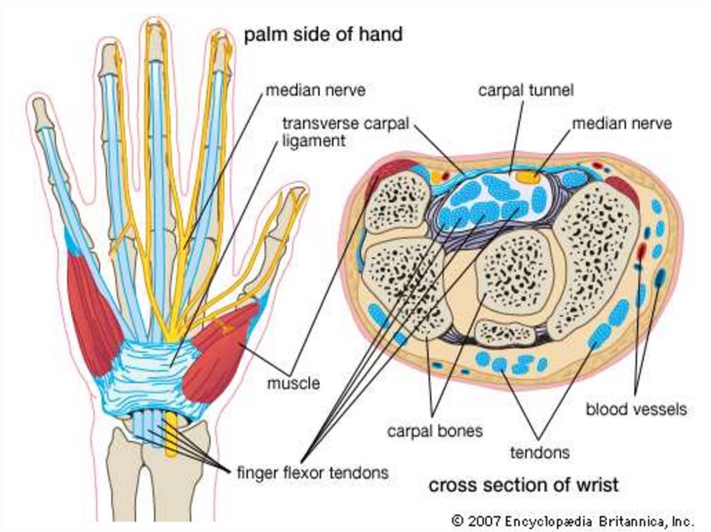
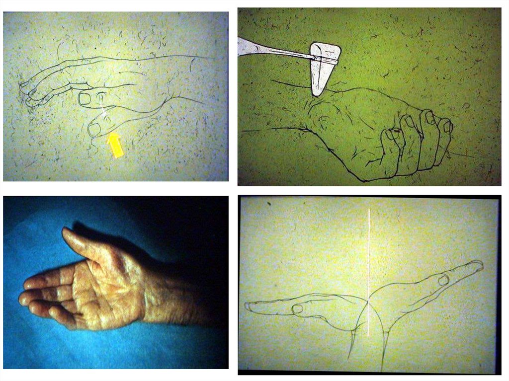
What IsCarpal Tunnel
Syndrome ?
100.
101.
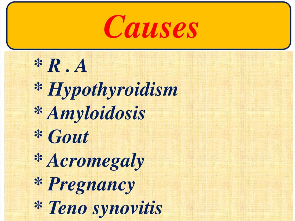
Causes*R.A
* Hypothyroidism
* Amyloidosis
* Gout
* Acromegaly
* Pregnancy
* Teno synovitis
102.
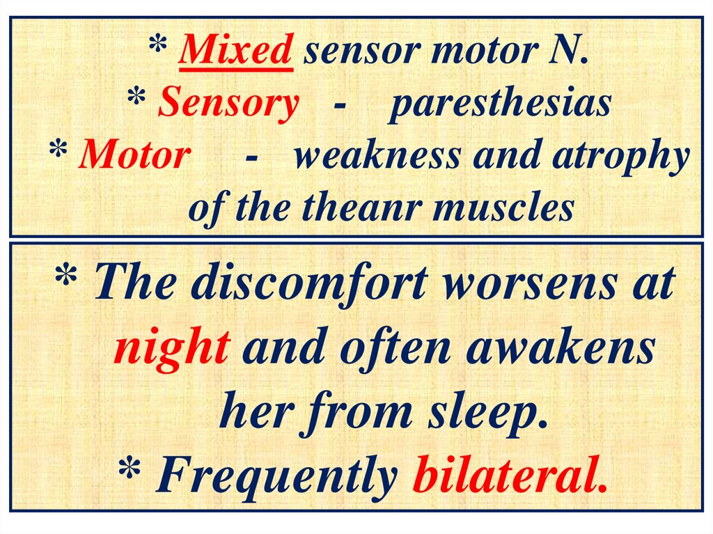
* Mixed sensor motor N.* Sensory - paresthesias
* Motor - weakness and atrophy
of the theanr muscles
* The discomfort worsens at
night and often awakens
her from sleep.
* Frequently bilateral.
103.
104.
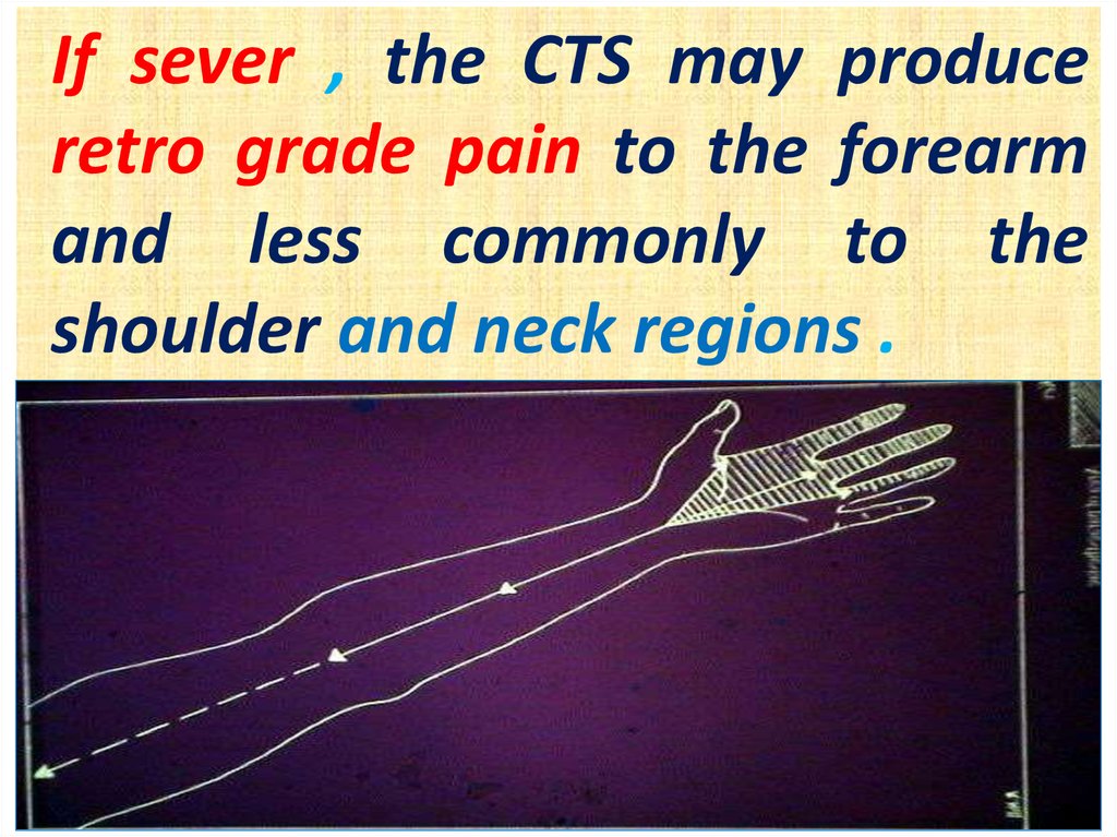
If sever , the CTS may produceretro grade pain to the forearm
and less commonly to the
shoulder and neck regions .
105.
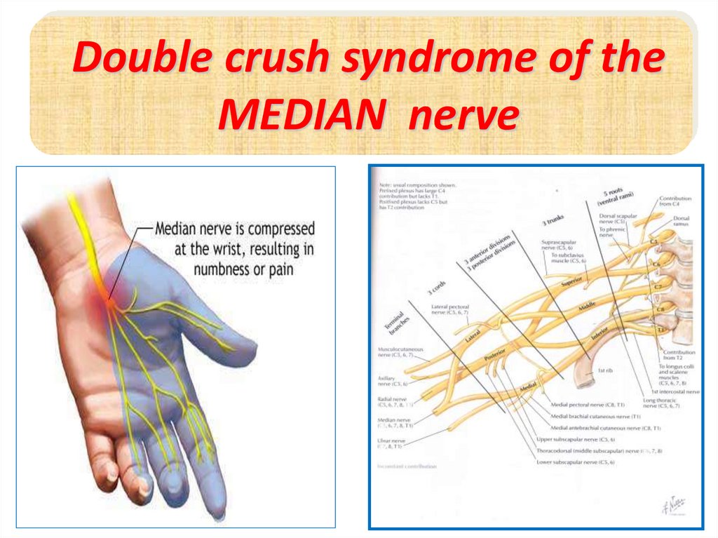
Double crush syndrome of theMEDIAN nerve
106.
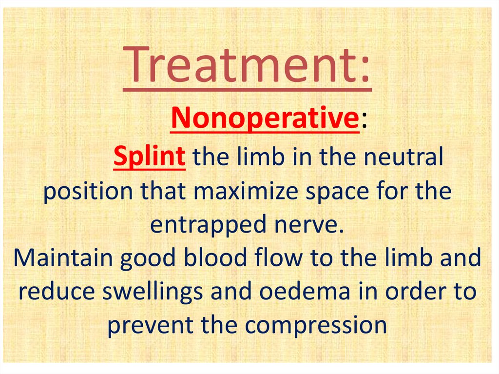
Treatment:Nonoperative:
Splint the limb in the neutral
position that maximize space for the
entrapped nerve.
Maintain good blood flow to the limb and
reduce swellings and oedema in order to
prevent the compression
107.
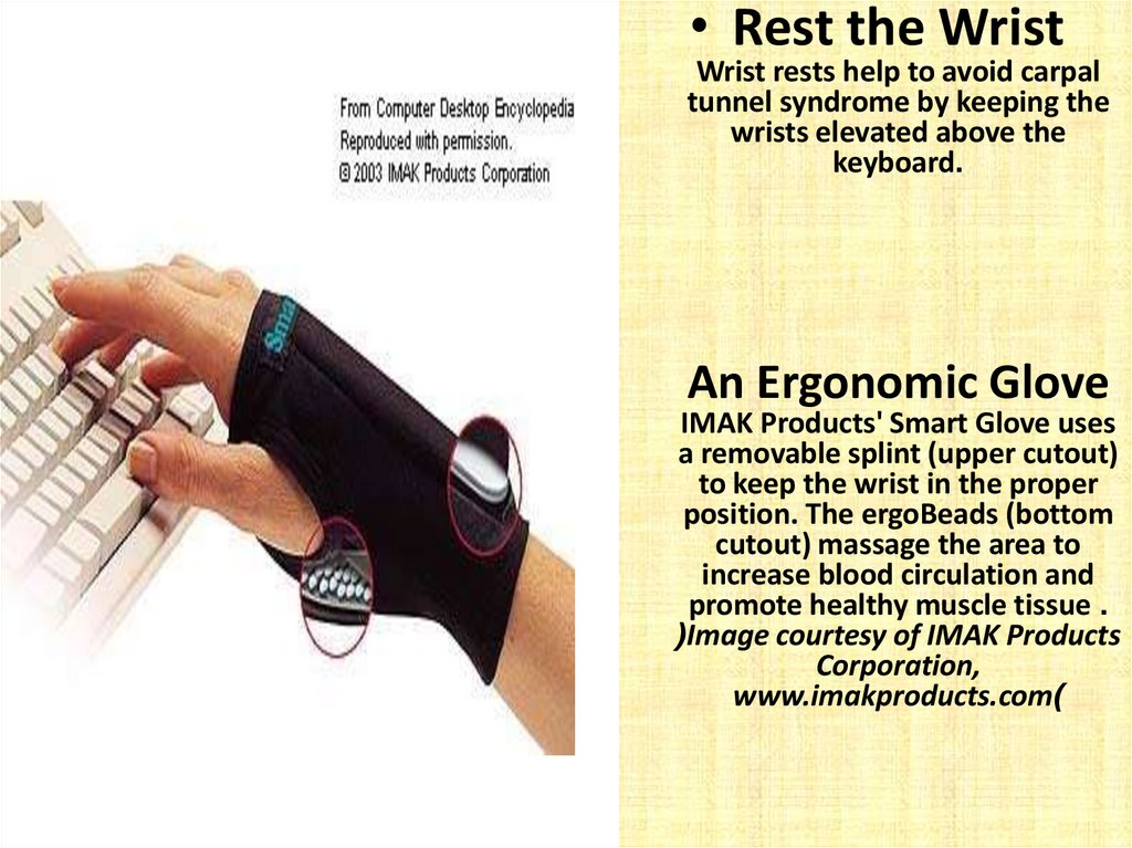
• Rest the WristWrist rests help to avoid carpal
tunnel syndrome by keeping the
wrists elevated above the
keyboard.
An Ergonomic Glove
IMAK Products' Smart Glove uses
a removable splint (upper cutout)
to keep the wrist in the proper
position. The ergoBeads (bottom
cutout) massage the area to
increase blood circulation and
promote healthy muscle tissue .
(Image courtesy of IMAK Products
Corporation,
www.imakproducts.com)
108.
Modify activityand avoid positions that can be a
source of trauma.
Reduce inflammation
and
consider the use of ice,NSAIDs and
corticosteroid injection
in structures around the nerves
that may be inflammed
109.
110.
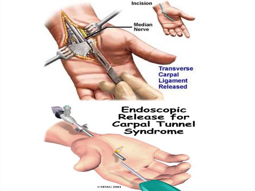
Operative:If despite of nonoperative
treatment,there is evidence of continuing
axonal degeneration in the entrapped
nerve,surgical decompression of
the nerve is considered
111.
112.
*surgeryproduces good results
in cases caused by
ganglion , some
selected causes of
truma and R .A .
113.
114.
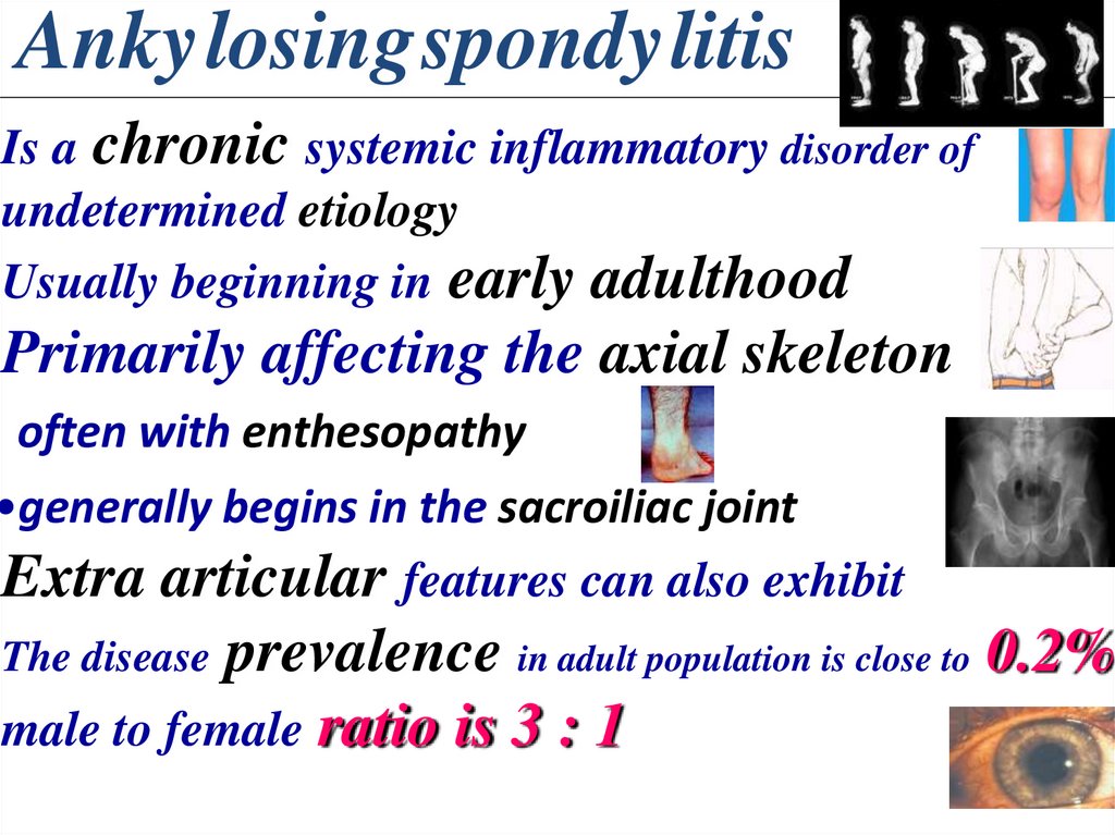
AnkylosingspondylitisIs a chronic systemic inflammatory disorder of
undetermined etiology
Usually beginning in early adulthood
Primarily affecting the axial skeleton
often with enthesopathy
•generally begins in the sacroiliac joint
Extra articular features can also exhibit
prevalence in adult population is close to 0.2%
male to female ratio is 3 : 1
The disease
115.
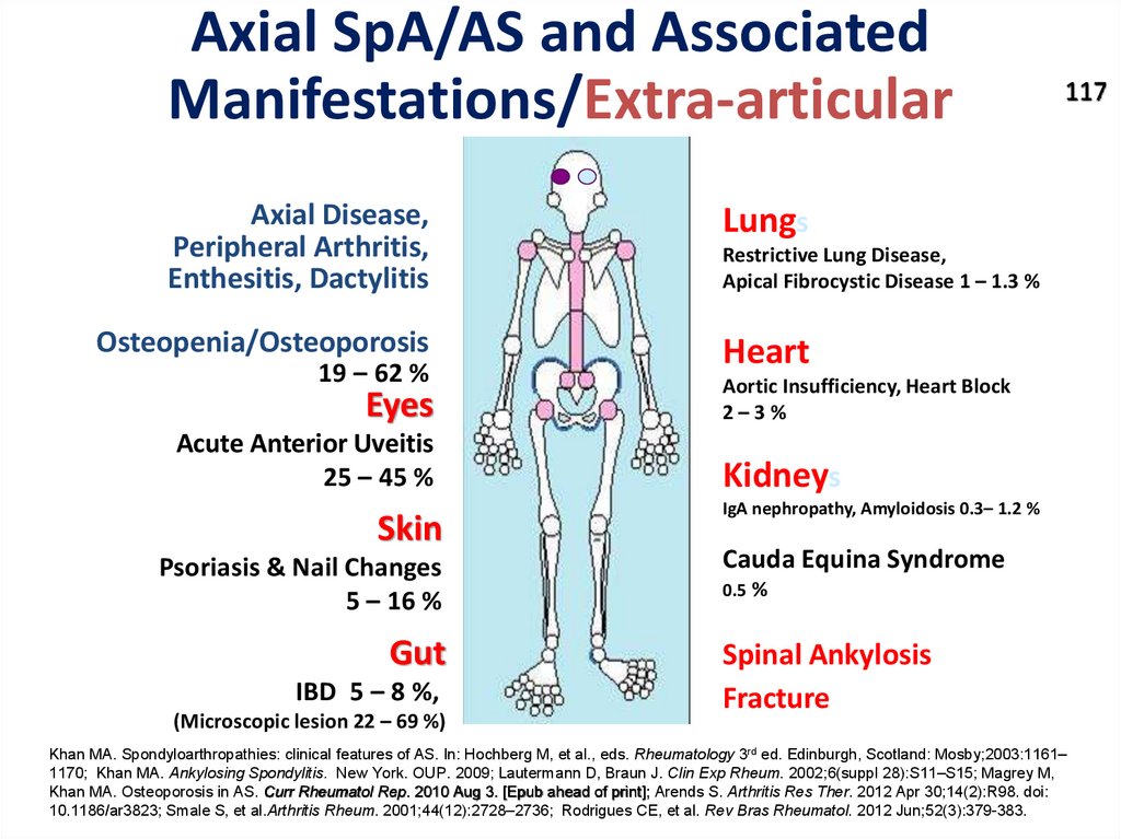
Axial SpA/AS and AssociatedManifestations/Extra-articular
Axial Disease,
Peripheral Arthritis,
Enthesitis, Dactylitis
Lungs
Osteopenia/Osteoporosis
Heart
19 – 62 %
Eyes
Acute Anterior Uveitis
25 – 45 %
Skin
Psoriasis & Nail Changes
5 – 16 %
Gut
IBD 5 – 8 %,
(Microscopic lesion 22 – 69 %)
117
Restrictive Lung Disease,
Apical Fibrocystic Disease 1 – 1.3 %
Aortic Insufficiency, Heart Block
2–3%
Kidneys
IgA nephropathy, Amyloidosis 0.3– 1.2 %
Cauda Equina Syndrome
0.5 %
Spinal Ankylosis
Fracture
Khan MA. Spondyloarthropathies: clinical features of AS. In: Hochberg M, et al., eds. Rheumatology 3rd ed. Edinburgh, Scotland: Mosby;2003:1161–
1170; Khan MA. Ankylosing Spondylitis. New York. OUP. 2009; Lautermann D, Braun J. Clin Exp Rheum. 2002;6(suppl 28):S11–S15; Magrey M,
Khan MA. Osteoporosis in AS. Curr Rheumatol Rep. 2010 Aug 3. [Epub ahead of print]; Arends S. Arthritis Res Ther. 2012 Apr 30;14(2):R98. doi:
10.1186/ar3823; Smale S, et al.Arthritis Rheum. 2001;44(12):2728–2736; Rodrigues CE, et al. Rev Bras Rheumatol. 2012 Jun;52(3):379-383.
116.
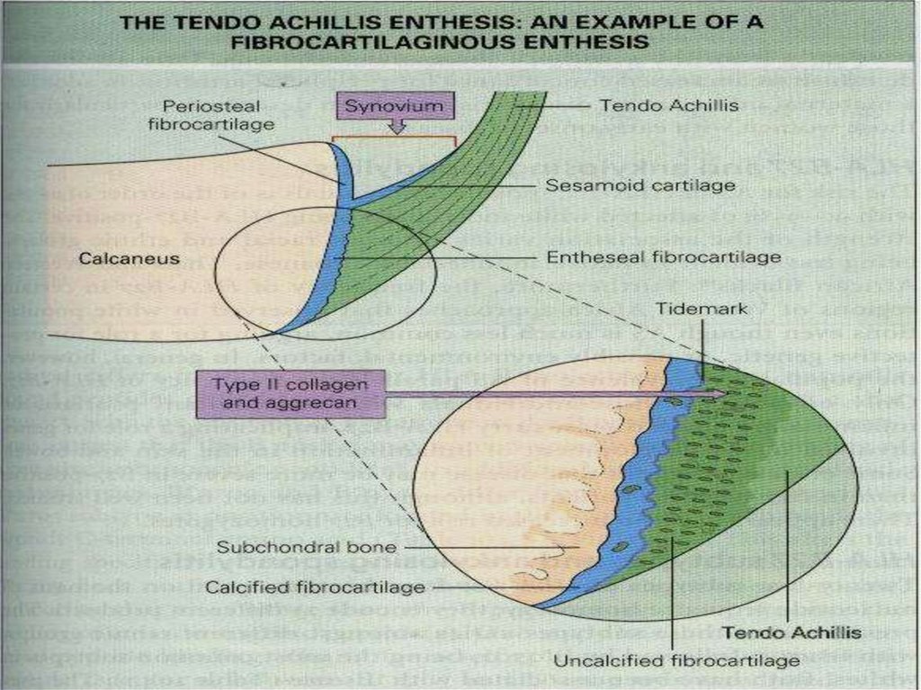
ETIOLOGY, PATHOGENESIS ANDPATHOLOGY
• Key initial inflammatory lesions occur at
FIBROCARTILAGINOUS ENTHESES rich in
aggrecans and type II collagen
(e.g. intervertebral disc, sacroiliac joint,
symphysis pubis and root of aorta)
117.
118.
120119.
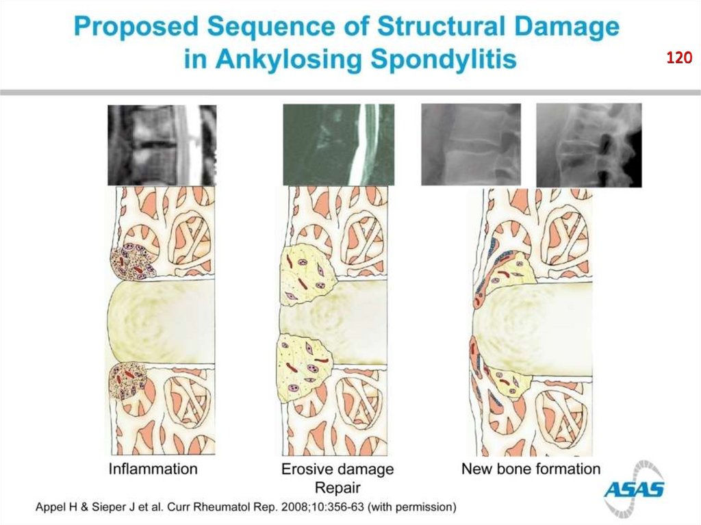
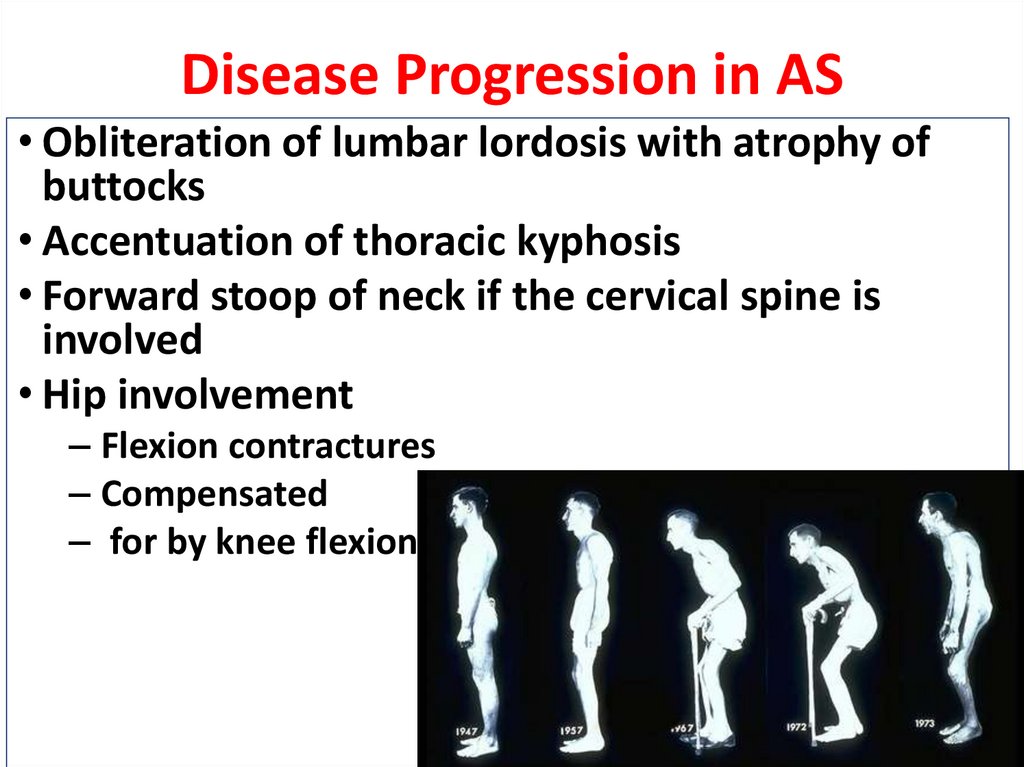
Disease Progression in AS• Obliteration of lumbar lordosis with atrophy of
buttocks
• Accentuation of thoracic kyphosis
• Forward stoop of neck if the cervical spine is
involved
• Hip involvement
– Flexion contractures
– Compensated
– for by knee flexion
120.
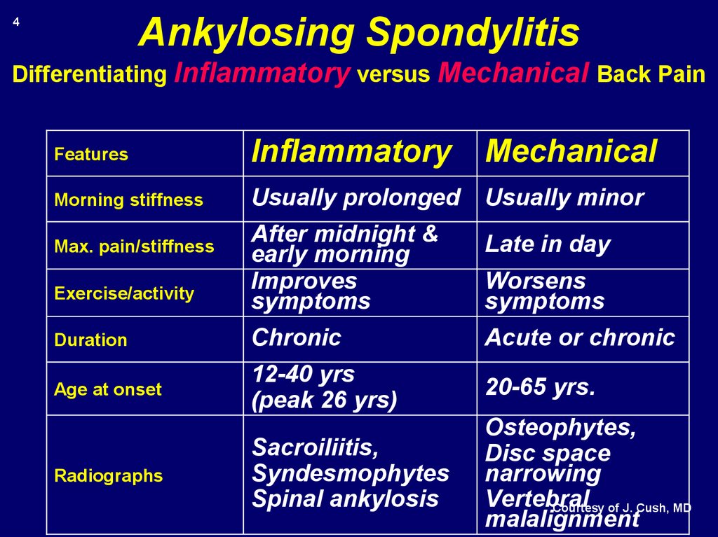
Ankylosing Spondylitis4
Differentiating Inflammatory versus Mechanical Back Pain
Features
Inflammatory
Mechanical
Morning stiffness
Usually prolonged
Usually minor
After midnight &
early morning
Improves
symptoms
Late in day
Duration
Chronic
Acute or chronic
Age at onset
12-40 yrs
(peak 26 yrs)
20-65 yrs.
Sacroiliitis,
Syndesmophytes
Spinal ankylosis
Osteophytes,
Disc space
narrowing
Vertebral
Courtesy of J. Cush, MD
malalignment
Max. pain/stiffness
Exercise/activity
Radiographs
Worsens
symptoms
121.
Inflammatory Back Pain according to experts*123
• Insidious onset
In fact, the
confirmation of IBP
• Pain at night
(with improvement upon getting up
is *the most widely
** *Mnemonic “IPAIN” or “iPAIN”
starting
*
• Age at onsetaccepted
<40 years
point for assessing
the potential
• Improvement with exercise
presence of an
• No improvement axial
with rest
spondyloarthritis
such
asRudwaleit
AS.1M, et al. ARD. 2009; 68(6):777-83.
*Sieper J, et al. Ann Rheum Dis. 2009;
68(6):784-8.
**Ozgocmen S, Akgul O, Khan MA. Mnemonic for ASAS criteria. J Rheumatol. 2010 Sep;37(9):1978-9.
122.
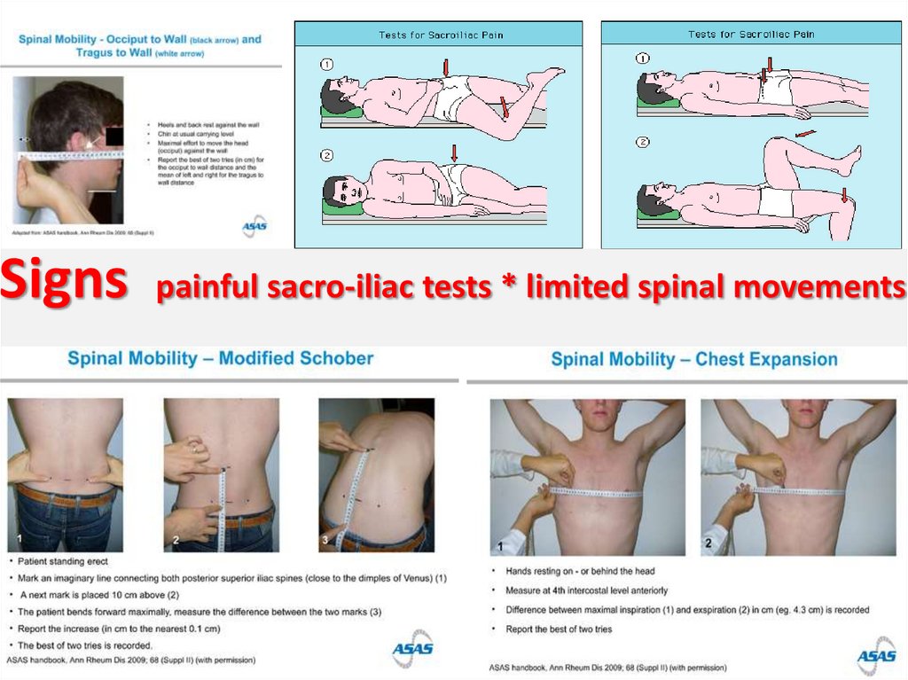
Signspainful sacro-iliac tests * limited spinal movements
123.
Ankylosing Spondylitis• Key Features:
–Low back pain
–Prolonged AM stiffness
–Nocturnal stiffness
–Improves with exercise
–Xrays with signs of sacroiliitis
–-Ensethopathy
–-Positive Family History.
124.
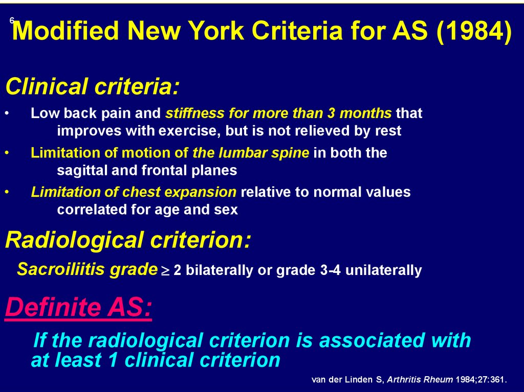
6Modified New York Criteria for AS (1984)
Clinical criteria:
Low back pain and stiffness for more than 3 months that
improves with exercise, but is not relieved by rest
Limitation of motion of the lumbar spine in both the
sagittal and frontal planes
Limitation of chest expansion relative to normal values
correlated for age and sex
Radiological criterion:
Sacroiliitis grade 2 bilaterally or grade 3-4 unilaterally
Definite AS:
If the radiological criterion is associated with
at least 1 clinical criterion
van der Linden S, Arthritis Rheum 1984;27:361.
125.
Age of Onset and Diagnosis in ASAnkylosing Spondylitis is a
disease charactrized by
Age of
Onset
Age at Diagnosis
early onset&
delayed diagnosis
Clearly there is a
Males
(N=920)
Females
(N=476)
Age in years
Feldtkeller E, et al. Rheumatol Int. 2003;23:61-6
significant gap
between
Onset and diagnosis
(8-9 years)
Khan M Arthritis Rheum Dis 2000;61(Suppl III):iii3-iii7.
126.
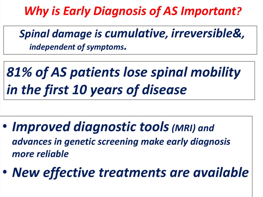
Why is Early Diagnosis of AS Important?Spinal damage is cumulative, irreversible&,
independent of symptoms.
81% of AS patients lose spinal mobility
in the first 10 years of disease
• Improved diagnostic tools (MRI) and
advances in genetic screening make early diagnosis
more reliable
• New effective treatments are available
Khan M Arthritis Rheum Dis 2000;61(Suppl III):iii3-iii7.
127.
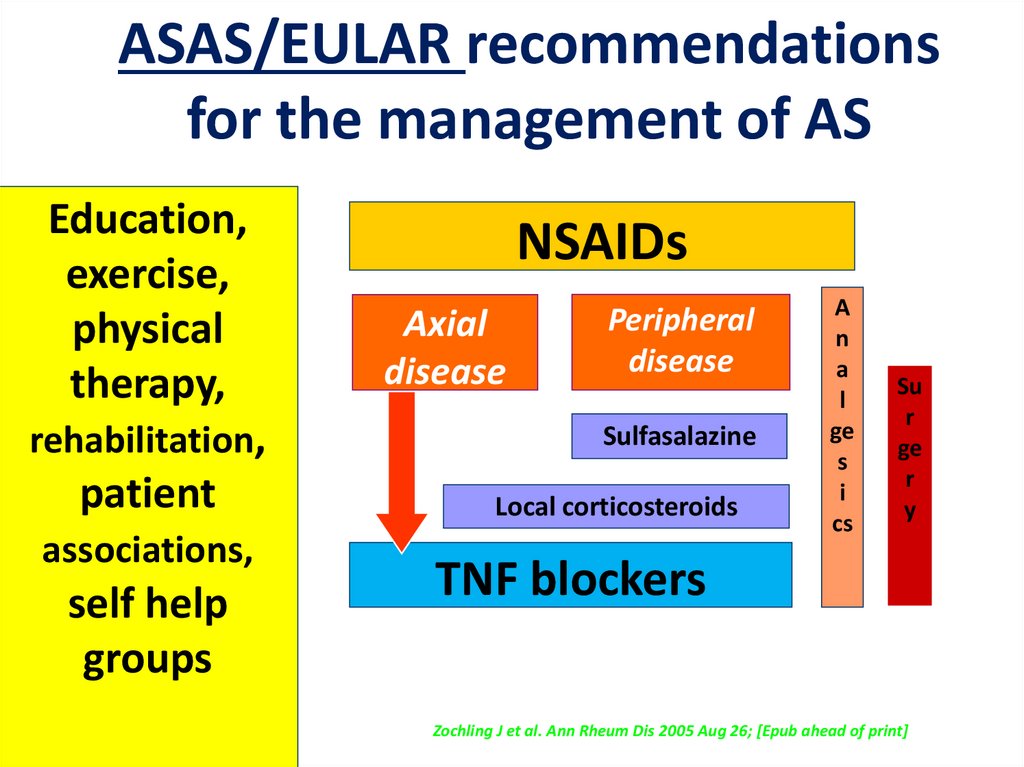
ASAS/EULAR recommendationsfor the management of AS
Education,
exercise,
physical
therapy,
rehabilitation,
patient
associations,
self help
groups
NSAIDs
Axial
disease
Peripheral
disease
Sulfasalazine
Local corticosteroids
A
n
a
l
ge
s
i
cs
Su
r
ge
r
y
TNF blockers
Zochling J et al. Ann Rheum Dis 2005 Aug 26; [Epub ahead of print]
128.
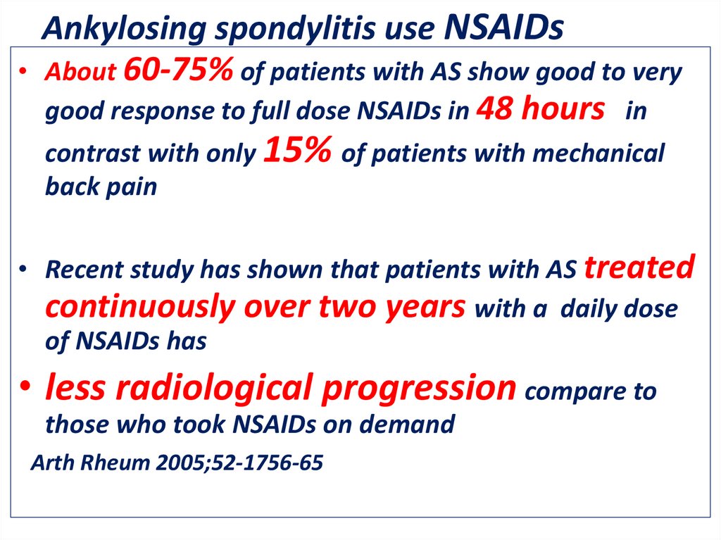
Ankylosing spondylitis use NSAIDs
About 60-75% of patients with AS show good to very
good response to full dose NSAIDs in 48 hours , in
contrast with only 15% of patients with mechanical
back pain.
• Recent study has shown that patients with AS treated
continuously over two years with a daily dose
of NSAIDs has
• less radiological progression compare to
those who took NSAIDs on demand
Arth Rheum 2005;52-1756-65
129.
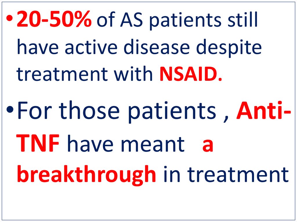
•20-50% of AS patients stillhave active disease despite
treatment with NSAID..
•For those patients , AntiTNF have meant a
breakthrough in treatment
130.
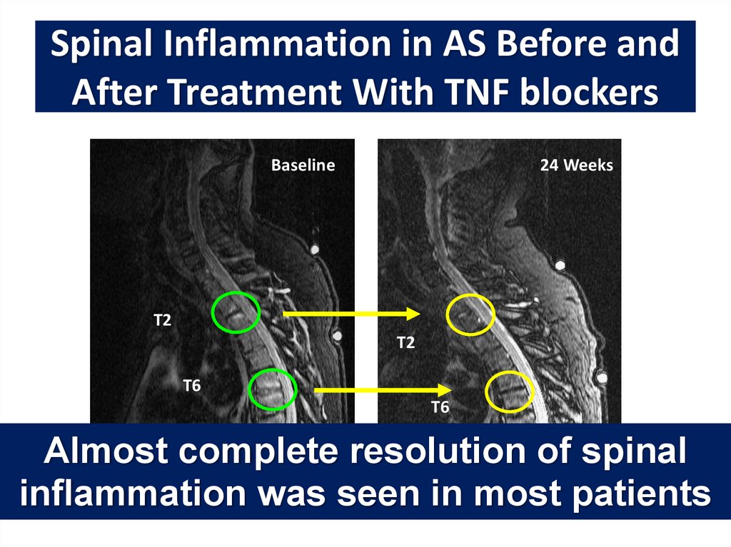
Spinal Inflammation in AS Before andAfter Treatment With TNF blockers
Baseline
24 Weeks
T2
T2
T6
T6
T9
Almost complete
resolution
of spinal
T9
inflammation was seen in most patients
STIR
Braun J, et al. Arthritis Rheum. 2006;54:1646-1652.
STIR
ASSERT MRI Study
131.
PRINCIPLES OF MANGEMENT OF AS1- No cure , but most patients can
be well managed
2- Early diagnosis is very important
3- Education to increase compliance
4- Appropriate use of antirheumatic drugs
,primarily (NSAIDs)and appropriate use of
biologic therapy
132.
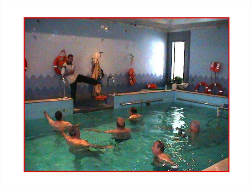
5- Continuity of care6- Daily exercise very
important (e.g., swimming )
7- Sleep on firm mattress
8-Avoidance of smoking
& trauma
133.
A full explanationof the disease , its
course , possible
complications, its
manegment&
prognosis is
essential to
achieve
appropriate
compliance by
the patient
134.
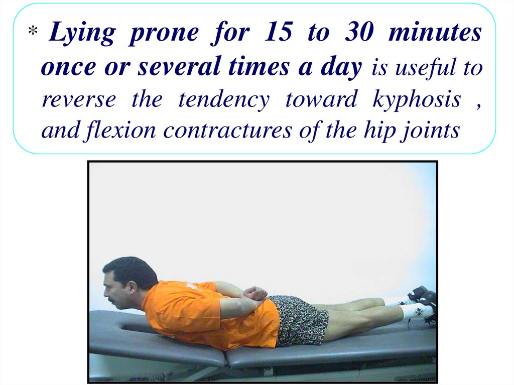
*Lying prone for 15 to 30 minutes
once or several times a day is useful to
reverse the tendency toward kyphosis ,
and flexion contractures of the hip joints
135.
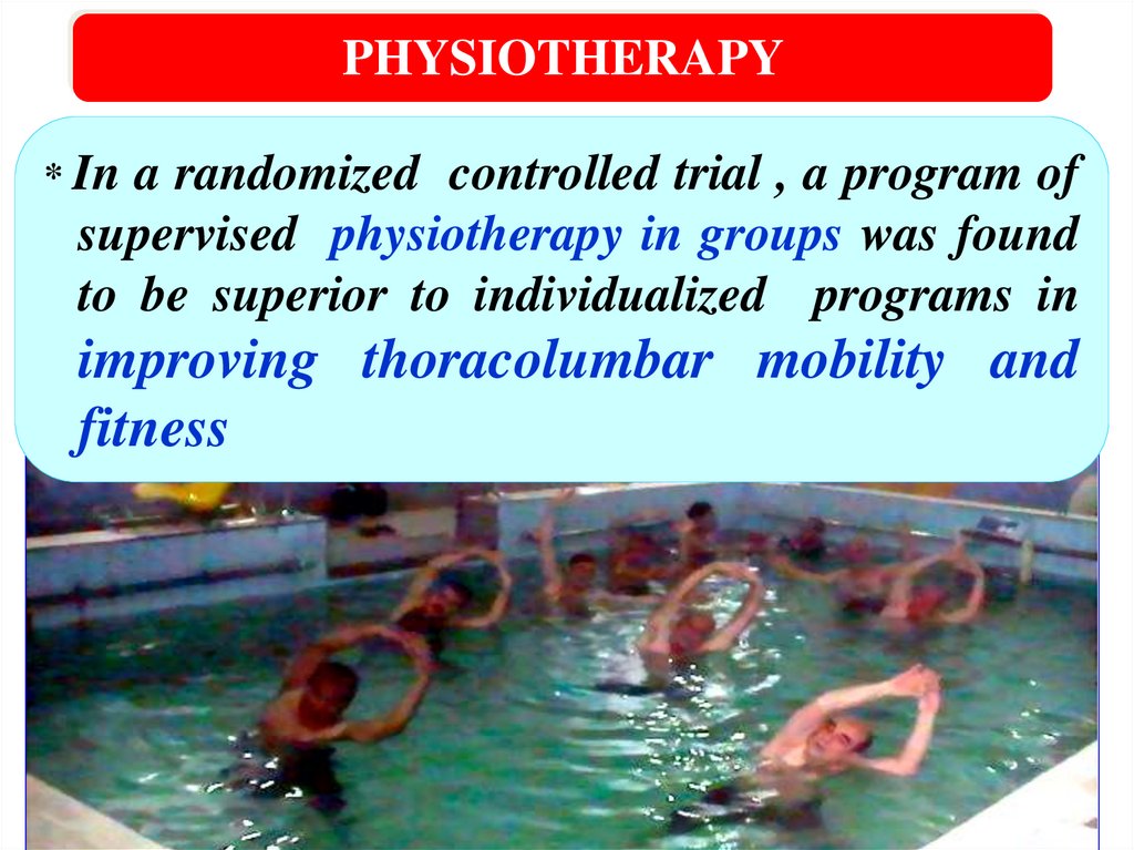
PHYSIOTHERAPY* In
a randomized controlled trial , a program of
supervised physiotherapy in groups was found
to be superior to individualized programs in
improving thoracolumbar mobility and
fitness
136.
A New Therapeutic ApproachIs Needed for AS
Early diagnosis is critical!
Eeeeeer
Early, aggressive therapy &early
introduction
of biologic
Early use
`of biologic
can yield
Lead to:
–Less structural damage
–Better function
–Remission
137.
138.
OsteoporosisPROF/
REDA AWAD
139.
Osteoporosis("porous bones",
from Greek: ὀστέον/
osteon meaning "bone" and
πόρος/poros meaning "pore")
140.
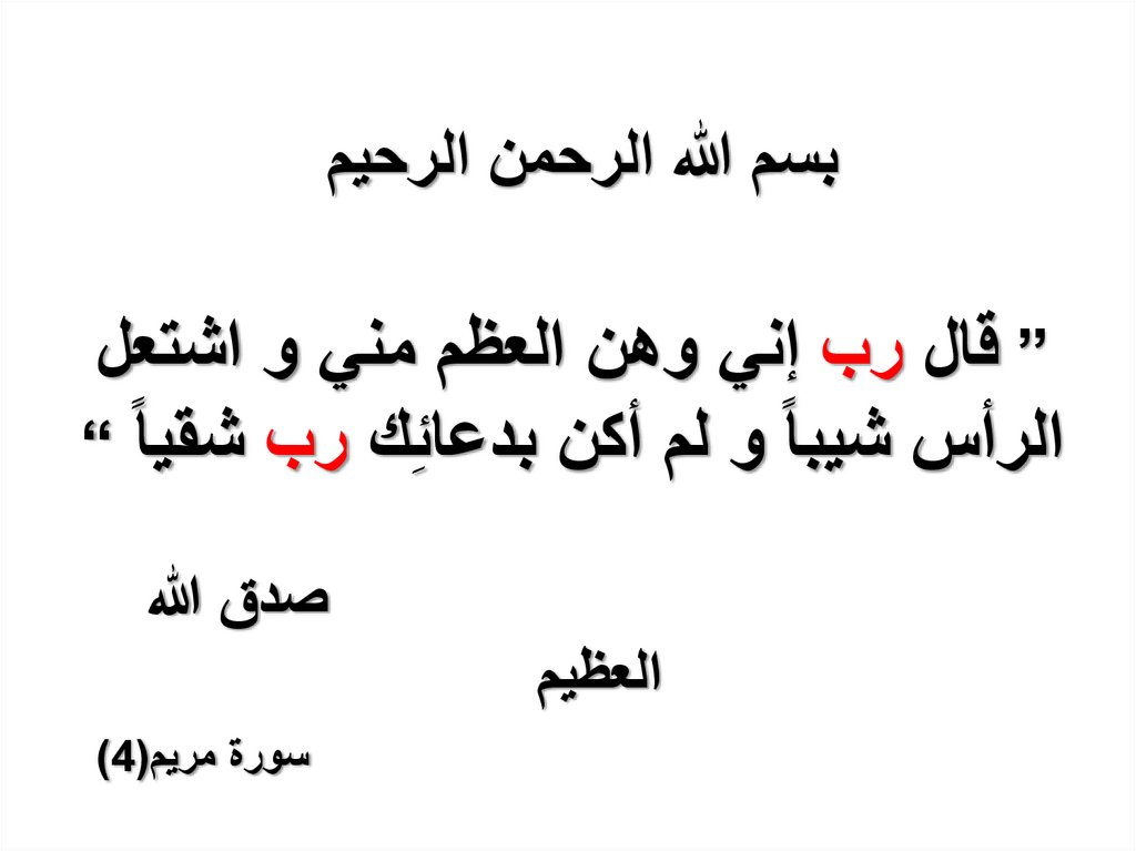
بسم هللا الرحمن الرحيم” قال رب إني وهن العظم مني و اشتعل
الرأس شيبا ً و لم أكن بدعا ِئك رب شقيا ً “
صدق هللا
العظيم
سورة مريم()4
141.
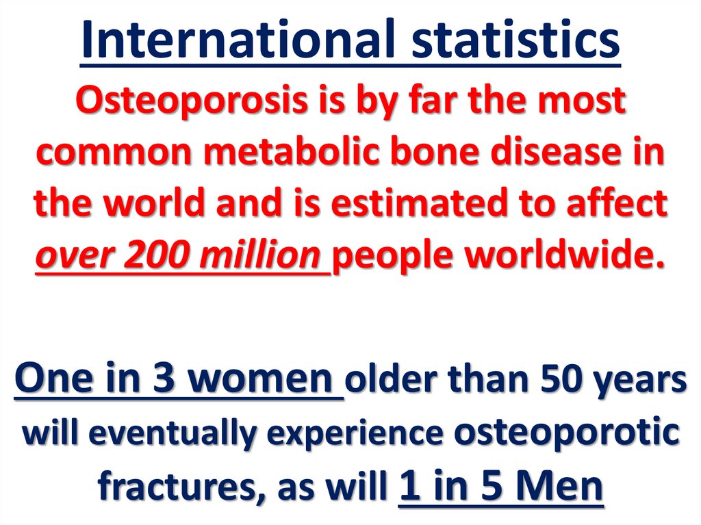
International statisticsOsteoporosis is by far the most
common metabolic bone disease in
the world and is estimated to affect
over 200 million people worldwide.
One in 3 women older than 50 years
will eventually experience osteoporotic
fractures, as will 1 in 5 Men
142.
143.
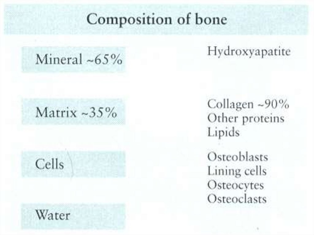
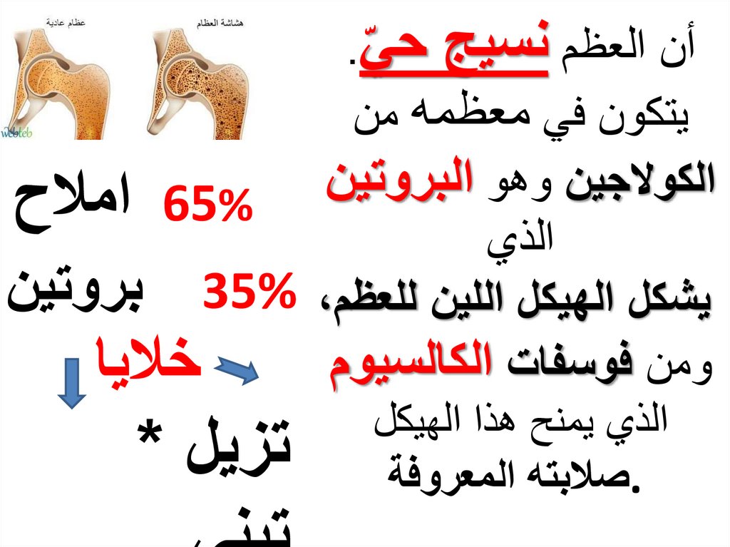
أن العظم نسيج حي.يتكون في معظمه من
البروتين
وهو
الكوالجين
65%امالح
الذي
يشكل الهيكل اللين للعظم 35% ،بروتين
ومن فوسفات الكالسيوم
خاليا
الذي يمنح هذا الهيكل
.صالبته المعروفة
تزيل *
144.
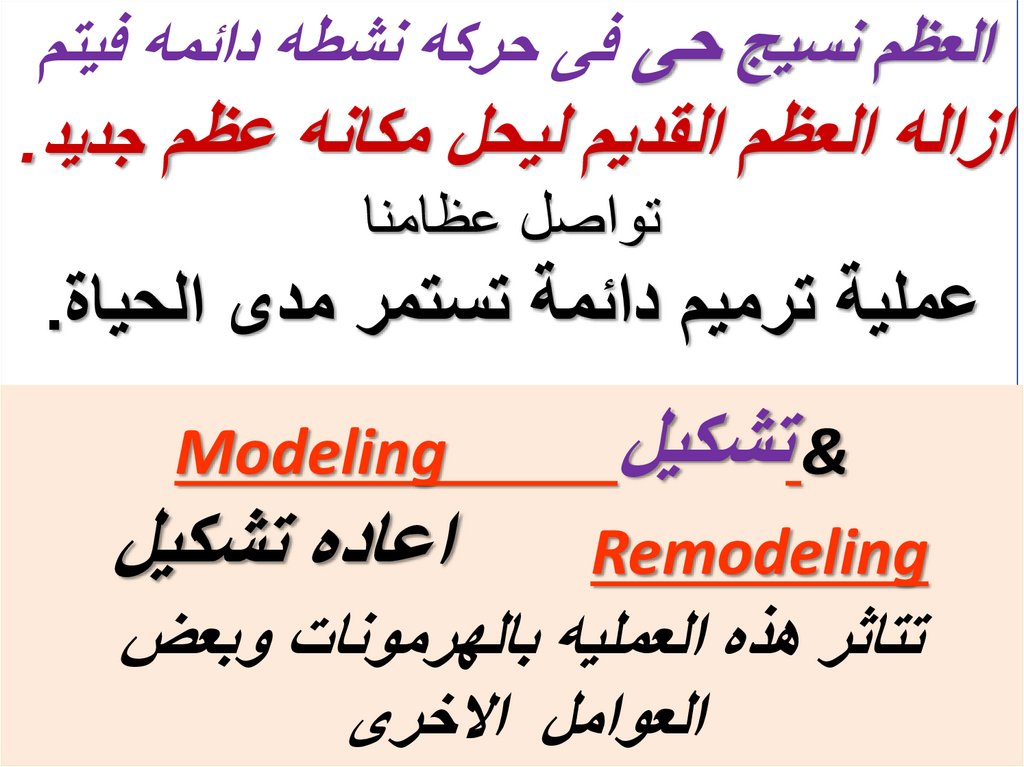
العظم نسيج حى فى حركه نشطه دائمه فيتمازاله العظم القديم ليحل مكانه عظم جديد.
تواصل عظامنا
عملية ترميم دائمة تستمر مدى الحياة.
& تشكيل
Remodeling
Modeling
اعاده تشكيل
تتاثر هذه العمليه بالهرمونات وبعض
العوامل االخرى
145.
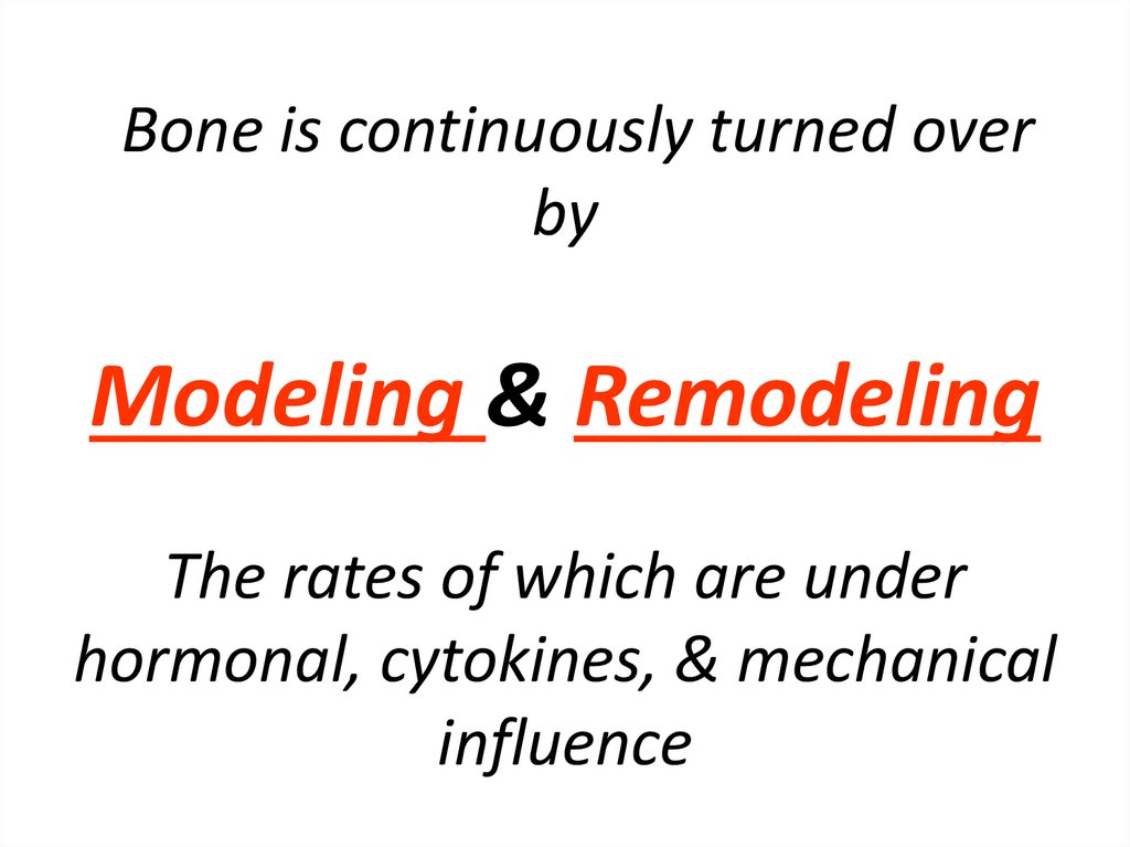
Bone is continuously turned overby
Modeling & Remodeling
The rates of which are under
hormonal, cytokines, & mechanical
influence
146.
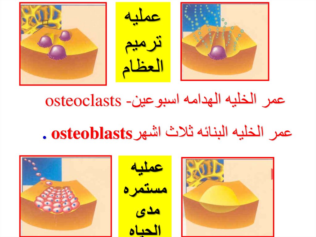
عمليهترميم
العظام
عمر الخليه الهدامه اسبوعينosteoclasts -
عمر الخليه البنائه ثالث اشهر. osteoblasts
.
عمليه
مستمره
مدى
الحياه
147.
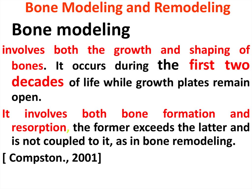
Bone Modeling and RemodelingBone modeling
involves both the growth and shaping of
bones. It occurs during the first two
decades of life while growth plates remain
open.
It involves both bone formation and
resorption, the former exceeds the latter and
is not coupled to it, as in bone remodeling.
[ Compston., 2001]
148.
هدم بطئهدم سريع
بناء سريع
هدم عادى
أكثر من
تكوين العظم
علىسرعة
تكون
والنمو،
الطفولة
إنخالل
فترة
مثالية في
كتلة عظم
يحصل
الذي ال
الفرد
العظمي
ذلك يبدأ
العظم .وبعد
سرعة إزالته
النسيج وحتى
بإزالةالوالدة
الجسم منذ
التي تبدأ
القصوى
بناء
القديم بسرعة أكبر من سرعة توليد النسيج العظمي الجديد
سن الثالثين تقريبا قد يتعرض لهشاشه العظام
149.
Once the skeleton has reachedmaturity, regeneration continues in
the form of a periodic replacement
of old bone with new at the same
location. This process is called
remodeling,
and is responsible for the
((complete
regeneration of the
adult skeleton every 10 years)).
[ Compston., 2001 ]
150.
In the uninjured adult skeleton, allosteoclasts and osteoblasts belong to a unique
temporary structure, known as:
Basic Multicellular Unit ( BMU )
The BMU, approximately 1–2 mm
long and 0.2 – 0.4 mm wide, comprises
a team of osteoclasts in the front, a
team of osteoblasts in the rear, a
central vascular capillary, a nerve
supply, and associated connective
tissue .
151.
In healthy human adults, 3–4million BMUs are initiated per year
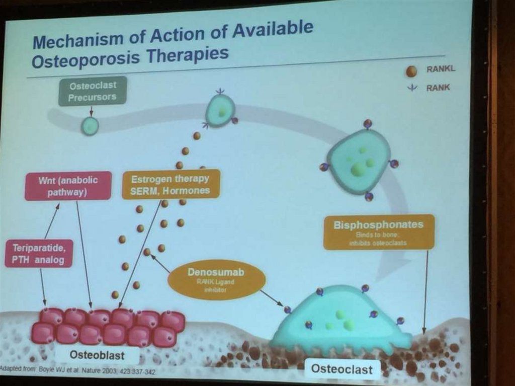
and at least
One million are
operating at any moment .
Each BMU begins at a particular place
and time toward a target, which is a
region of bone in need of replacement.
[ Manolagas., 2000]
152.
الحاله الطبيعيهحاله الهشاشه
153.
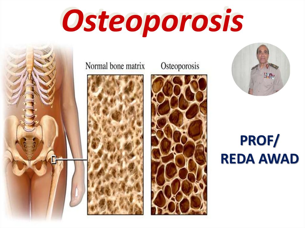
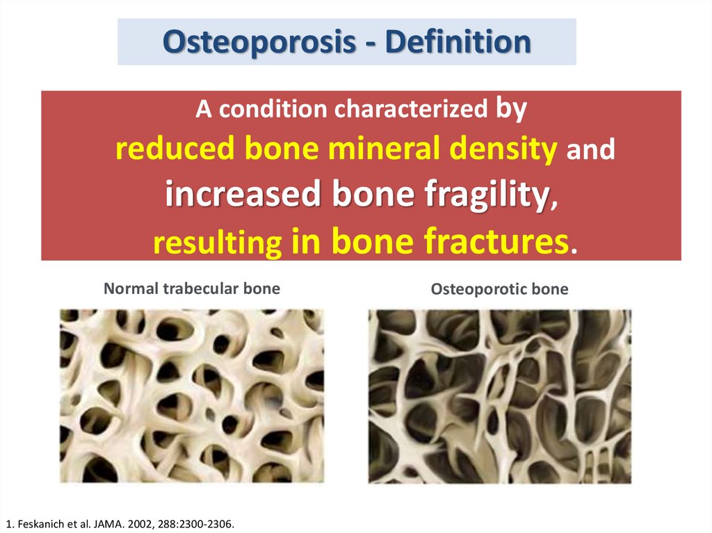
Osteoporosis - DefinitionA condition characterized by
reduced bone mineral density and
increased bone fragility,
resulting in bone fractures.
Normal trabecular bone
1. Feskanich et al. JAMA. 2002, 288:2300-2306.
Osteoporotic bone
154.
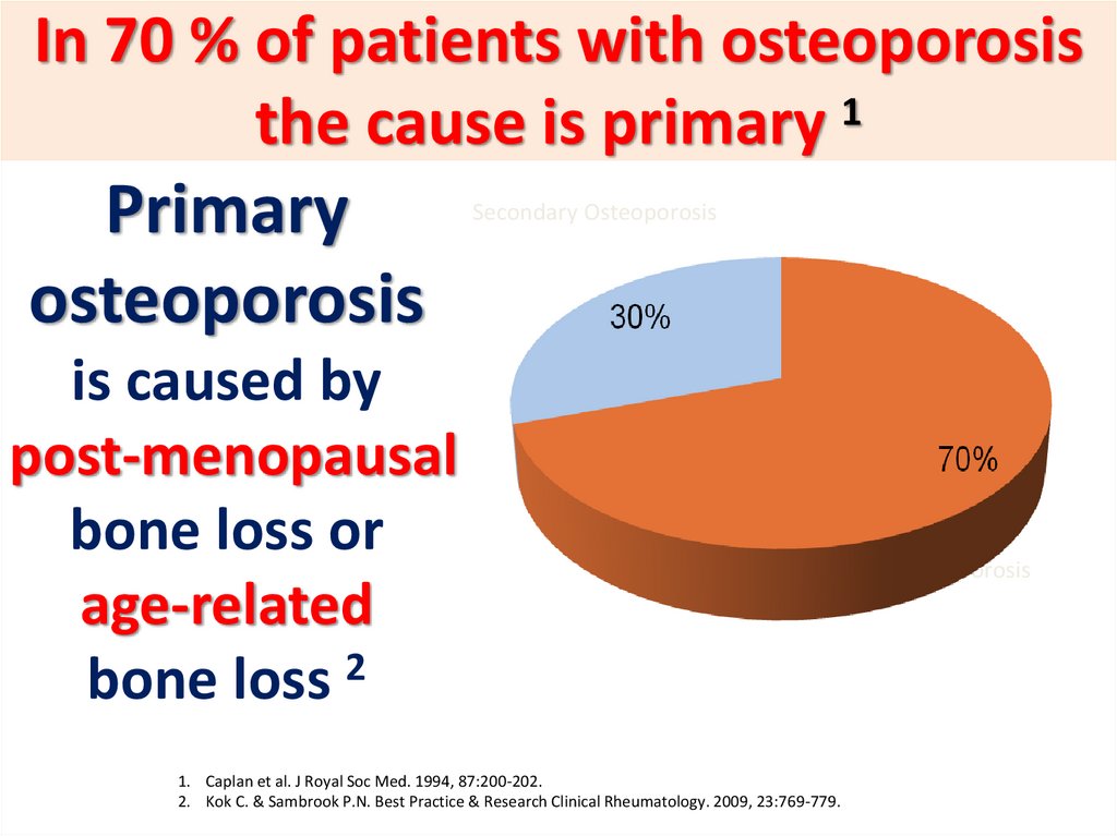
In 70 % of patients with osteoporosisthe cause is primary 1
Primary
osteoporosis
is caused by
post-menopausal
bone loss or
age-related
bone loss 2
Secondary Osteoporosis
Primary Osteoporosis
1. Caplan et al. J Royal Soc Med. 1994, 87:200-202.
2. Kok C. & Sambrook P.N. Best Practice & Research Clinical Rheumatology. 2009, 23:769-779.
155.
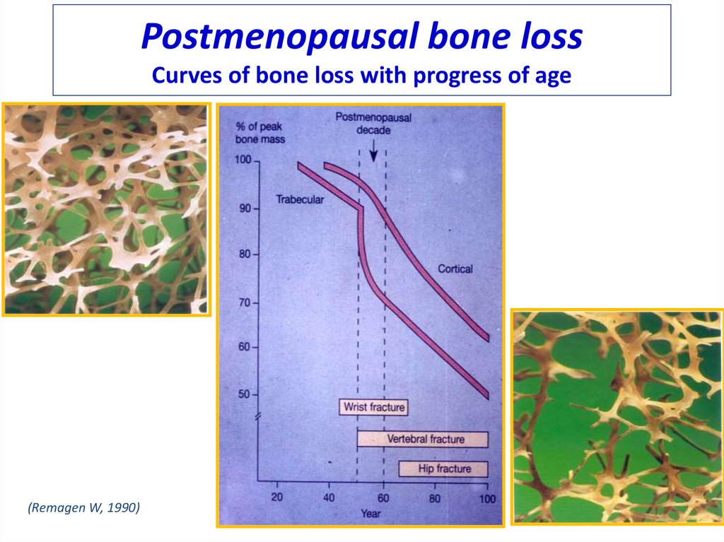
Postmenopausal bone lossCurves of bone loss with progress of age
(Remagen W, 1990)
156.
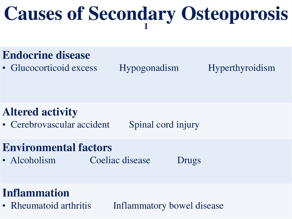
Causes of SecondaryOsteoporosis
1
Endocrine disease
• Glucocorticoid excess
Hypogonadism
Hyperthyroidism
Altered activity
• Cerebrovascular accident
Spinal cord injury
Environmental factors
• Alcoholism
Coeliac disease
Drugs
Inflammation
• Rheumatoid arthritis
Inflammatory bowel disease
157.
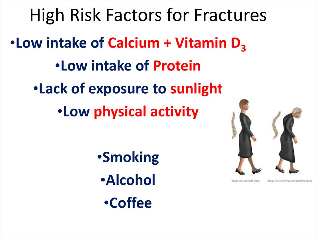
High Risk Factors for Fractures•Low intake of Calcium + Vitamin D3
•Low intake of Protein
•Lack of exposure to sunlight
•Low physical activity
•Smoking
•Alcohol
•Coffee
158.
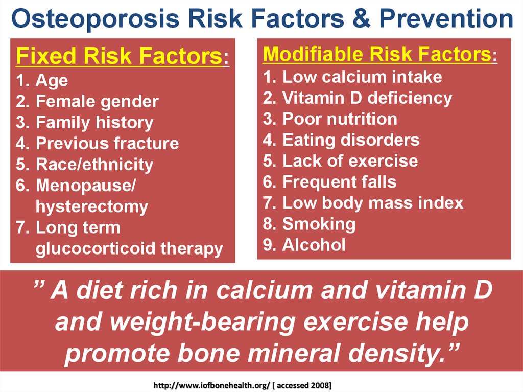
Osteoporosis Risk Factors & PreventionFixed Risk Factors:
Modifiable Risk Factors:
1. Age
2. Female gender
3. Family history
4. Previous fracture
5. Race/ethnicity
6. Menopause/
hysterectomy
7. Long term
glucocorticoid therapy
1. Low calcium intake
2. Vitamin D deficiency
3. Poor nutrition
4. Eating disorders
5. Lack of exercise
6. Frequent falls
7. Low body mass index
8. Smoking
9. Alcohol
” A diet rich in calcium and vitamin D
and weight-bearing exercise help
promote bone mineral density.”
http://www.iofbonehealth.org/ [ accessed 2008]
159.
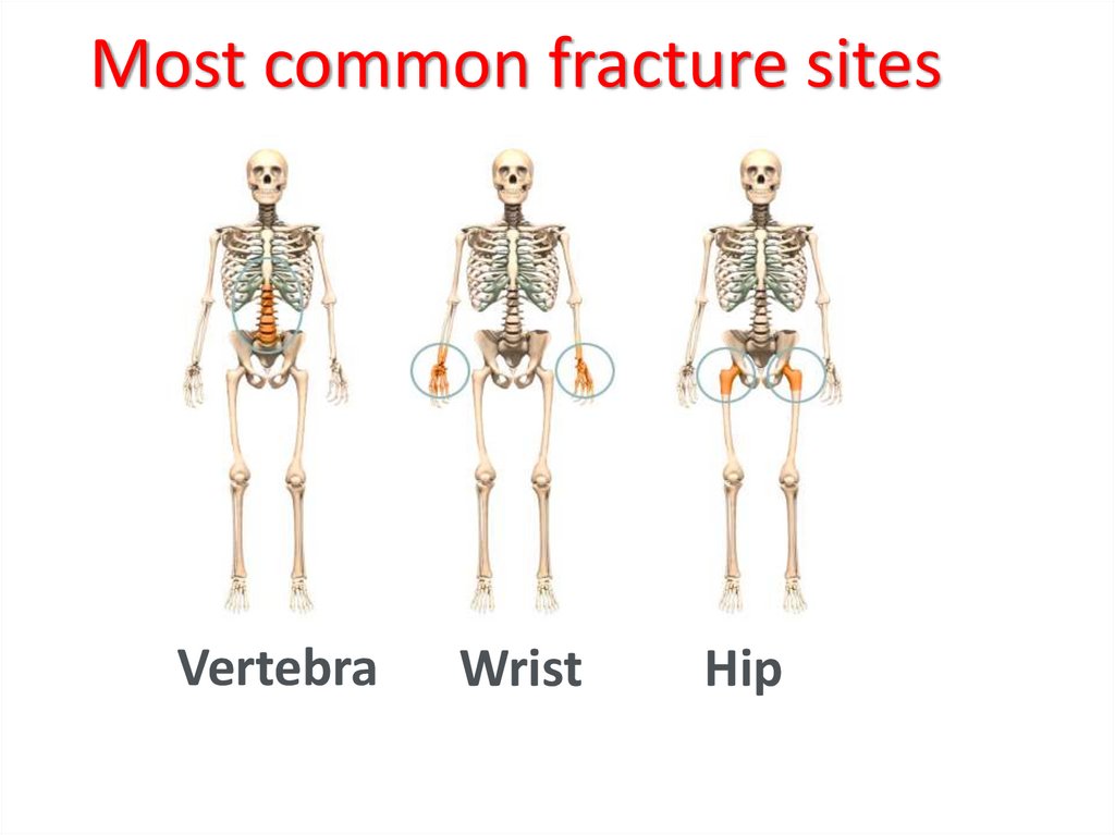
Most common fracture sitesVertebra
Wrist
Hip
160.
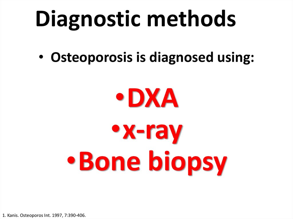
Diagnostic methods• Osteoporosis is diagnosed using:
•DXA
•x-ray
•Bone biopsy
1. Kanis. Osteoporos Int. 1997, 7:390-406.
161.
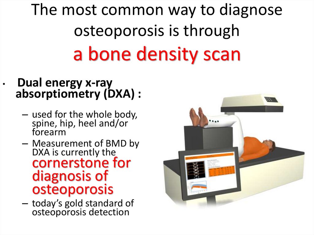
The most common way to diagnoseosteoporosis is through
a bone density scan
Dual energy x-ray
absorptiometry (DXA) :
– used for the whole body,
spine, hip, heel and/or
forearm
– Measurement of BMD by
DXA is currently the
cornerstone for
diagnosis of
osteoporosis
– today’s gold standard of
osteoporosis detection
162.
T-scoreT-score is useful for the expression of BMD
( bone mineral density)
= measured BMD – mean young adult BMD
young adult SD
• T-score indicates the difference between
a patient’s BMD and the ideal BM
achieved by a young adult
1. Blake & Fogelman. Curr Pharm Des. 2002, 8:1885-1905.
163.
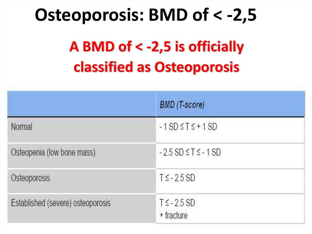
Osteoporosis: BMD of < -2,5A BMD of < -2,5 is officially
classified as Osteoporosis
164.
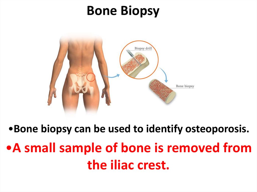
Bone Biopsy•Bone biopsy can be used to identify osteoporosis.
•A small sample of bone is removed from
the iliac crest.
165.
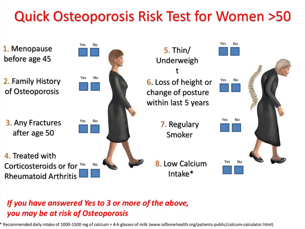
Quick Osteoporosis Risk Test for Women >501. Menopause
before age 45
Yes
No
2. Family History
of Osteoporosis
Yes
No
3. Any Fractures
after age 50
Yes
No
4. Treated with
Corticosteroids or for Yes
Rheumatoid Arthritis
No
5. Thin/
Underweigh
t
6. Loss of height or
change of posture
within last 5 years
7. Regulary
Smoker
8. Low Calcium
Intake*
Yes
No
Yes
No
Yes
Yes
No
No
If you have answered Yes to 3 or more of the above,
you may be at risk of Osteoporosis
* Recommended daily intake of 1000-1500 mg of calcium = 4-6 glasses of milk (www.iofbonehealth.org/patients-public/calcium-calculator.html)
166.
Diagnosing osteoporosisis usually a combination of
BMD,
Age,
previous fractures
history of falls
&
167.
Osteoporosis:Underdiagnosis
168.
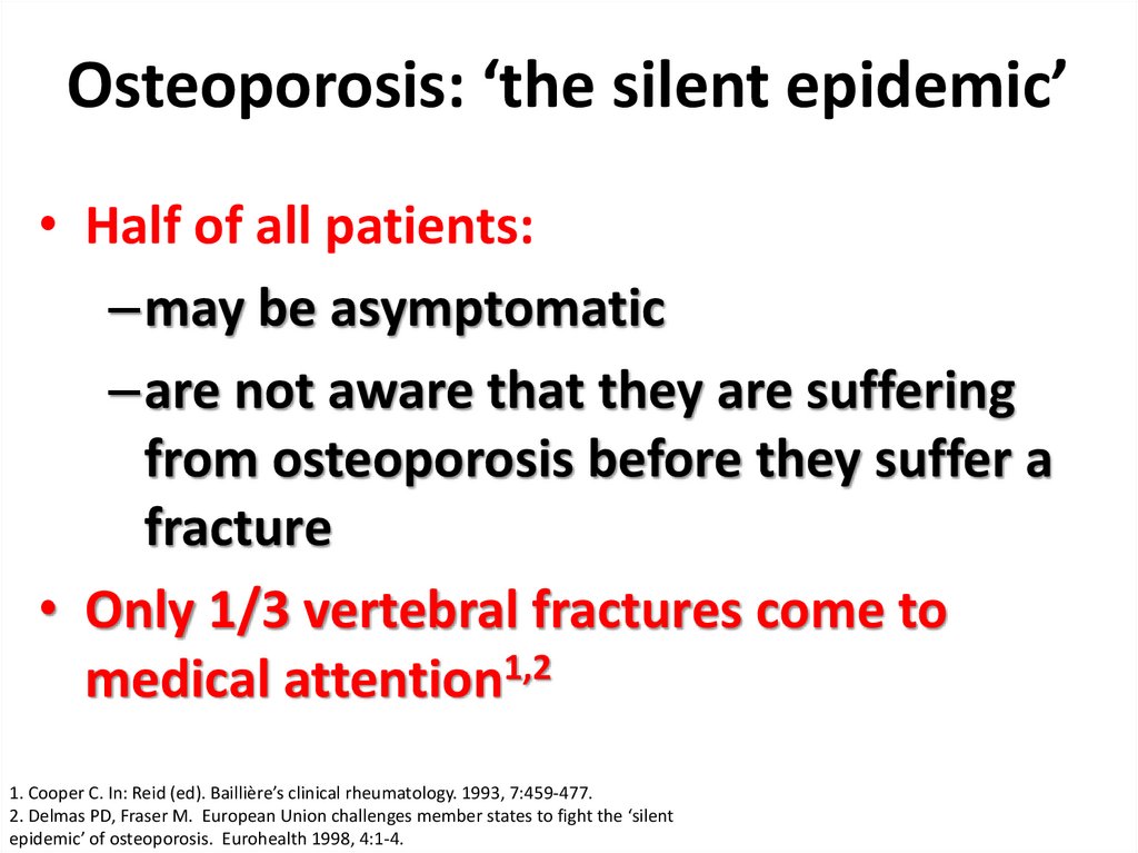
Osteoporosis: ‘the silent epidemic’• Half of all patients:
–may be asymptomatic
–are not aware that they are suffering
from osteoporosis before they suffer a
fracture
• Only 1/3 vertebral fractures come to
medical attention1,2
1. Cooper C. In: Reid (ed). Baillière’s clinical rheumatology. 1993, 7:459-477.
2. Delmas PD, Fraser M. European Union challenges member states to fight the ‘silent
epidemic’ of osteoporosis. Eurohealth 1998, 4:1-4.
169.
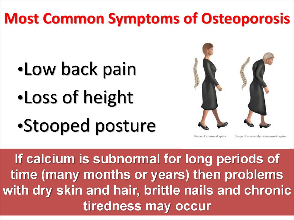
Most Common Symptoms of Osteoporosis•Low back pain
•Loss of height
•Stooped posture
If calcium is subnormal for long periods of
time (many months or years) then problems
with dry skin and hair, brittle nails and chronic
tiredness may occur
170.
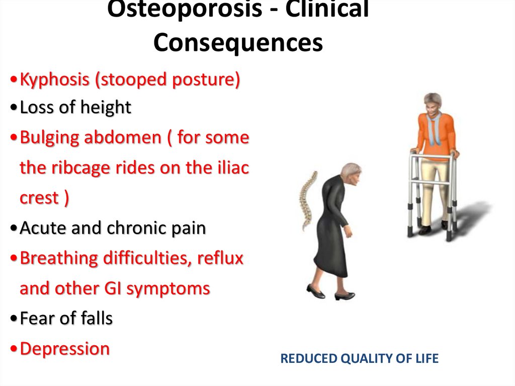
Osteoporosis - ClinicalConsequences
•Kyphosis (stooped posture)
•Loss of height
•Bulging abdomen ( for some
the ribcage rides on the iliac
crest )
•Acute and chronic pain
•Breathing difficulties, reflux
and other GI symptoms
•Fear of falls
•Depression
REDUCED QUALITY OF LIFE
171.
Osteoporosis:Treatment Options &
Guidelines
172.
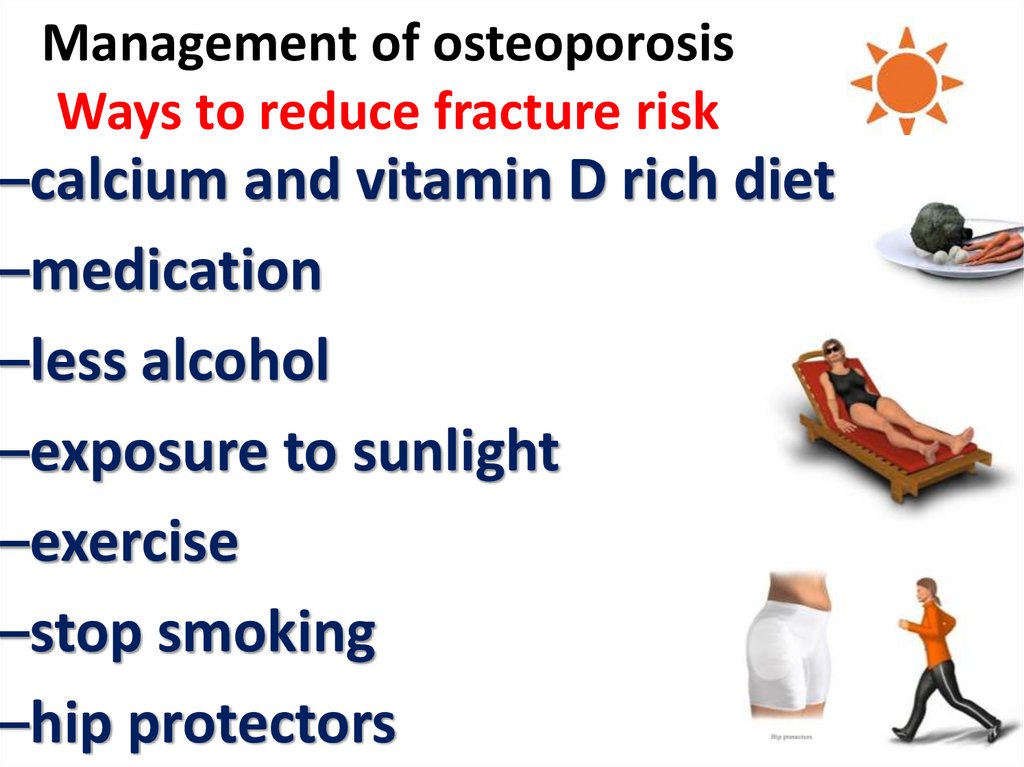
Management of osteoporosisWays to reduce fracture risk
–calcium and vitamin D rich diet
–medication
–less alcohol
–exposure to sunlight
–exercise
–stop smoking
–hip protectors
173.
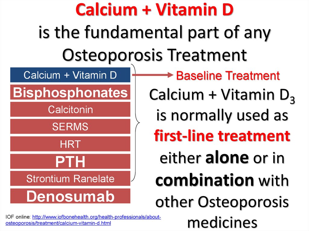
Calcium + Vitamin Dis the fundamental part of any
Osteoporosis Treatment
Baseline Treatment
Calcium + Vitamin D
Bisphosphonates
Calcitonin
SERMS
HRT
PTH
Strontium Ranelate
Denosumab
Calcium + Vitamin D3
is normally used as
first-line treatment
either alone or in
combination with
other Osteoporosis
medicines
IOF online: http://www.iofbonehealth.org/health-professionals/aboutosteoporosis/treatment/calcium-vitamin-d.html
174.
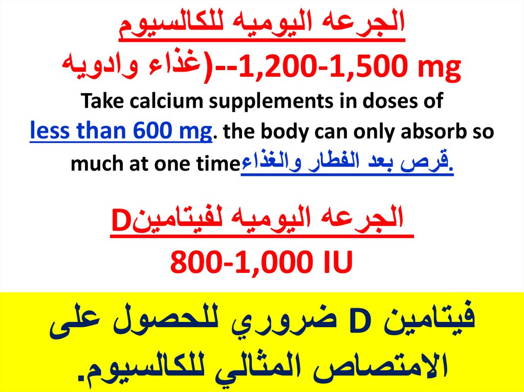
الجرعه اليوميه للكالسيوم(--1,200-1,500 mgغذاء وادويه
Take calcium supplements in doses of
less than 600 mg. the body can only absorb so
.قرص بعد الفطار والغذاءmuch at one time
الجرعه اليوميه لفيتامينD
800-1,000 IU
فيتامين Dضروري للحصول على
االمتصاص المثالي للكالسيوم.
175.
176.
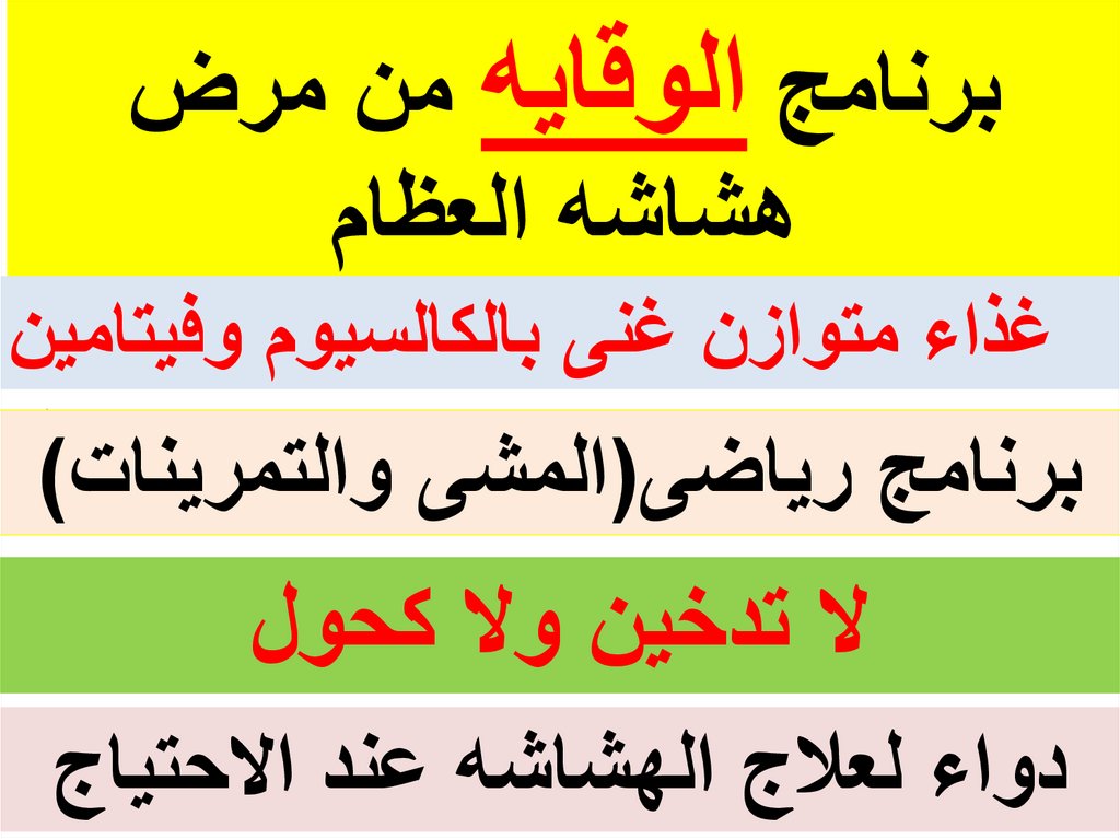
برنامج الوقايه من مرضهشاشه العظام
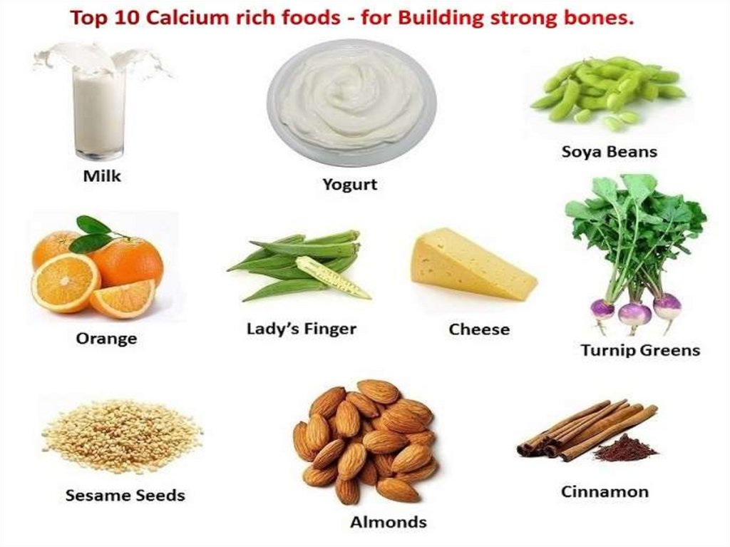
غذاء متوازن غنى بالكالسيوم وفيتامين
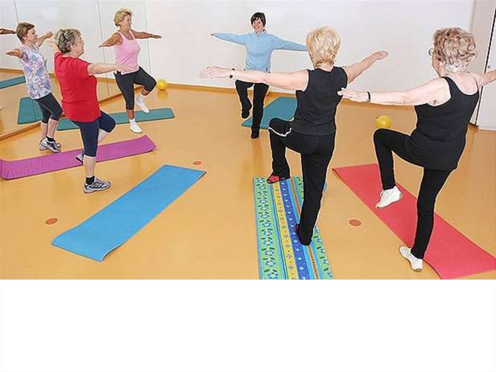
د
برنامج رياضى(المشى والتمرينات)
ال تدخين وال كحول
دواء لعالج الهشاشه عند االحتياج
177.
منع السقوط1تاكد من االبصار الجيد
(تصحيح النظر) -
---واالضاءه جيده
2تجنب االدويه المنومه
3
ازاله معوقات المشى من المنزل(السجاد –االثاث)
4الحذاء مريح ومثبت جيدا بالقدم
5استعمال سواند الحائط عند اللزوم