 Маркетинг
Похожие презентации:


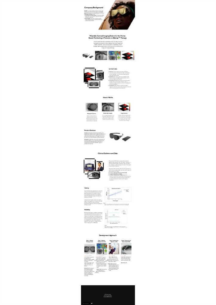
Company Background
1.
Company BackgroundSight.ED™ is a clinically validated, wearable electronic sight
aid that enables patients with severe sight loss to see clearly
again, while at the same time, providing AI-powered continuous
remotesight monitoring including:
1. Visual acuity and contrast testing: an inbuilt high- resolution
display allow the user to perform a visual acuity test from
the comfort of their home.
2. Retinal Imaging: in-built retinal imag ing and analytics
system detects the chang es in the retinal thickness a s a
proxy for disease deterioratio
Wearable Corneal Imaging Device for the HomeBased Monitoring of Patients on Blenrep™ Therapy
A novel method that allows quantifying corneal clouding cau sed by
keratopathy an d monitor d i sease progression over time. A quick an d
reliable, acceptable, an d accessi b l e mean s of quantifying corneal
clouding, objective quanti=cation of the h az e seen by clinicians via an
i mage p ro cessi ng technology.
KEY FEATURES:
Precision: Multiple i m a g e s of e a ch eye with different
illumination a n g l e s a n d frequencies of lig ht including nearInfrared waveleng th illumination penetrating throug h a n y
severe level of corneal haze.
Scalability: doesn't rely on in-clinic visits to support clinical
decision-making (initiating/reducing treatment) a s well a s
a d va n ce d research a nd clinical trials.
Consistency: Wearable device allows for the =xed distance
between the eye a n d the sensor distance a s well a s
consistent low lig ht environment to m a x i m i se i m a g e quality
a n d minimise variability.
Accessibility: Rapid, simple to u s e a n d non-invasive
diag nostics allows for unsupervised u s e by patients with even
the m os t severe physical or mental disabilities.
How it Works
Image Analysis
Reliable Eye Imaging
Wearable IR Camera
P rop ria tary i m a g e a n a lysis a lg orith m is th en
Th e u s e of mu ltip le wa velen g th s (incl. n ea r-
Hea d -worn , h ig hly sop h istica ted iris
recog n ition ca me r a th at is s m a l l a n d
a d a p ted to a ch ieve ra p id , relia ble a n d
sta n d a rd ised ob jective i m a g i n g in a wid e
va riety of p a tien ts wh ile m i n im is i n g
in fra red ) a llows p en etra tion of even severe
a p p lied to p re-p rocessed i m a g e s from th e
levels of corn ea l h a ze; th e iris c a me r a
iris sen sor; P ix el in ten sity m a p is g en era ted
ra p id ly ta kes severa l i m a g e s th a t a re th en
a n d corn ea l op a ci= ca tion m e a s u r e (COM)
c o m b in e d a n d pre-p rocessed for a n a lysis.
score is g en era ted ; th e c h a n g e in corn ea l
op a ci= ca tion is m e a s u r e d over time.
a rtefa ctual interferen ce in i ma g e q uality.
Product Evolution
Form Factor: Once we have validated the clinical eQcacy of the
proposed system (Stage 3) using a VR headset a s a platform for the
imag ing sensor we will commence (Stage 4) engineering work to
miniaturise and re=ne the device into a hardware solution with a
form factor of a small and comfortable pair of electronic g lasses.
Architecture: Wearable device (a) with an IRIS imag ing system (b)
on board equipped with a pair of high-resolution displays for the
Visual Acuity and Contrast Sensitivity test (c) connected via a
smartphone app (d) to the secure cloud where imag es are
processed and analysed (d).
Clinical Evidence and Data
The application of IRIS biometric s yste ms for op h t hal mi c
diag nostics w a s pioneered in 2 011 by Professor Tariq As l a m - a
Consultant Ophthalmolog ist at Manchester Royal Eye Hospital
a n d a Professor of Ophthalmolog y a n d Interface Technolog ies
at the University of Manchester.
The studies initially published in 2012 a n d further publication up
to 2019 ( a s well a s on g oi ng work) h a ve s h ow n that their alg orithm
a n a l ys i ng corneal IRIS i m a g e s :
Provided a n objective measure of corneal clouding
Ha s good repeatability a n d reliability
Was faster, more practical a n d objective than a full
op h t ha l mi c examination at quanti=cation of corneal clouding
(including subjective a s se ssme nt of corneal clouding with a
slit-lamp a n d u se of more complex corneal i m a g i n g
equipment)
Validity:
Fig ure 2 demonstrates in g raphical form a clear a n d
strong relationship between the values of corneal
opacity score obtain by our alg orithm a n d the
clinical g rading of participants. Linear reg ression
( S P S S V.14) con=rmed a strong relationship with a
coeQcient of p<0.0001 .
In addition to the 1 8 eyes of patients with anterior
s e g m e nt patholog y who were analysed by the COM
measure, a n additional 1 8 normal eyes were
a s s e s se d by the alg orit hms. All 1 8 normal eye s g a ve
Figure 1:
Plot o f corneal o p aci catio n measure (COM ) score against clinical g rad in g o f corneal opacity. This
g u re is p ro d uced in colour in the o n lin e journaldplease visit the website to v iew the colour g u re.
COM score s of z ero.
Reliability:
Seventeen subjects eyes (included four normal eyes)
h a d i m a g i ng repeated by the s a m e examiner on two
occa s i on s , at least a n hour apart to a s s e s s the
reliability of the COM measure. A Blande-Altman plot
(=g ure 2) demonstrates no evident systematic b i a s
a n d narrow coeQcient of repeatability of 1 . 2 1 (95%
limits of ag reement). The intraclass correlation
coeQcient (mixed model) for averag e m e a s ure s
( S P S S V.15.0) w a s 0 . 9 9 7 (0.989e0.999).
Figure 2:
Blande-Altman p lo t o f mean o f corneal o p aci cation measure (COM ) rep eat measures
against d ifference. This g u re is p ro d uced in colour in the o n lin e journaldplease visit the
website to v iew the colour g u re.
Development Approach
Stage 1: Working
Prototype Model
DU R AT I ON : 4 -
5
M o n th s
B
U DG E T : £ 3 6 0 ,0 0 0
The =rst stage of the project will
the development of a
wearable IRIS scanner integrated
standard VR. And iterative
and
the imag ing
system until the highest quality
imag e representations of
patients with disease are
captured
Decision Gate: The prototype is
able to repeatedly take highquality/precision/repeatable
Stage 2: Algorithm
Development (n=20)
DU R AT I ON : 2 -
3
M o n th s
B
U DG E T : £ 1 8 0 ,0 0 0
Stage 3: Validating the
Algorithm (n=90)
DU R AT I ON : 6 M o n th s
DU R AT I ON : T B D
B UDGE T : T B D
B UDGE T : T B D
Acquire a bigger sample of
Iteratively Adjust the imag ing be
imag es to enhance and adjust
system and an imag e analysis
software to automatically detect
the sensitivity and reliability of into a
grade corneal abnormalities.
the diagnostics system and adjustment of
We will use a a small number of
imag e analysis software.
patients with Blenrep corneal
disease (20 patients) and healthy
volunteers in order to develop
the engineering and software
analysis
Decision Gate: Demonstrate that
the proposed system and
corneal imag es. SuQcient quality
algorithm are able to detect for
the analytics algorithm to
keratopathy
process. Varied enough to
combine and compensate for
any artefacts
Decision Gate: Demonstrate
relationship between the camera
automatic pathology score as
well as remote expert analysis of
imag es with grading obtained by
clinical grading of participants.
(n=90)
20 Air Street, London
Phone: +44 7873 942426
E-mail: stan@givevision.net
Вернут ь с я к ред ак т ирован ию
Stage 4: Prepare for the
Clinical Trial (n=TBD)
Engineering and development of
the pilot-ready medical device
(incl cert) in parallel with design
and set up of the clinical s tudy
Decision Gate: TBD