


 Медицина
Похожие презентации:
 fungal infections")


")
Pathogenic fungus
1.
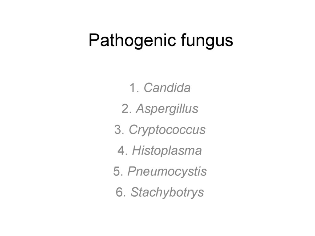
Pathogenic fungus1. Candida
2. Aspergillus
3. Cryptococcus
4. Histoplasma
5. Pneumocystis
6. Stachybotrys
2.
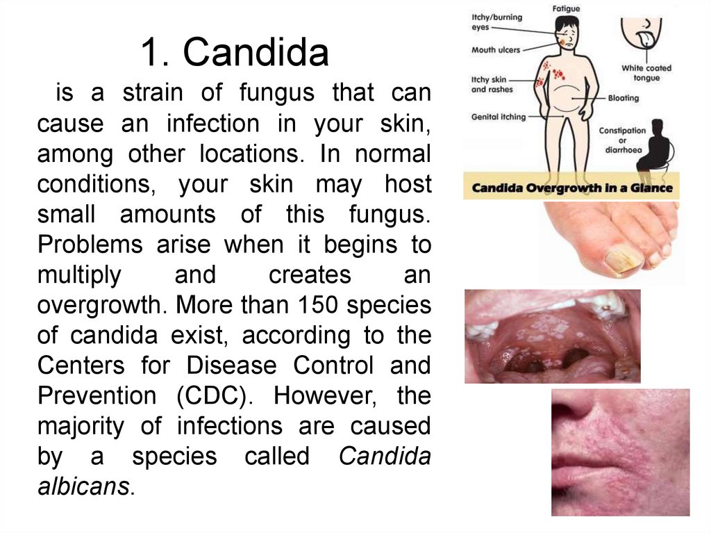
1. Candidais a strain of fungus that can
cause an infection in your skin,
among other locations. In normal
conditions, your skin may host
small amounts of this fungus.
Problems arise when it begins to
multiply
and
creates
an
overgrowth. More than 150 species
of candida exist, according to the
Centers for Disease Control and
Prevention (CDC). However, the
majority of infections are caused
by a species called Candida
albicans.
3.
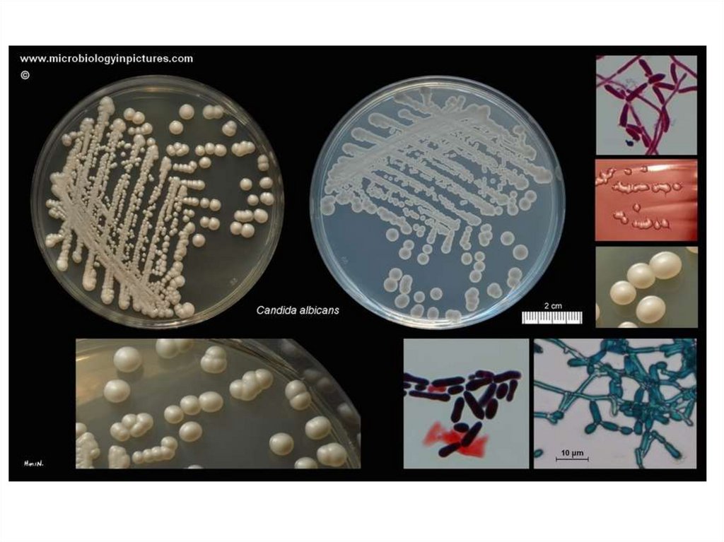
Structure of CandidaMicroscopy of Candida albicans
Fungi belong to the group of organisms called
eukaryotes that are organisms with complex cells in
which the cell has a nucleus. Animals and plants
share this center nucleus among their cell types,
bacteria don’t.
The cellular wall of fungi is composed of
mannoproteins and chitin, which itself is composed of
cellulose and hemicellulose. Chitin is vegetable in
nature and what gives the cell its rigidity.
4.
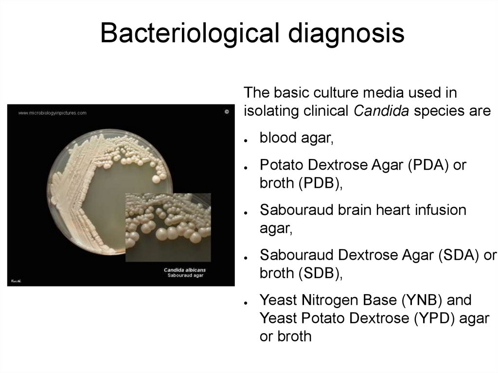
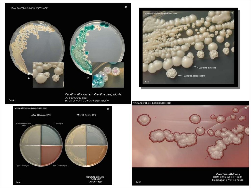
Bacteriological diagnosisThe basic culture media used in
isolating clinical Candida species are
blood agar,
Potato Dextrose Agar (PDA) or
broth (PDB),
Sabouraud brain heart infusion
agar,
Sabouraud Dextrose Agar (SDA) or
broth (SDB),
Yeast Nitrogen Base (YNB) and
Yeast Potato Dextrose (YPD) agar
or broth
5.
6.
7.
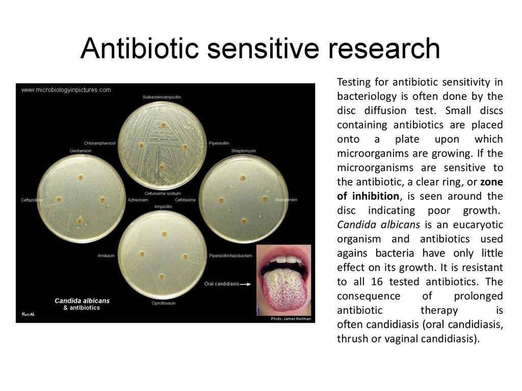
Antibiotic sensitive researchTesting for antibiotic sensitivity in
bacteriology is often done by the
disc diffusion test. Small discs
containing antibiotics are placed
onto a plate upon which
microorganims are growing. If the
microorganisms are sensitive to
the antibiotic, a clear ring, or zone
of inhibition, is seen around the
disc indicating poor growth.
Candida albicans is an eucaryotic
organism and antibiotics used
agains bacteria have only little
effect on its growth. It is resistant
to all 16 tested antibiotics. The
consequence
of
prolonged
antibiotic
therapy
is
often candidiasis (oral candidiasis,
thrush or vaginal candidiasis).
8.
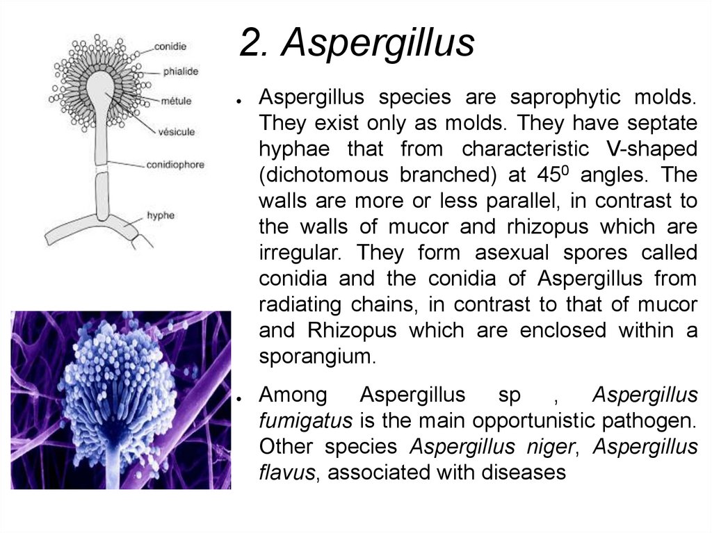
2. AspergillusAspergillus species are saprophytic molds.
They exist only as molds. They have septate
hyphae that from characteristic V-shaped
(dichotomous branched) at 450 angles. The
walls are more or less parallel, in contrast to
the walls of mucor and rhizopus which are
irregular. They form asexual spores called
conidia and the conidia of Aspergillus from
radiating chains, in contrast to that of mucor
and Rhizopus which are enclosed within a
sporangium.
Among
Aspergillus
sp ,
Aspergillus
fumigatus is the main opportunistic pathogen.
Other species Aspergillus niger, Aspergillus
flavus, associated with diseases
9.
Diseases caused by Aspergillus speciesis commonly called Aspergillosis.
Respiratory diseases
Aspergillus asthma. It is an allergic bronchopulmonary aspergillosis due to
hypersensitivity to Aspergillus antigen i.e. inhaled air-bone conidia. The fungus grows in
the lumen of bronchioles and produces plugs of mycelium and mucous that may block
the lumen.
Aspergilloma fungus ball. It is often called fungus ball in which fungus colonizes in the
pre-existing cavities, often in the case of TB cavity i.e. in aspergilloma inhaled conidia
germination in a pulmonary cavity and grows into fungus ball.
Disseminated (systemic) Aspergillosis
In this case, the fungus first establishes in the lungs tissues and then disseminates to
different organs such as a brain, kidney, heart and other organs particularly in
immunocompromised patients.
Superficial infections
Aspergillus flavus and Aspergillus fumigatus colonize paranasal sinuses(sinusitis),
external ear(otomycosis) and in some case eyes(mycotic keratitis)
10.
Lab diagnosisMaterial for research: Sputum and
biopsy materials are generally used as
preferred specimens.
×40
×100
Direct microscopy
Direct microscopy in KOH mount of
exudates shows on pigments septate
mycelium (3-5mm)in diameter of the
fungus with characteristic
branching(dichotomously branched)
at 450 angles. Gram`s staining may
give gram positive reaction. In a case
of a fluorescence microscope,
calcofluor white stain may be used for
direct observation of Aspergillus
species.
11.
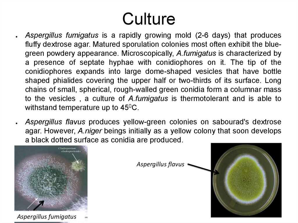
CultureAspergillus fumigatus is a rapidly growing mold (2-6 days) that produces
fluffy dextrose agar. Matured sporulation colonies most often exhibit the bluegreen powdery appearance. Microscopically, A.fumigatus is characterized by
a presence of septate hyphae with conidiophores on it. The tip of the
conidiophores expands into large dome-shaped vesicles that have bottle
shaped phialides covering the upper half or two-thirds of its surface. Long
chains of small, spherical, rough-walled green conidia form a columnar mass
to the vesicles , a culture of A.fumigatus is thermotolerant and is able to
withstand temperature up to 450C.
Aspergillus flavus produces yellow-green colonies on sabourad's dextrose
agar. However, A.niger beings initially as a yellow colony that soon develops
a black dotted surface as conidia are produced.
Aspergillus flavus
Aspergillus fumigatus
12.
Skin testIntradermal skin test to Aspergillus antigen extracts
is useful for patients suspected of allergic bronchial
pulmonary Aspergillosis.
13.
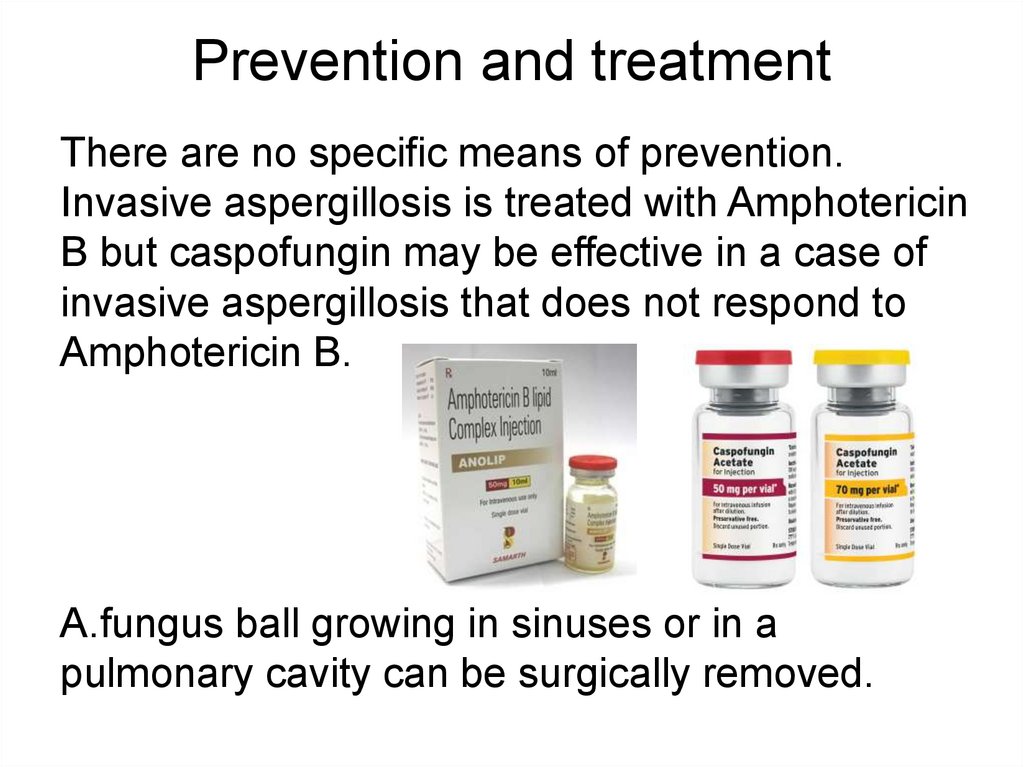
Prevention and treatmentThere are no specific means of prevention.
Invasive aspergillosis is treated with Amphotericin
B but caspofungin may be effective in a case of
invasive aspergillosis that does not respond to
Amphotericin B.
A.fungus ball growing in sinuses or in a
pulmonary cavity can be surgically removed.