


 История
Похожие презентации:


Pathological Anatomy Department
1.
International Medical FacultyPathological Anatomy Department
Head of the Department - PhD Kriventsov M.A.
2.
1. Introduction1. Pathological anatomy and anatomic pathologist
2. History
3. Tasks of the pathological anatomy
4. Biopsy, operational material, autopsy
2. Damage (alteration) Definition, factors, adaptation limits.
1. Morphology of the damage (alteration)
2. Definition, classification, mechanisms of degenerations
3. Parenchymal (intracellular) degenerations
4. Stromal vascular (extracellular) degenerations
5. Disorders of hemoglobin derived pigments (porphyria, jaundice, hemosiderosis)
6. Melanin
7. Calcinosis
3.
Pathological anatomy is the science that studiesthe structural bases of the disease at different
levels of morphological organization
Anatomic pathologist (pathomorphologist) is a
doctor who deals with the identification of disease
based on the normal structure of the human body
anatomy
4.
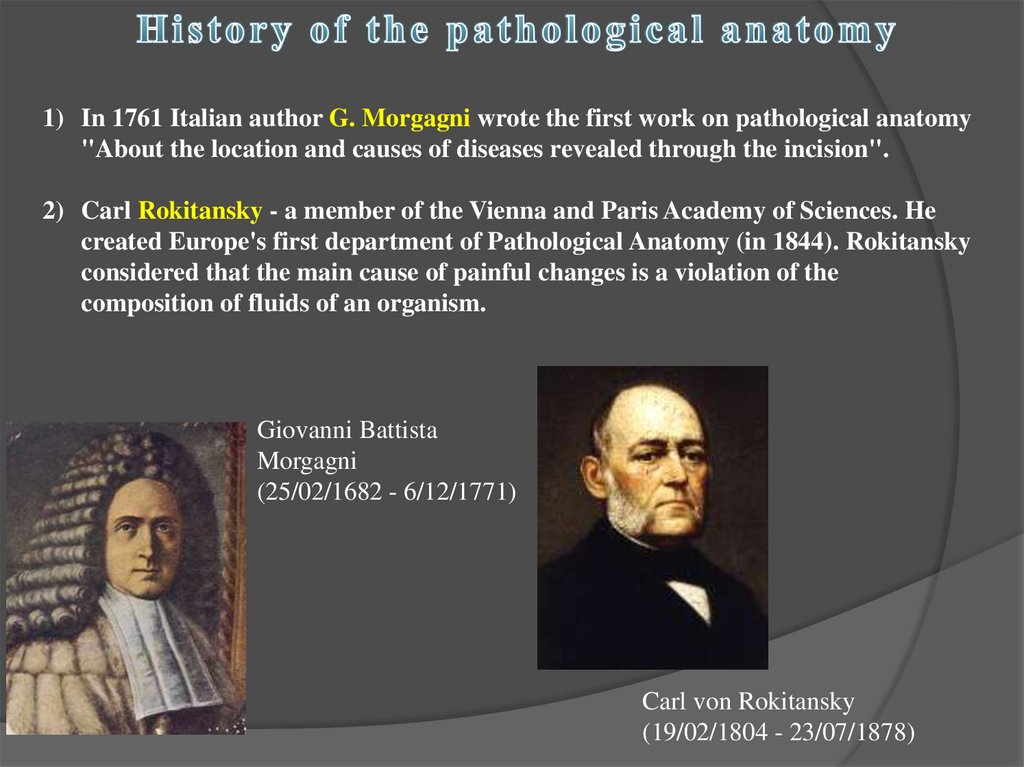
1) In 1761 Italian author G. Morgagni wrote the first work on pathological anatomy"About the location and causes of diseases revealed through the incision".
2) Carl Rokitansky - a member of the Vienna and Paris Academy of Sciences. He
created Europe's first department of Pathological Anatomy (in 1844). Rokitansky
considered that the main cause of painful changes is a violation of the
composition of fluids of an organism.
Giovanni Battista
Morgagni
(25/02/1682 - 6/12/1771)
Carl von Rokitansky
(19/02/1804 - 23/07/1878)
5.
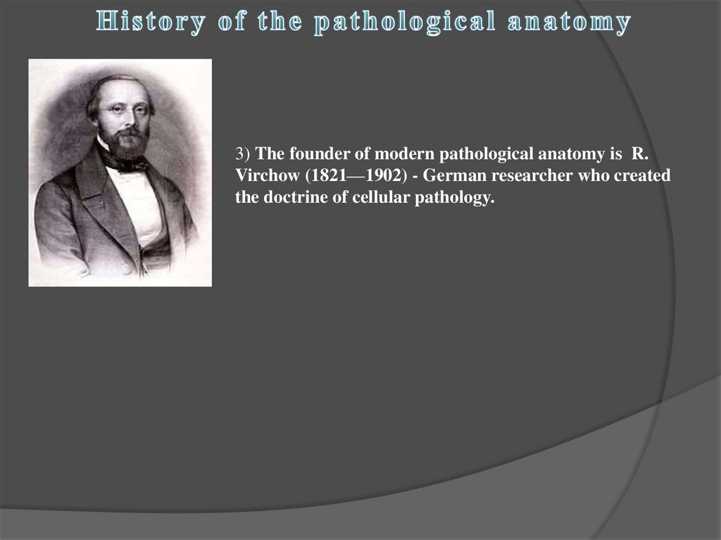
3) The founder of modern pathological anatomy is R.Virchow (1821—1902) - German researcher who created
the doctrine of cellular pathology.
6.
Theoretic tasks of the pathological anatomy:1) Study of the etiology, pathogenesis, morphology
and morphogenesis of the diseases;
2) Study of pathomorphism of the diseases (medical,
natural);
3) Study of outcomes and complications of the
diseases;
4) Study of the mechanism of death (tanatogenesis);
5) Evaluation of the functioning and state of
damaged organs.
7.
Practical tasks of the pathological anatomy:1) Control of accuracy and timeliness of clinical
diagnosis;
2) Training of the attending physician;
3) Establishing clinical diagnosis in vivo (during the
patient's life);
4) Monitoring the effectiveness of treatment
(repeated biopsy);
5) Statistical records.
8.
Approaches in the pathological anatomy1) Post mortal study (autopsy);
2) In vivo (during the life) study (biopsy, operational
material);
3) Experiment.
Methods of the pathological anatomy
•Macroscopic
•Microscopic (light microscope)
•Electron microscope
•Cytochemistry
•Histochemistry
•Immunohistochemistry (IHC)
9.
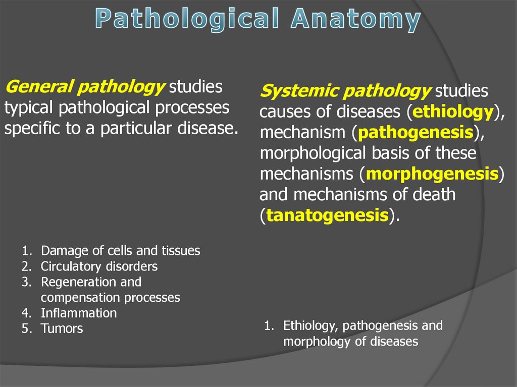
General pathology studiestypical pathological processes
specific to a particular disease.
1. Damage of cells and tissues
2. Circulatory disorders
3. Regeneration and
compensation processes
4. Inflammation
5. Tumors
Systemic pathology studies
causes of diseases (ethiology),
mechanism (pathogenesis),
morphological basis of these
mechanisms (morphogenesis)
and mechanisms of death
(tanatogenesis).
1. Ethiology, pathogenesis and
morphology of diseases
10.
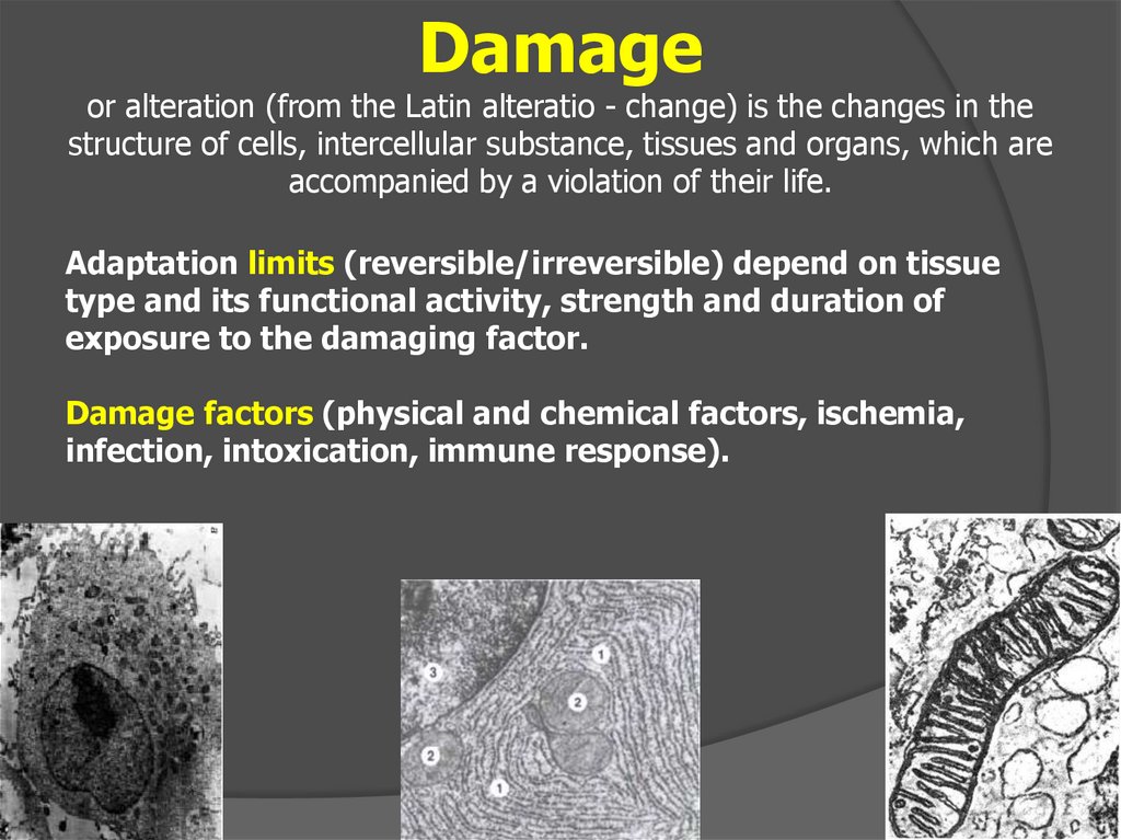
Damageor alteration (from the Latin alteratio - change) is the changes in the
structure of cells, intercellular substance, tissues and organs, which are
accompanied by a violation of their life.
Adaptation limits (reversible/irreversible) depend on tissue
type and its functional activity, strength and duration of
exposure to the damaging factor.
Damage factors (physical and chemical factors, ischemia,
infection, intoxication, immune response).
11.
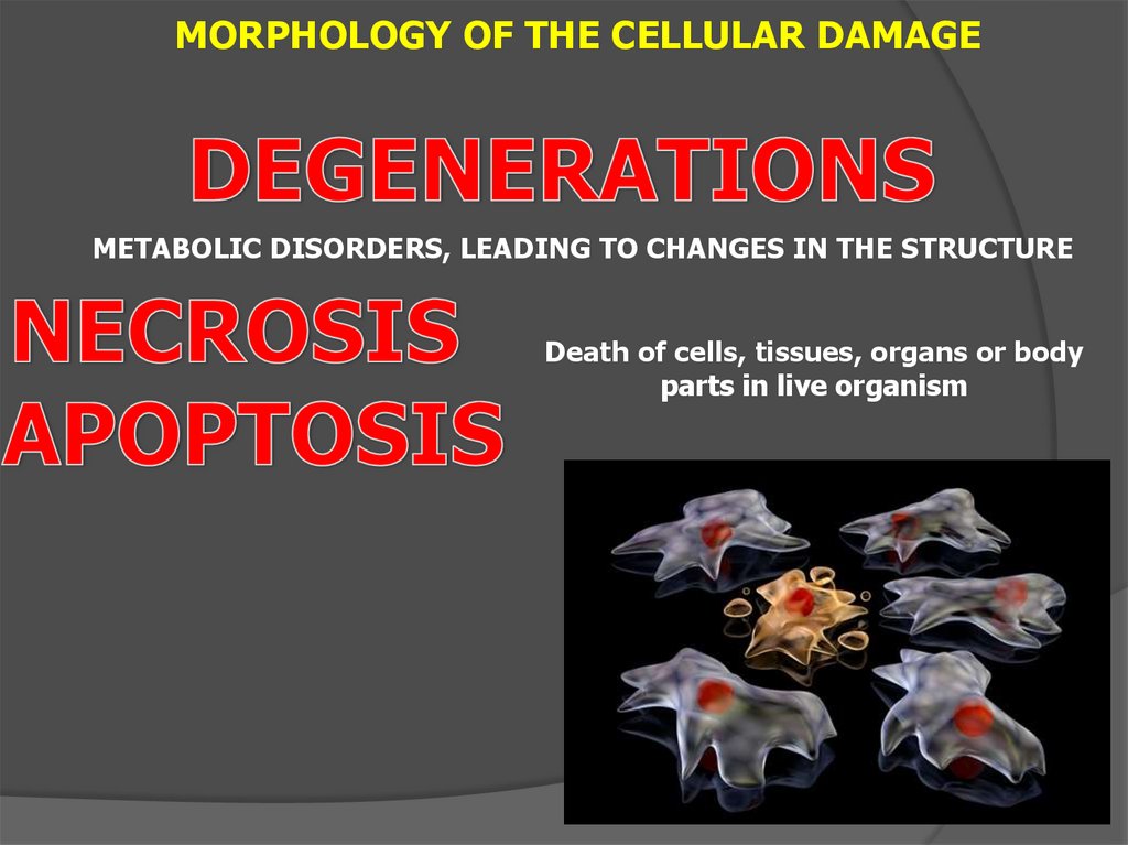
MORPHOLOGY OF THE CELLULAR DAMAGEMETABOLIC DISORDERS, LEADING TO CHANGES IN THE STRUCTURE
Death of cells, tissues, organs or body
parts in live organism
12.
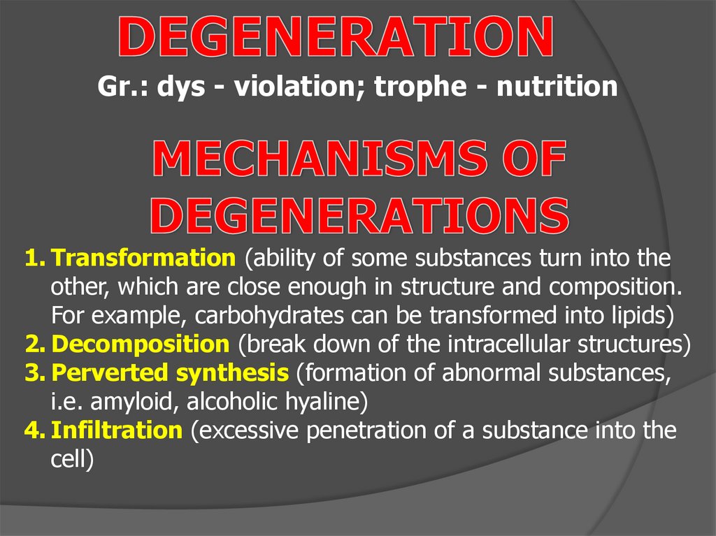
Gr.: dys - violation; trophe - nutrition1. Transformation (ability of some substances turn into the
other, which are close enough in structure and composition.
For example, carbohydrates can be transformed into lipids)
2. Decomposition (break down of the intracellular structures)
3. Perverted synthesis (formation of abnormal substances,
i.e. amyloid, alcoholic hyaline)
4. Infiltration (excessive penetration of a substance into the
cell)
13.
I. By localization1. Intracellular (parenchymal);
2. Extracellular (stromal vascular, mesenchymal);
3. Mixed
II. By extent
1. General (systemic).
2. Local.
III. By etiology
1. Acquired
2. Hereditary
IV. By type of metabolic disorders
1. Protein;
2. Lipid (fat);
3. Carbohydrate;
4. Minerals.
14.
1.2.
3.
4.
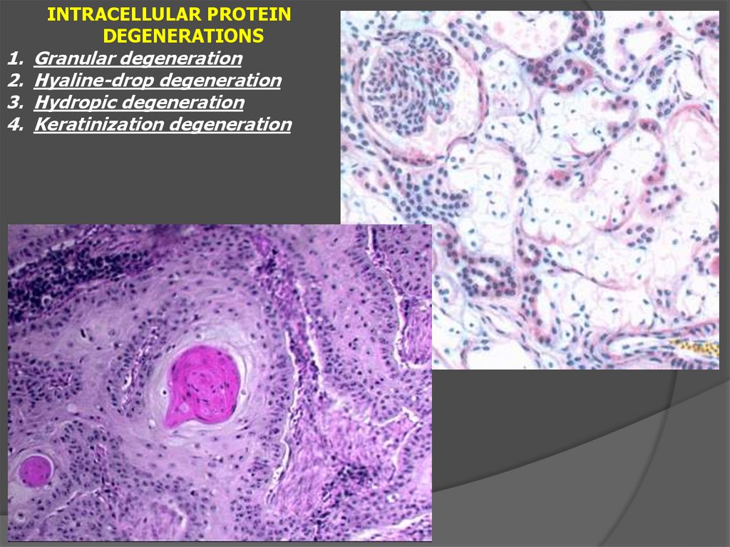
INTRACELLULAR PROTEIN
DEGENERATIONS
Granular degeneration
Hyaline-drop degeneration
Hydropic degeneration
Keratinization degeneration
15.
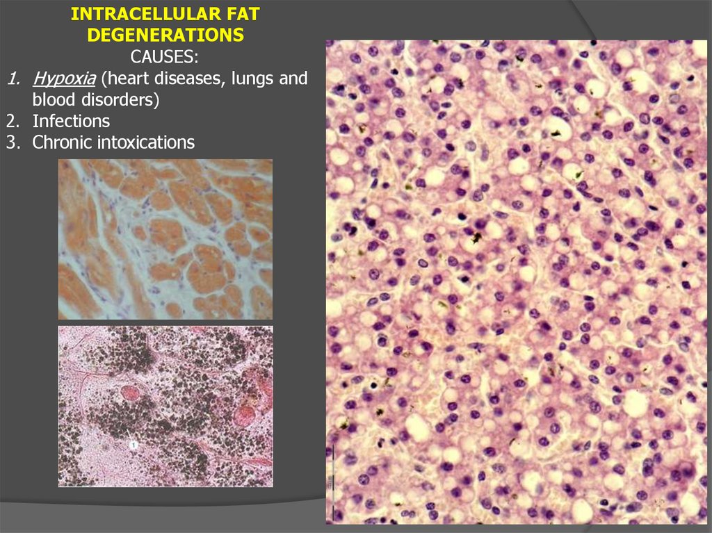
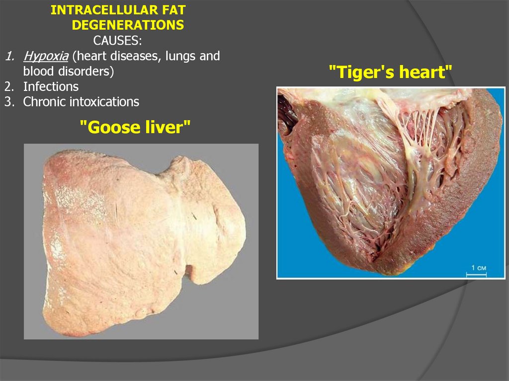
INTRACELLULAR FATDEGENERATIONS
CAUSES:
1. Hypoxia (heart diseases, lungs and
blood disorders)
2. Infections
3. Chronic intoxications
16.
INTRACELLULAR FATDEGENERATIONS
CAUSES:
1. Hypoxia (heart diseases, lungs and
blood disorders)
2. Infections
3. Chronic intoxications
"Goose liver"
"Tiger's heart"
17.
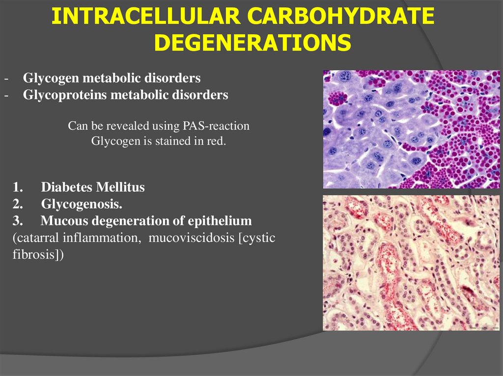
INTRACELLULAR CARBOHYDRATEDEGENERATIONS
-
Glycogen metabolic disorders
Glycoproteins metabolic disorders
Can be revealed using PAS-reaction
Glycogen is stained in red.
1. Diabetes Mellitus
2. Glycogenosis.
3. Mucous degeneration of epithelium
(catarral inflammation, mucoviscidosis [cystic
fibrosis])
18.
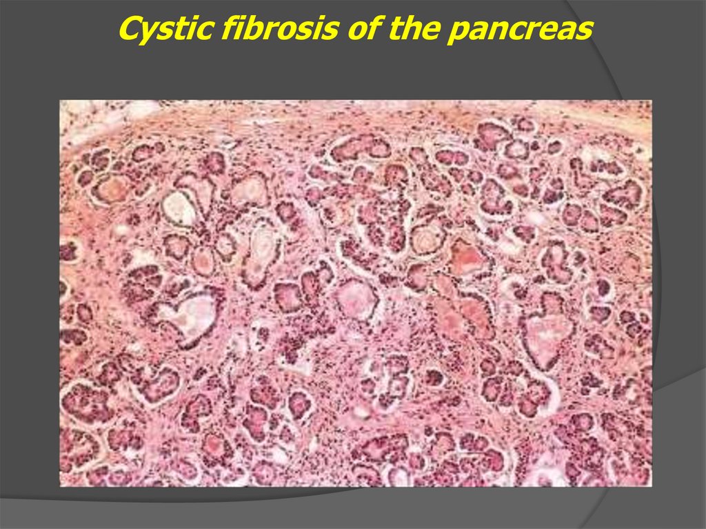
Cystic fibrosis of the pancreas19.
EXTRACELLULAR PROTEIN DEGENERATIONS- Mucoid swelling
- Fibrinoid swelling
- Hyalinosis
- Amyloidosis
20.
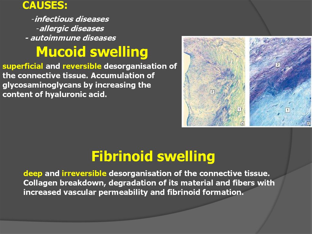
CAUSES:-infectious diseases
-allergic diseases
- autoimmune diseases
Mucoid swelling
superficial and reversible desorganisation of
the connective tissue. Accumulation of
glycosaminoglycans by increasing the
content of hyaluronic acid.
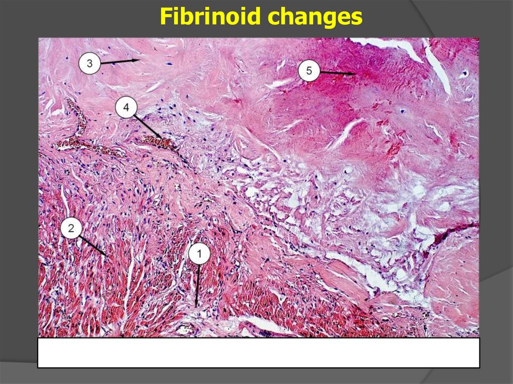
Fibrinoid swelling
deep and irreversible desorganisation of the connective tissue.
Collagen breakdown, degradation of its material and fibers with
increased vascular permeability and fibrinoid formation.
21.
Fibrinoid changes22.
HyalinosisDegradation of connective tissue is accompanied by
increased vascular permeability, degradation of collagen
fibers and precipitation of plasma proteins.
Hyaline is the substance of complex chemical composition
consisted of fibrin, immunoglobulins and proteins.
1. Simple hyaline
2. Complex hyaline
3. Lipohyaline
23.
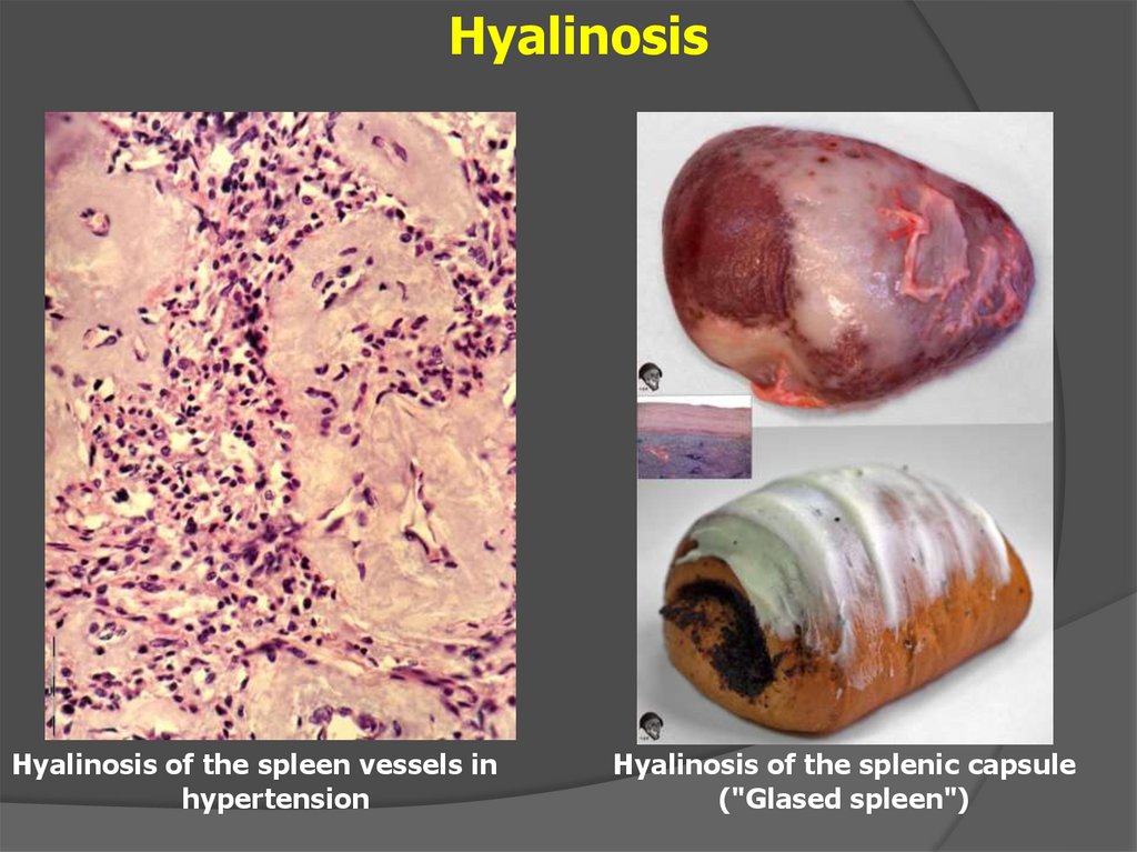
HyalinosisHyalinosis of the spleen vessels in
hypertension
Hyalinosis of the splenic capsule
("Glased spleen")
24.
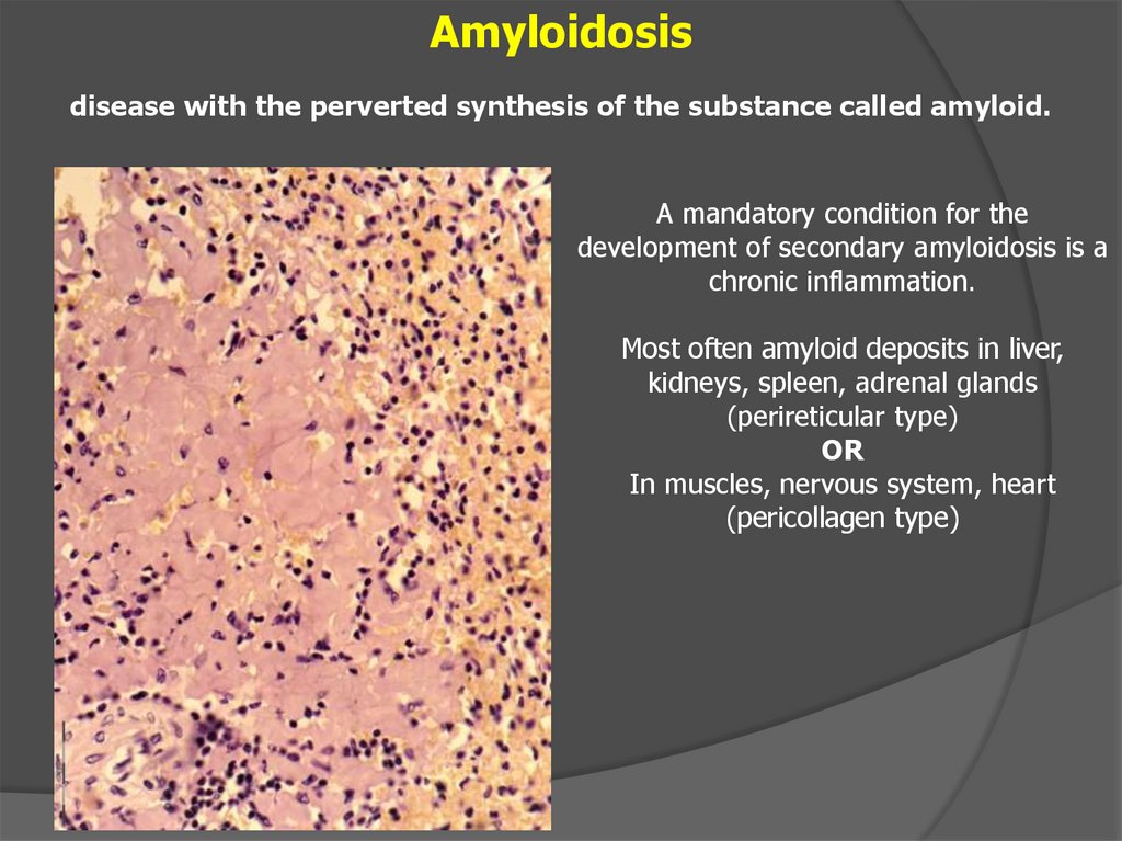
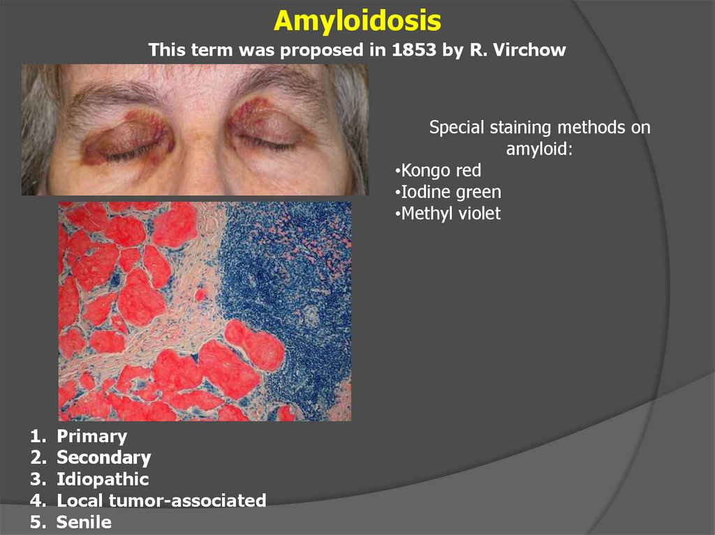
Amyloidosisdisease with the perverted synthesis of the substance called amyloid.
A mandatory condition for the
development of secondary amyloidosis is a
chronic inflammation.
Most often amyloid deposits in liver,
kidneys, spleen, adrenal glands
(perireticular type)
OR
In muscles, nervous system, heart
(pericollagen type)
25.
AmyloidosisThis term was proposed in 1853 by R. Virchow
Special staining methods on
amyloid:
•Kongo red
•Iodine green
•Methyl violet
1.
2.
3.
4.
5.
Primary
Secondary
Idiopathic
Local tumor-associated
Senile
26.
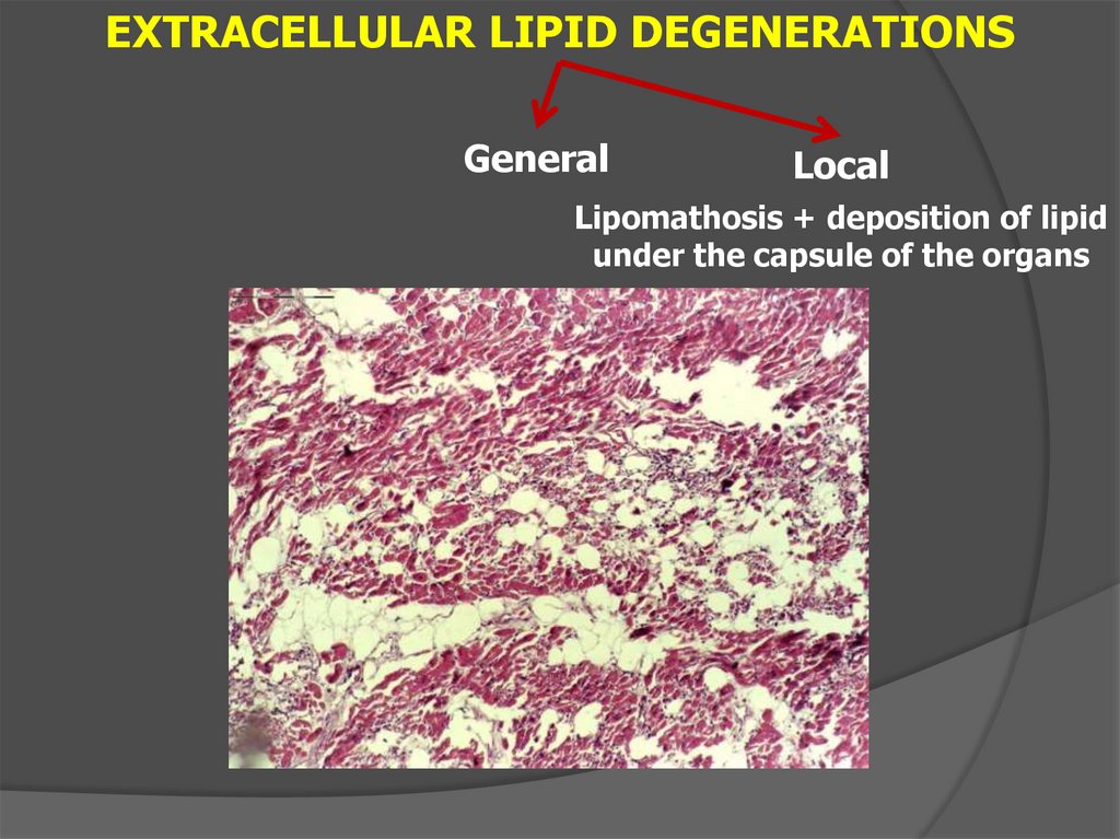
EXTRACELLULAR LIPID DEGENERATIONSGeneral
Local
Lipomathosis + deposition of lipid
under the capsule of the organs
27.
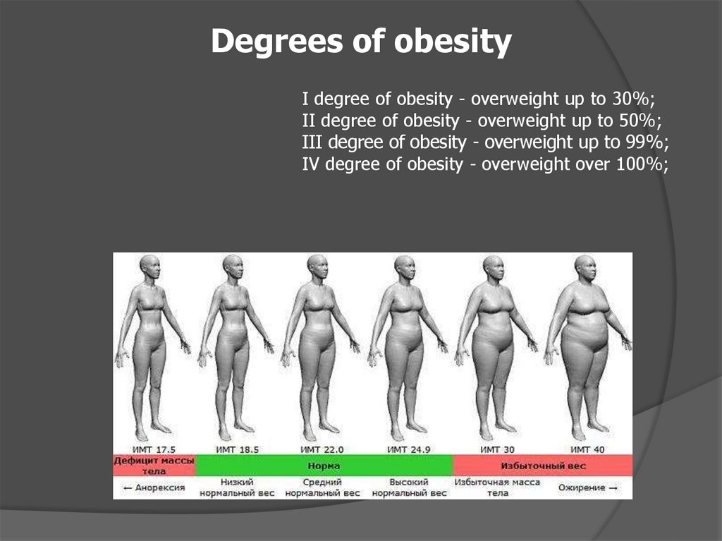
Degrees of obesityI degree of obesity - overweight up to 30%;
II degree of obesity - overweight up to 50%;
III degree of obesity - overweight up to 99%;
IV degree of obesity - overweight over 100%;
28.
Obesity1. Hyperplastic
2. Hypertrophic
3. Mixed
General
Symmetrical
Non symmetrical
Upper
Middle
Lower
29.
ATHEROSCLEROSISchronic disease characterized by abnormalities in
lipid and protein metabolism, which is manifested by
the deposition of lipid complexes in the vascular wall
30.
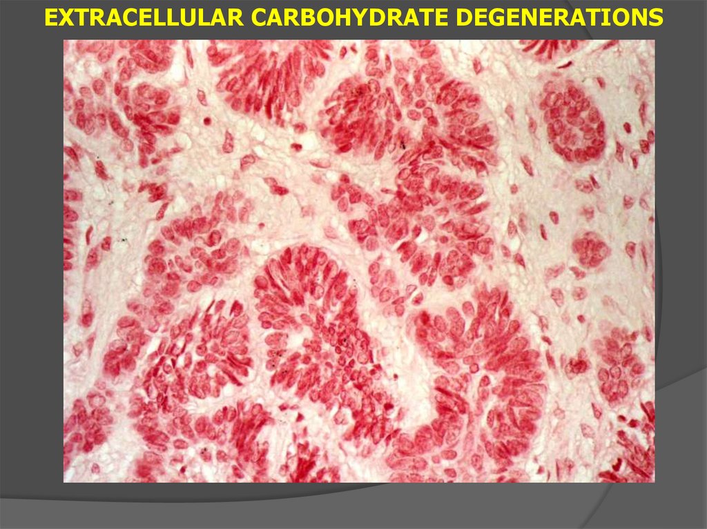
EXTRACELLULAR CARBOHYDRATE DEGENERATIONS31.
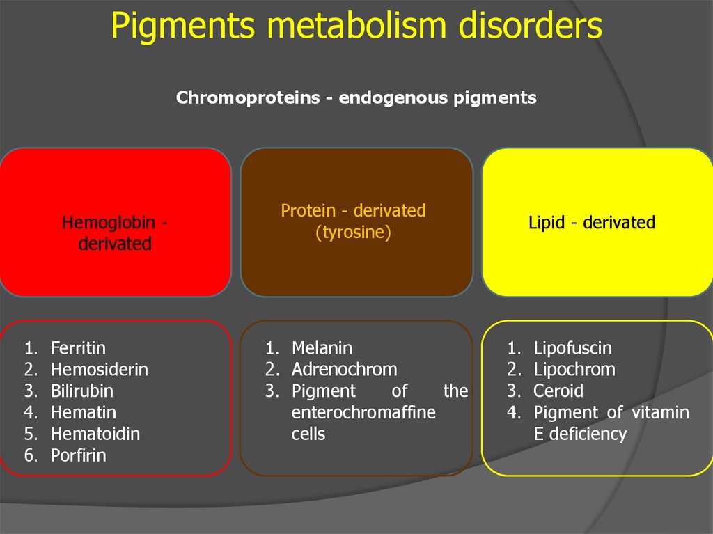
Pigments metabolism disordersChromoproteins - endogenous pigments
Hemoglobin derivated
1.
2.
3.
4.
5.
6.
Ferritin
Hemosiderin
Bilirubin
Hematin
Hematoidin
Porfirin
Protein - derivated
(tyrosine)
1. Melanin
2. Adrenochrom
3. Pigment
of
the
enterochromaffine
cells
Lipid - derivated
1.
2.
3.
4.
Lipofuscin
Lipochrom
Ceroid
Pigment of vitamin
E deficiency
32.
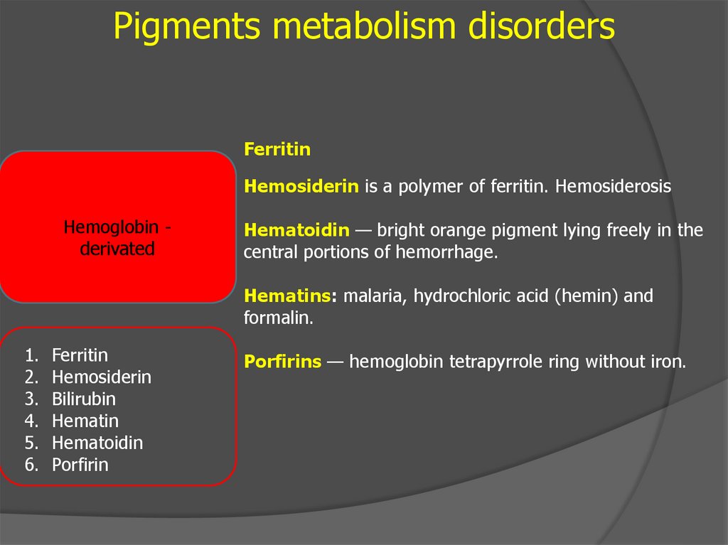
Pigments metabolism disordersFerritin
Hemosiderin is a polymer of ferritin. Hemosiderosis
Hemoglobin derivated
Hematoidin — bright orange pigment lying freely in the
central portions of hemorrhage.
Hematins: malaria, hydrochloric acid (hemin) and
formalin.
1.
2.
3.
4.
5.
6.
Ferritin
Hemosiderin
Bilirubin
Hematin
Hematoidin
Porfirin
Porfirins — hemoglobin tetrapyrrole ring without iron.
33.
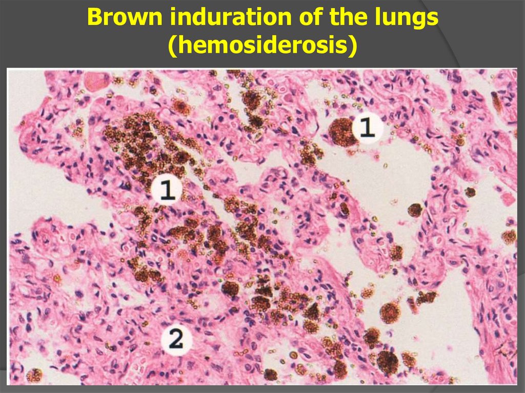
Brown induration of the lungs(hemosiderosis)
34.
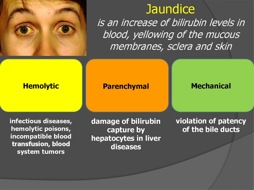
Jaundiceis an increase of bilirubin levels in
blood, yellowing of the mucous
membranes, sclera and skin
Hemolytic
Parenchymal
Mechanical
infectious diseases,
hemolytic poisons,
incompatible blood
transfusion, blood
system tumors
damage of bilirubin
capture by
hepatocytes in liver
diseases
violation of patency
of the bile ducts
35.
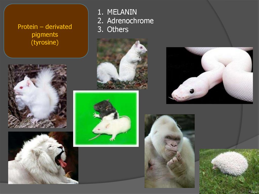
Protein – derivatedpigments
(tyrosine)
1. MELANIN
2. Adrenochrome
3. Others