


 История
Похожие презентации:


Bacteria – the causative agents of respiratory tract diseases
1. BACTERIA – THE CAUSATIVE AGENTS OF RESPIRATORY TRACT DISEASES
Department of Microbiology,Virology & Immunology
Ass. Prof. E. O. Kravtsova
2. DIPHTHERIA
• Is acute infectious disease caused byCorynebacterium diphtheriae and
characterized by a primary lesion, usually in
the upper respiratory tract, and more
generalized symptoms resulting from the
spread of bacterial toxin throughout the
body.
3. DIPHTHERIA
• The disease was first described in the 5thcentury BC by Hippocrates.
• The bacteria was discovered in 1882 by Edwin
Klebs.
• F.Loeffler- 1884 –isolate in pure culture
• E.Roux -1888- separate the toxin
• G.Ramon – 1923- diphtheria toxoid
4. Cor. diphtheriae
5. Cor. diphtheriae
• are Gram-positive, rod-shaped bacteria, stainsmore intensely at its ends (volutin).
They have the characteristic of forming irregular,
club-shaped or V-shaped arrangements in normal
growth. The cytoplasm is granular.
They do not form spores.
Outer layer of the cell wall forming a
microcapsula.
Nonmotile.
6. Neisser s stain
7.
C. diphtheriae is a gram-positive, non-motile rodsor somewhat pleomorphic organism. The clubshaped forms are long and slender with swollen
ends, especially when stained with methylene blue
or Neisser's stain (this reveals intensely stained
volutin granules). On a slide, these microbes often
arrange in pairs at acute angles to each other (V, L,
X-arrangements). The non-pathogenic and
opportunistic corynebacteria (diphtheroids) are
rods arranged in parallel (“fence-like”) clusters.
These bacteria possess irregular swellings only at
one end of the cell. NOTE
8. Physiology of Cor. diphtheriae
Aerobes or facultative anaerobes
T opt = 37º C
Grows readily on media with protein, sugar.
Serum agar
Blood agar
Roux's media (coagulated horse serum)
Loeffler's media (serum, sugar broth)
9.
C. diphtheriae grows much more readily oncoagulated serum agar, on whose slope there is a
creamy growth within 12 h. On blood tellurite agar
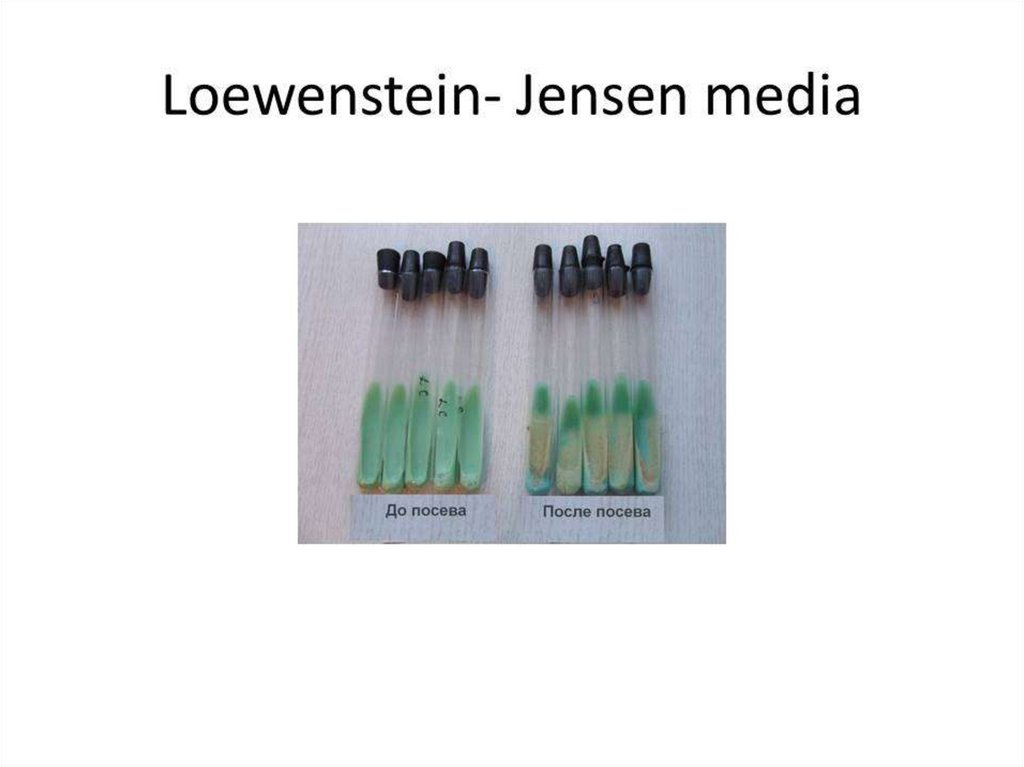
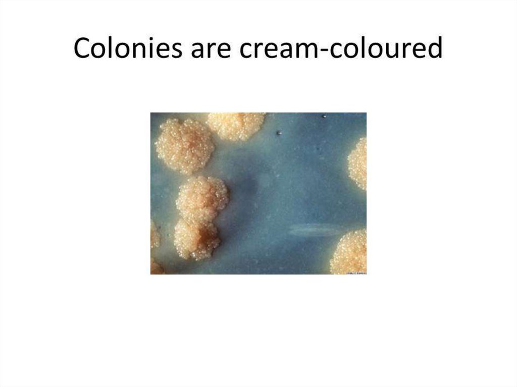
(Klauberg agar) the three biotypes of C. diphtheriae
- gravis, mitis, and intermedius - form different
colonies. The gravis type forms relatively large,
grayish flat, rough colonies with radial lines and
wavy edges. Colonies of the mitis type are small,
lustrous, black, with a smooth surface. The three
biotypes also differ biochemically. If typical colonies
are obtained, a pure culture is then identified by
fermentative and toxigenic properties.
10. Physiology of Cor. diphtheriae
Glucose «+»
Do not coagulate milk
Do not break down urea
Indol «-» H2S «+»or «-»
Reduce nitrates to nitrites
Potassium tellurite is also reduced
11. Biovars of Cor. diphtheriae
BiovarsGRAVIS
MITIS
Morphology
Short rods with granules
Long, curved rods with
granules
Colony on tellurite blood
agar
Large, rough, greyish with
crenated edges. Rosettelike.
Small, smooth, shiny, black
Growth in broth
Surface pellicle
Diffuse turbidity
Glycogen and starch
fermentation
Positive
Negative
Toxicity
Highly toxic
Less toxic
12. Tellurite agar
GravisMitis
13.
The gravis type form relatively large, grayish flat,rough colonies with radial lines and a wavy
edges.
Colonies of the mitis type are small, black, with
a smooth surface.
14. Antigens
• K – antigen , type specific, thermolabile,surface protein.
• O – antigen , group specific, thermostabile,
somatic polysaccharide.
• The diphtheria exotoxin is a complex of more
than 20 antigens.
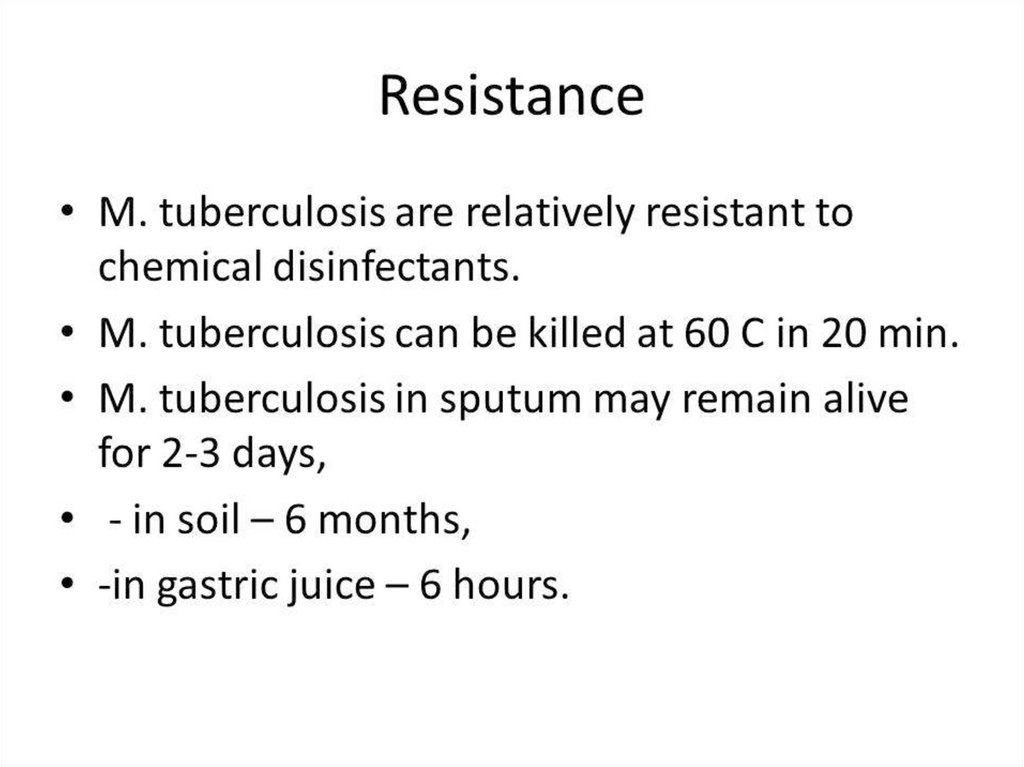
15. Resistance
• Cor. diphtheriae are relatively resistant toharmful environmental factors.
• They survive for 1 year on coagulated serum,
for 2 months at room t, for several days on
children's toys.
• Cor. diphtheriae are killed by t=60-100ºC and
by 1% phenol in 10 min.
16. Diphtheria toxin
Diphtheria toxin• Is a protein with 2 subunits:
• A – have enzymatic activity
• B- is responsible for binding the toxin to the cells
• Is complex of more than 20 antigens
• Is lable, destroyed easily by exposure to heat,
light, O2.
Diphtheria toxin can cause myocarditis,
polyneuritis, and other systemic toxic effects.
17. Diphtheria toxin
Diphtheria toxin• The toxigenicity of the Cor. diphtheriae depends
on the presence in it of corynephages (tox+),
which act as the genetic determinant controlling
toxin production.
• The diphtheria toxin acts by inhibiting protein
synthesis.
• The toxin causes local necrotic changes and the
resulting fibrinous exudate, together with the
disintegrating epithelial cells, leucocytes,
erythrocytes and bacteria.
18. Diphtheria toxin
19.
Virulence Factors and Pathogenesis of Diphtheria The serious effects ofdiphtheria in man are caused by the diphtheria exotoxin, and only
testing the organism for toxigenicity can definitively determine
whether it is pathogenic or not. The diphtheria exotoxin is a
polypeptide, which attaches to the cell membrane, allowing to enter
the cells, where it catalyses a reaction that stops the synthesis of the
polypeptide chains and the most profound effects are on the
myocardium, peripheral nerves, and kidneys. C diphtheriae may have
different portals of entry: they can colonize the pharynx (espessially
the tonsillar regions), the larynx, the nose, the conjunctiva and
occasionally the genital tract and the skin (wounds). After adhesion the
microbes multiply locally without invading deeper tissues or spreading
through the body. The microbe destroys epithelial cells with ulcer
formation. This is covered with a necrotic exudate forming a “false
membrane.”
20. DIPHTHERIA
• Sources of infection are patients and carriers.• The disease is transmitted by an air-droplet
route.
• Transmission by various objects (toys) and
foodstuffs (milk) contaminated with Cor.
diphtheriae is also possible.
21. Diphtheria. Clinical Manifestations
The typical membrane on the throat or on otherparts of the body, e.g., the skin, is the result of an
inflammatory reaction to the presence of
multiplying C.diphtheriae. This film soon becomes
dark and malodorous, and bleeding occurs on
attempting to remove it from tonsils surface. When
the larynx is involved, it can result in lifethreatening respiratory obstruction. The microbe
produces exotoxin that passes into the bloodstream
and the lymphatics. This toxemia causes severe
generalized effects: myocarditis, polyneuritis,
nephritis and suprarenal failure.
22. Clinical forms
• 1. Anterior nasal diphtheria, in which the membraneappears inside the nostrils. Almost no toxin is absorbed
from this site, so there is no danger to life and
complications are rare.
• 2. Tonsillar diphtheria - the most common type, in which
the infection is limited mostly to the tonsillar region. Most
patients recover if properly treated with diphtheria
antitoxin.
• 3. Nasopharyngeal diphtheria - the most often fatal form,
in which the tonsillar infection spreads to the nose and
throat structures, sometimes completely covering them
with the membrane and causing toxemia.
23. Tonsillar diphtheria
24. Clinical forms
• 4. Laryngeal diphtheria - usually resultingfrom the spread of the infection downward
from the nasopharynx to the larynx.
• 5. Extra-respiratory diphtheria, consisting of
those forms of the infection that affect parts
of the body other than the respiratory tract –
ex. the skin and wound.
25. Diphtheria. Microbiological Diagnosis
SPECIMENS. Swabs from the nose, throat, orother suspected lesions must be obtained.
26. Diphtheria. Microbiological Diagnosis
Bacterioscopical examinationPreparation of smears (Gram and Neisser
staining) • Gram-positive rods.
• The club-shaped long and slender cells with
swollen ends, cells arranged in pairs at acute
angles to each other (V, L, X-arrangements).
27. Diphtheria. Microbiological Diagnosis
Bacteriological examination1. Inoculation of blood- tellurite agar (Klauberg
medium)
2. Subculture on coagulated serum medium
3. Identification of pure culture and
differentiation from the diphtheroids
4. Determination of toxigenicity (IHA, ELISA,
precipitation test)
28. Diphtheria. Treatment and Prevention
Only diphtheria antitoxin can neutralize diphtheria exotoxin, and it cando so only before the toxin reaches and damages tissue cells.
Therefore, it must be given as soon as possible after C. diphtheriae
begins to multiply in a patient's throat, on clinical suspicion, and
before bacteriological confirmation. C. diphtheriae is nearly always
sensitive to penicillin or to erythromycin. These will rid the patients of
the organisms. Almost carriers can be cleared with antibiotics.
Active immunity.
The diphtheria toxoid is usually given, along with the tetanus and
pertussis vaccine, as DPT (diphtheria -pertussis-tetanus vaccine) to
infants between 3 and 6 months old. The usual course is three doses. A
booster dose of the diphtheria tetanus (DT) vaccine is given at school
entry. Passive immunity. Contacts of a patient may be protected by
antitoxin. This may be useful when there is a danger of cross-infection
in a ward from a missed case, or in home contacts of a patient.
29.
• Diphtheria toxoid (for stimulating antitoxinproduction)
• AB – tetracycline, erythromycin
• antitoxin
30.
• Pertussis-diphtheria vaccine• Diphtheria toxoid
• Pertussis-diphtheria-tetanus vaccine
31.
32.
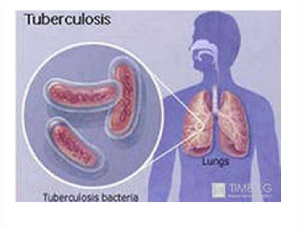
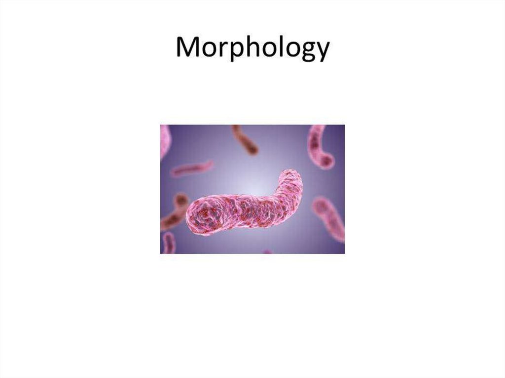
The genus Mycobacterium belongs to the familyMycobacteriaceae. Mycobacterium contains about
50 species that are normally environmental
saprophytes, although some species cause
opportunistic disease of animals and man. The
group of pathogenic mycobacteria includes
M.leprae that causes leprosy, and the tubercle
bacilli. There are three species of tubercle bacilli:
M.tuberculosis, the human tubercle bacillus; M.
bovis, the bovine tubercle bacillus; M. africanum, a
rather heterogeneous type found in Equatorial
Africa.
33.
34.
35.
36.
37.
38.
39.
40.
41.
42.
43.
44.
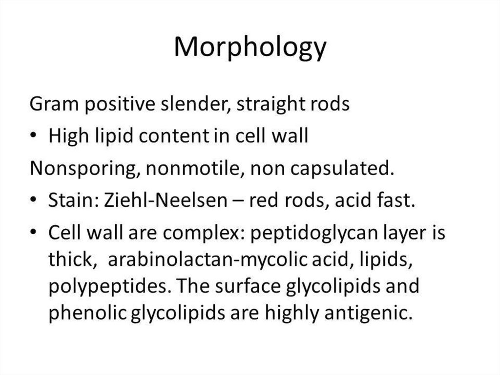
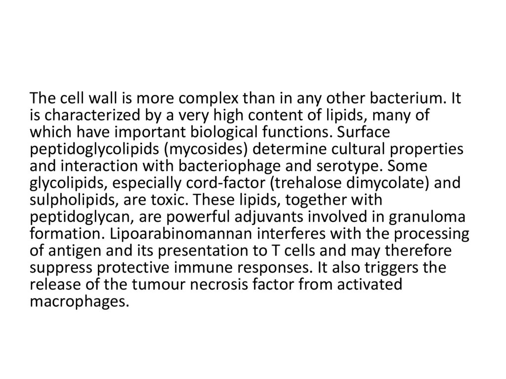
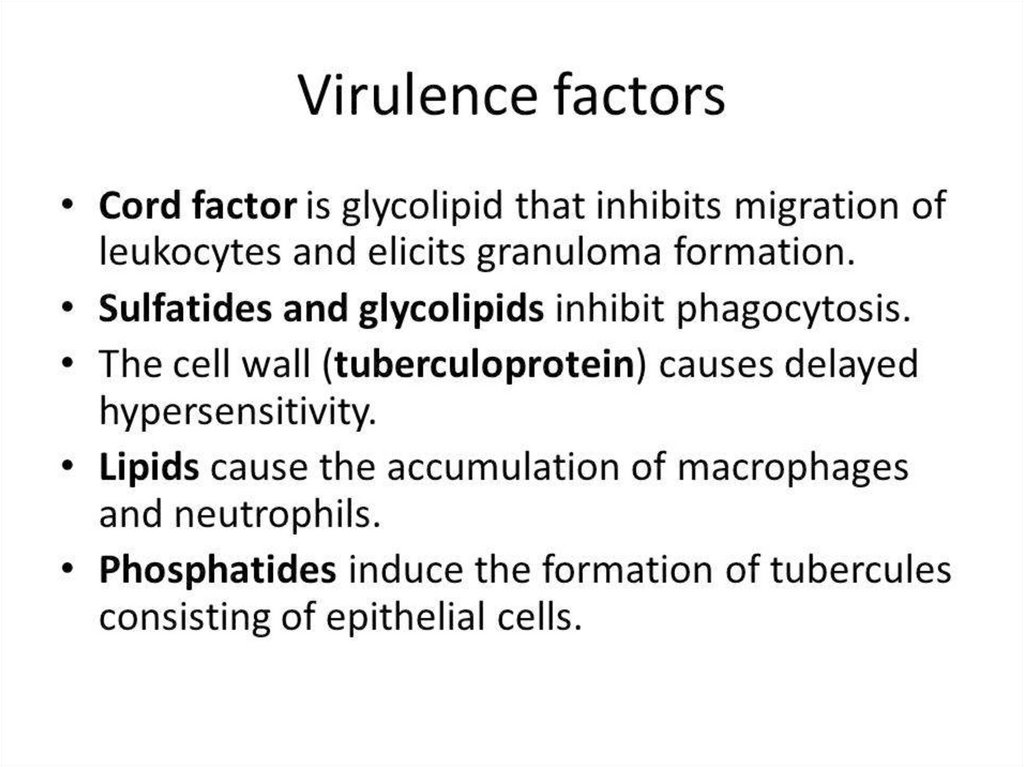
The cell wall is more complex than in any other bacterium. Itis characterized by a very high content of lipids, many of
which have important biological functions. Surface
peptidoglycolipids (mycosides) determine cultural properties
and interaction with bacteriophage and serotype. Some
glycolipids, especially cord-factor (trehalose dimycolate) and
sulpholipids, are toxic. These lipids, together with
peptidoglycan, are powerful adjuvants involved in granuloma
formation. Lipoarabinomannan interferes with the processing
of antigen and its presentation to T cells and may therefore
suppress protective immune responses. It also triggers the
release of the tumour necrosis factor from activated
macrophages.
45.
46.
47.
48.
49.
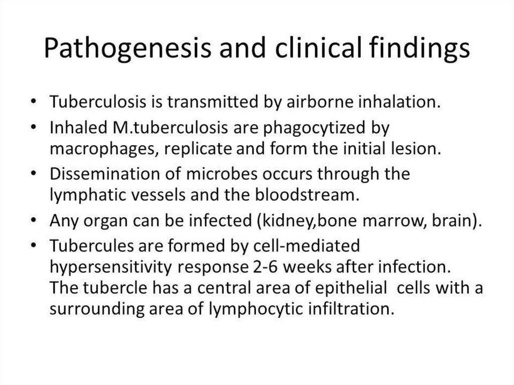
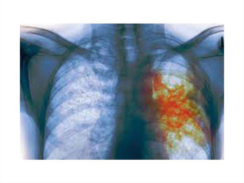
50. Tuberculosis. Clinical Manifestations
The initial lesion of tuberculosis occurs at the site ofimplantation of the bacillus (the lung, skin, or alimentary
tract). Bacteria are ingested by phagocytes, migrate to the
draining lymph nodes where secondary lesions develop.
The initial pulmonary lesion, the Ghon focus, together
with the lymphadenopathy, forms the primary complex .
The characteristic lesion of tuberculosis is the granuloma.
The primary complex often heals with calcification. Some
bacilli are disseminated through lymphatics and blood,
leading in some cases to non-respiratory tuberculosis:
lymph-node tuberculosis, genitourinary tuberculosis,
bone and joint disease, tuberculous meningitis,
abdominal tuberculosis, and tuberculous pericarditis.
51. Tuberculosis. Microbiological Diagnosis
SPECIMENS. Sputum should be collected intosterile wide-mouthed, screw-capped glass or
plastic pots. At least three sputum samples,
preferably early-morning samples, should be
collected. Bronchoscopy enables specimens to
be obtained from abnormal areas of the lung by
brushing, bronchoalveolar washing, and by
bronchial or transbronchial biopsy. In cases of
non-respiratory location of the tuberculosis
process, other specimens could be obtained.
52. Tuberculosis. Microbiological Diagnosis
PCR. The polymerase chain reaction should provide anextremely specific, sensitive, and rapid diagnosis.
Mycobacteria are detectable in clinical specimens by PCR and
by the demonstration of tuberculostearic acid by mass
spectroscopy. However, these techniques are not yet widely
available.
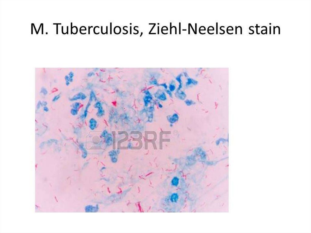
BACTERIOSCOPICAL EXAMINATION. After drying and heat
fixing, the smear is stained by the Ziehl-Neelsen method
(Acid-Fast staining). Smears may be stained and examined by
fluorescence microscopy. Both methods depend on the acidfastness of mycobacteria. Microscopy is also used to detect
acid-fast bacilli in the urine, pleural, peritoneal, and
cerebrospinal fluid after centrifugation, and in homogenates
or histological sections of tissue.
53. Tuberculosis. Microbiological Diagnosis
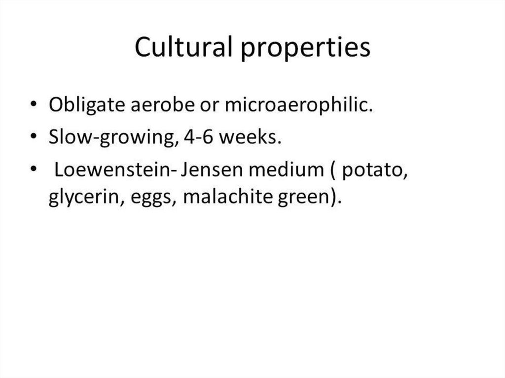
BACTERIOLOGICAL EXAMINATION. As tubercle bacilli grow very slowly,they are readily overgrown by fungi or other bacteria in the specimen.
This may be avoided by treating the specimen with an agent, usually
an acid or alkali that will preferentially kill organisms other than
mycobacteria. The Lowenstein-Jensen medium is the most widely
used. It is solid and contains eggs, glycerol, and mineral salts.
Specimens are incubated at 35oC-40oC for 4-8 weeks. The following
tests allow division of the tubercle bacilli into individual species:
reduction of nitrate to nitrite (nitratase test), oxygen preference
(aerobic strains grow on the surface of semisolid agar media, while
micro-aerophilic strains form a band deep in the medium),
susceptibility to different chemicals. On glycerol-containing media,
colonies of M. tuberculosis are usually large and heaped up and
produce nicotinamide while those of M. bovis are small and flat.
54. Tuberculosis. Microbiological Diagnosis
PRICE METHOD. This is the rapid technique of the bacteriologicaldiagnosis of tuberculosis. Samples of sputum, pus, urine, lavage
waters, etc. are spread in a thick layer on the sterile slide glass. After
the preparation is dried and manipulated with sulfuric acid, it is placed
into a vial with citrate blood. After 3 to 4 days of incubation, the
preparation is retrieved, fixed, and then stained with the Ziehl-Neelsen
stain. Microcolonies in the preparation appear as rope-like
microcolonies termed cords. Cord formation is regarded as a marker of
the presence of a specific “cord-factor” in the microcapsule of the
pathogen.
BIOLOGICAL EXAMINATION. M. tuberculosis causes disease in a wide
range of mammals. This microbe is virulent for the guinea-pig. M. bovis
causes infection in cattle and badgers and, less frequently, in deer and
other wild or feral mammals. Experimentally, M. bovis is highly virulent
to rabbits.
55. Tuberculosis. Microbiological Diagnosis
ALLERGIC SKIN TEST (the Mantoux test). In the Mantouxtest, tuberculin, the solution containing a known number
of international units of mycobacterial antigen - purified
protein derivative (PPD) - is injected intradermally. The
diameter of the induration is read 48 to 72 h later. The
induration of up to 10 mm-15 mm is usually regarded as
positive and may indicate previous exposure to
mycobacterial antigens through infection with one of the
tubercle bacilli or to BCG vaccination. (That is because of
type IV hypersensitivity which develops three to eight
weeks after the primary infection). Tuberculin reactivity
does not correlate with protective immunity.
56. Tuberculosis. Microbiological Diagnosis
SEROLOGICAL EXAMINATION. The immuneresponse in tuberculosis is predominantly cellmediated, and detection of antibody rising is not
of great importance in diagnosis. Thus,
tuberculin conversion usually occurs 3 to 8
weeks from the time of infection.
57.
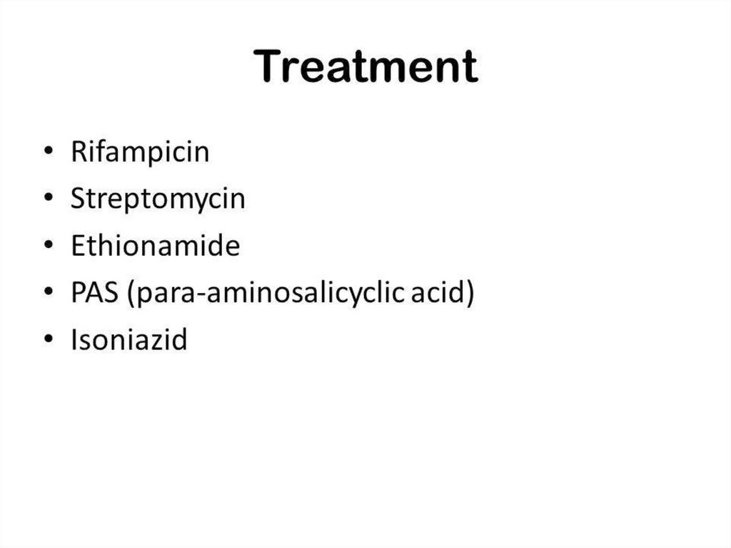
The three key first-line drugs used for previouslyuntreated patients are isoniazid, rifampicin, and
pyrazinamide. Ethambutol and streptomycin are
valuable additional drugs. Reserve drugs, which
may be used when first-line treatment has
failed, are ethionamide or prothionamide,
kanamycin, etc. All M. tuberculosis strains
contain drug-resistant mutants.
58.
59.
60.
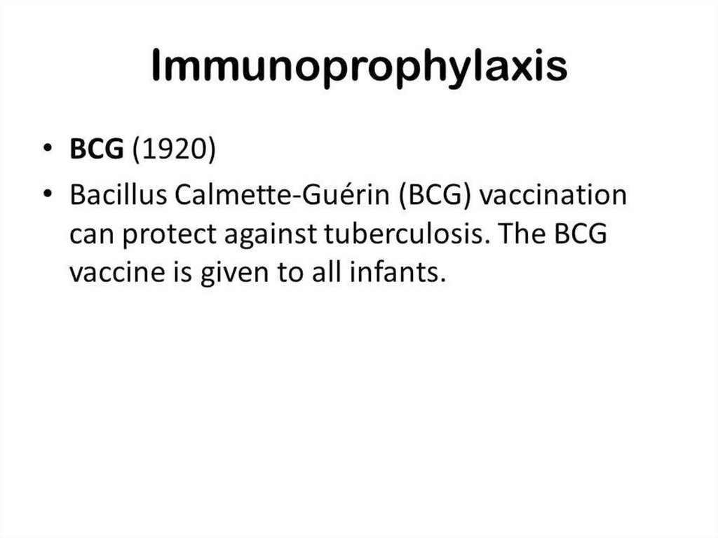
Mass BCG vaccination of neonates is valuable.The vaccine strain, Bacillus Calmette-Guerrin
(BCG), was supposedly derived from a strain of
M. bovis. It is now prepared as a freeze-dried
live vaccine for intradermal injection. The
booster vaccination should be performed only in
those negative on tuberculin testing (the
Mantoux test).