


 Медицина
Похожие презентации:


Мeningeal syndrome in clinic of infectious diseases. Meningococcal infection
1.
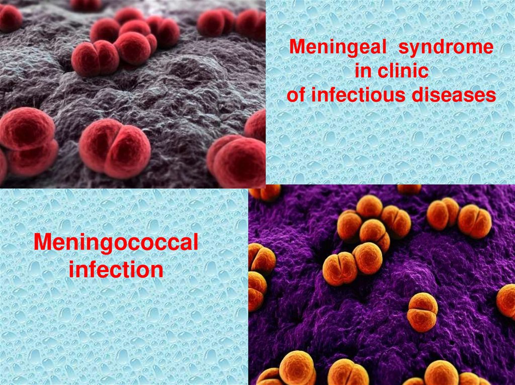
Мeningeal syndromein clinic
of infectious diseases
Meningococcal
infection
2.
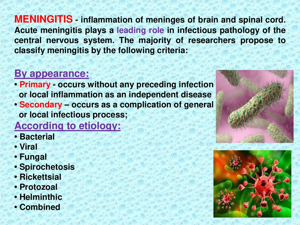
MENINGITIS - inflammation of meninges of brain and spinal cord.Acute meningitis plays a leading role in infectious pathology of the
central nervous system. The majority of researchers propose to
classify meningitis by the following criteria:
By appearance:
• Primary - occurs without any preceding infection
or local inflammation as an independent disease
• Secondary – occurs as a complication of general
or local infectious process;
According to etiology:
• Bacterial
• Viral
• Fungal
• Spirochetosis
• Rickettsial
• Protozoal
• Helminthic
• Combined
3.
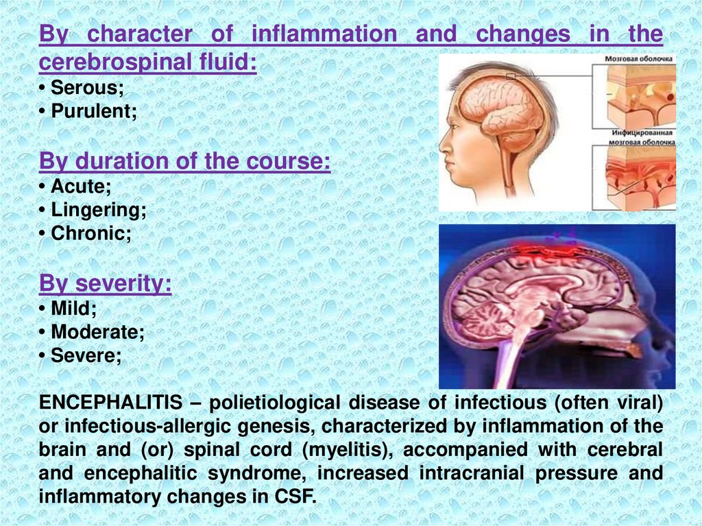
By character of inflammation and changes in thecerebrospinal fluid:
• Serous;
• Purulent;
By duration of the course:
• Acute;
• Lingering;
• Chronic;
By severity:
• Mild;
• Moderate;
• Severe;
ENCEPHALITIS – polietiological disease of infectious (often viral)
or infectious-allergic genesis, characterized by inflammation of the
brain and (or) spinal cord (myelitis), accompanied with cerebral
and encephalitic syndrome, increased intracranial pressure and
inflammatory changes in CSF.
4.
PATHOGENESISThe entrance gates for causative agent can be:
- nasopharynx,
- respiratory tract,
- intestine,
- primary focal inflammation;
Dissemination of the pathogen occurs by:
- hematogenous,
- lymphogenous,
- contact way.
3 pathogenetic factors play an important role
in the development of the disease:
- bacteremia,
- toxemia,
- macroorganism (initial immunity)
5.
- Destruction of bacteria,- releasing of endotoxins,
- development of toxemia,
- increased levels of biologically active substances,
-products of disturbed metabolism
Leads to
- increased permeability of cellular and vascular membranes,
blood-brain barrier;
- active penetration of bacteria and toxins in the CNS
with development of the pathological process.
- irritation of the vascular plexus of brain ventricles
(by bacteria, antigens, toxins and immune complexes);
- increase of cerebrospinal fluid production and intracranial
pressure.
6.
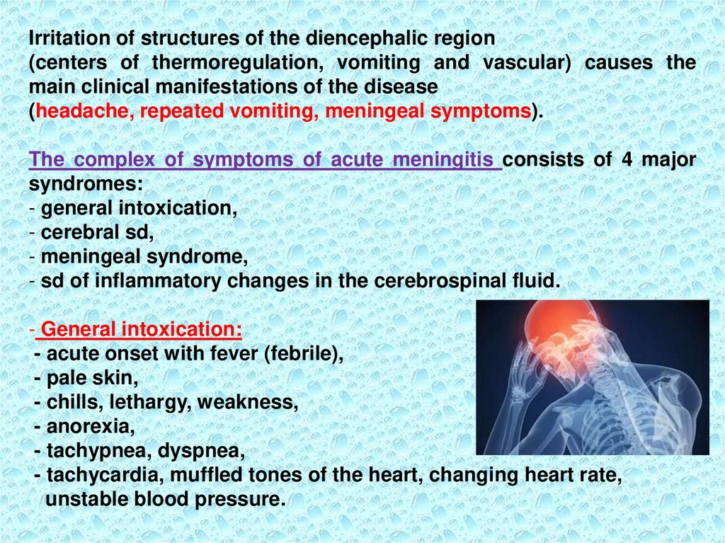
Irritation of structures of the diencephalic region(centers of thermoregulation, vomiting and vascular) causes the
main clinical manifestations of the disease
(headache, repeated vomiting, meningeal symptoms).
The complex of symptoms of acute meningitis consists of 4 major
syndromes:
- general intoxication,
- cerebral sd,
- meningeal syndrome,
- sd of inflammatory changes in the cerebrospinal fluid.
- General intoxication:
- acute onset with fever (febrile),
- pale skin,
- chills, lethargy, weakness,
- anorexia,
- tachypnea, dyspnea,
- tachycardia, muffled tones of the heart, changing heart rate,
unstable blood pressure.
7.
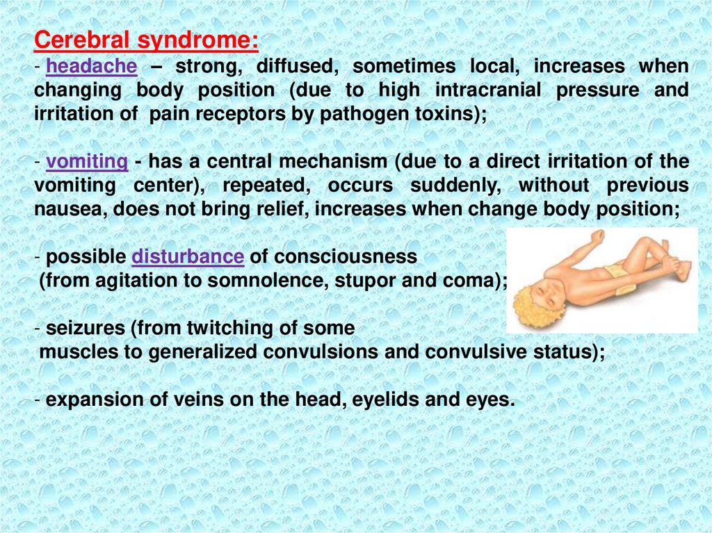
Cerebral syndrome:- headache – strong, diffused, sometimes local, increases when
changing body position (due to high intracranial pressure and
irritation of pain receptors by pathogen toxins);
- vomiting - has a central mechanism (due to a direct irritation of the
vomiting center), repeated, occurs suddenly, without previous
nausea, does not bring relief, increases when change body position;
- possible disturbance of consciousness
(from agitation to somnolence, stupor and coma);
- seizures (from twitching of some
muscles to generalized convulsions and convulsive status);
- expansion of veins on the head, eyelids and eyes.
8.
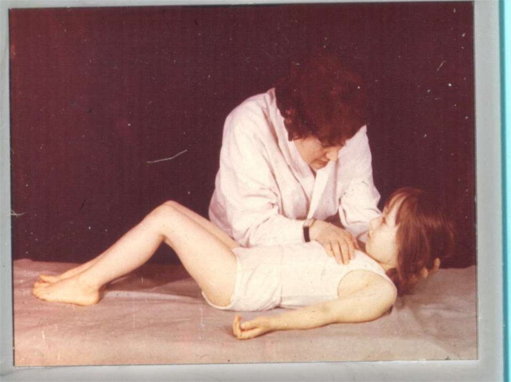
Meningeal syndrome - develops due to irritation ofcerebral
baroreceptors by high liquor pressure or viral/bacterial toxins.
Several clinical signs facilitate the diagnosis of meningitis
There are 4 main groups of symptoms:
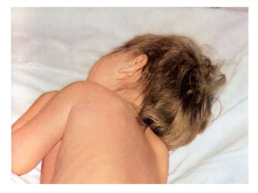
1-st group - the symptoms of tonic muscle tension:
- rigidity of occipital muscles - passive flexing the patient’s neck
(chin to chest) is difficult or impossible
(the severity is estimated by the distance
between the head and surface);
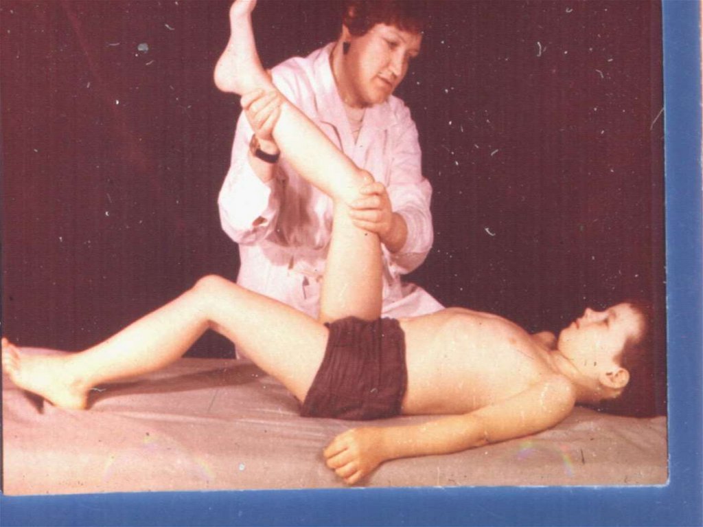
- Kernig‘s-sign - flexing the patient’s hip
90 degrees then extending the patient’s knee
causes pain;
- Brudzinski’s-sign - flexing the patient’s neck
causes flexion of the patient’s hips and knees;
Both of these signs are thought to be caused
by the irritation of motor nerve roots passing through
inflamed meninges as the roots are brought under
tension.
9.
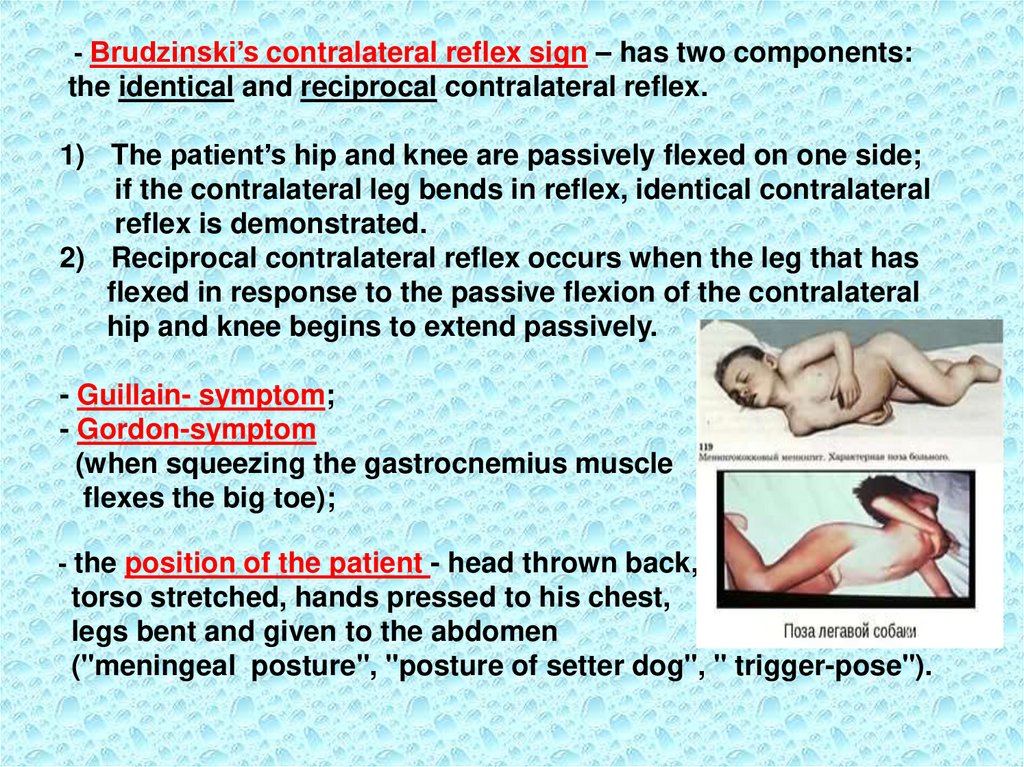
- Brudzinski’s contralateral reflex sign – has two components:the identical and reciprocal contralateral reflex.
1) The patient’s hip and knee are passively flexed on one side;
if the contralateral leg bends in reflex, identical contralateral
reflex is demonstrated.
2) Reciprocal contralateral reflex occurs when the leg that has
flexed in response to the passive flexion of the contralateral
hip and knee begins to extend passively.
- Guillain- symptom;
- Gordon-symptom
(when squeezing the gastrocnemius muscle
flexes the big toe);
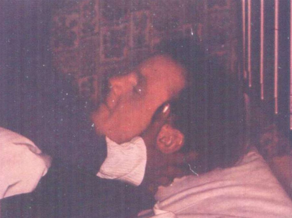
- the position of the patient - head thrown back,
torso stretched, hands pressed to his chest,
legs bent and given to the abdomen
("meningeal posture", "posture of setter dog", " trigger-pose").
10.
2-d group – symptoms of general hyperesthesia and hypersensitivityof the sensory organs (photophobia, hyperacusia) due to irritation of
the posterior roots, cells of the spinal nodes, receptors of meninges,
and dicreased level of sensitivity to various stimuli.
3-d group - reactive pain phenomena:
- Bekchterev-symptom;
- Lobzin-symptom (painful grimace when pressing on the eyeballs
through closed eyelids);
- Kerer‘s symptom (pain in points of the trigeminal nerve);
- Pulatov-symptom (tenderness to percussion of the skull);
- Mendel's symptom (pain when pressure on the frontal wall of the
external ear canal).
4-th group – changed dermal, pariosteal and tendon reflexes:
- Babinsky-reflex;
- Oppenheim-reflex;
- Gordon's reflex (pinching the gastrocnemius muscle);
- Sheffer’s reflex (compression of the Achilles tendon);
- Rossolimo-reflex.
11.
Syndrome of inflammatory changes in the cerebrospinal fluidThe investigation of CSF can establish the diagnosis of
meningitis, its character, intensity and dynamics of the process, the
effectiveness of treatment, course of illness and recovery.
Etiologic diagnosis of CNS infections is very important,
because the process is temporary and its effects can be dangerous
for the patient’s life.
Many meningitis may be treatable due to early diagnostics and
adequate treatment.
Leading role in the diagnosis of meningitis belongs to investigation of
cerebrospinal fluid.
There are 3 types of liquor - A, B,C
according to the pathological changes
CSF can be purulent, serous, serous-fibrinous, hemorrhagic and
xanthochromic.
12.
Inflammation of meninges is accompanied by thefollowing changes of CSF:
• Increased pressure - cerebrospinal fluid is flowing
streamly or by frequent drops.
• Change in transparency (cloudy) or color (white,
yellow-green etc.) depending on the etiology of the
process.
• Pleocytosis with a predominance of neutrophils,
lymphocytes, or mixed.
• Increase the protein content.
• Change in the level of glucose
and chlorides.
13.
Type A – turbid, mononuclear cells < 50, polymorphic( 90%) - 1000 –10000/l, protein - 0.5 – 2 g/l, glucose < ½ of glucose in blood.
Characteristic for:
a) bacterial meningitis caused by: Мeningicoccus, Haemophilus,
Streptococcus pneumonia, Listeria;
b) perforated abscess of the brain;
C) amoebic meningoencephalitis;
Type B – slightly turbid or transparent, sticky, mononuclear cells - 100
– 300/l, polymorphic 0 – 200/l , protein 0.5 – 3 g/l, glucose < 1/2 of the
blood glucose;
Typical for:
granulomatous meningitis – tuberculous, mycotic;
Type C - transparent or slightly turbid, mononuclear cells - 10 – 1001000/l, polymorphic - 0, protein - 0.4 - 0.8 g/l, glucose >1/2 of the blood
glucose;
Characteristic for:
a) parameningeal infection;
b) toxic encephalopathy;
c) viral infection;
14.
Differential-diagnostic signs of meningitis by CSFSIGN
NORMAL
MENINGISM
COLORLESS
TRANSPARENT
COLORLESS
TRANSPARENT
PRESSURE
(DROPS/MIN)
40-60
60-90
CYTOSIS (IN MKL)
2-10
2-12
L=80-85
N=3-5
L=80-85
N=3-5
2,2-3,3
2,2-3,3
0,22-0,33
0,22-0,45
-
-
HEADACHES
RELIEF
COLOR
TRANSPARENCY
CYTOGRAM (%)
RATIO OF LYMPHOCYTES AND
NEUTROPHILS
GLUCOSE (MMOL/L)
PROTEIN (G/L)
FIBRIN FILM
REACTION TO
PUNCTURE
15.
Differential-diagnostic signs of meningitis by CSFSIGN
SEROUS-VIRAL
SEROUS-BACTERIAL
COLOR
TRANSPARENCY
COLORLESS ,TRANSPARENT,
OPALESCENT
COLORLESS, OPALESCENT,
XANTHOCHROMIC
PRESSURE
(DROPS/MIN)
80-120
STREAM
CYTOSIS (IN MKL)
20-800
200-700
CYTOGRAM (%)
L=80-100
N=0-20
L=40-60
N=20-50
GLUCOSE (MMOL/L)
0,55-0,66
DICREASED ON THE 2-d
WEEK OF DISEASE
PROTEIN (G/L)
0,16-1,0
1,0-3,3
FIBRIN FILM
IN 3-5%
IN 30-40%
REACTION TO
PUNCTURE
EXPRESSED RELIEF, OFTEN
THE TURNING POINT OF THE
DISEASE
EXPRESSED SHORTTERM RELIEF
RATIO OF LYMPHOCYTES AND
NEUTROPHILS
16.
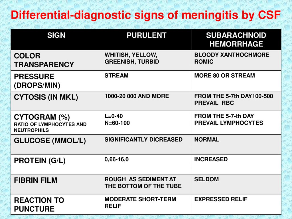
Differential-diagnostic signs of meningitis by CSFSIGN
PURULENT
SUBARACHNOID
HEMORRHAGE
COLOR
TRANSPARENCY
WHITISH, YELLOW,
GREENISH, TURBID
BLOODY XANTHOCHMORE
ROMIC
PRESSURE
(DROPS/MIN)
STREAM
MORE 80 OR STREAM
CYTOSIS (IN MKL)
1000-20 000 AND MORE
FROM THE 5-7th DAY100-500
PREVAIL RBC
CYTOGRAM (%)
L=0-40
N=60-100
FROM THE 5-7-th DAY
PREVAIL LYMPHOCYTES
GLUCOSE (MMOL/L)
SIGNIFICANTLY DICREASED
NORMAL
PROTEIN (G/L)
0,66-16,0
INCREASED
FIBRIN FILM
ROUGH AS SEDIMENT AT
THE BOTTOM OF THE TUBE
SELDOM
REACTION TO
PUNCTURE
MODERATE SHORT-TERM
RELIF
EXPRESSED RELIF
RATIO OF LYMPHOCYTES AND
NEUTROPHILS
17.
Primary purulent meningitis is of meningococcal,pneumococcal, Haemophilus influenzae etiology.
Secondary purulent meningitis developed as a
complication of sepsis, diseases of ENT-organs (otitis,
mastoiditis, mastoiditis, sinusitis), lungs, kidneys and
other organs and is most often caused by staphylococci,
streptococci, Pseudomonas aeruginosa etc., Fungi,
protozoa, helminths are rare pathogens.
18.
Diagnostic criteria of primary purulent meningitisMeningococcal infection – is acute respiratory infection,
antroponosis, with aerogenic mechanism of transmission,
pathogenically is characterized by the destruction of respiratory
epithelium, clinically - intoxication, rhinopharyngitis, affection of
CNS and sepsis-like state.
• Anamnesis
– winter-spring seasonality
(strong seasonality now is not seen);
- route of transmission is airborne droplets, realized through close
contact (within 2 hours at a distance of 30 – 50 cm);
- source of infection - is sick of any form of meningococcal disease
patient or carrier;
- risk group - children under 1 year, the elderly , persons with
chronic pathology of RT, ENT-organs, immunosuppression;
- frequency of incidence is 8-10 years.
19.
EPIDEMIOLOGY - anthroponosisSource: Epidemiological danger
- patients with generalised forms of disease
- patients with a nasopharyngitis
- carriers of N.m.
Duration of infectious period 3 - 4 weeks.
Patients are INFECTIOUS - during a prodromal and acute
phases of disease!!!
• Mortality in meningitis is 3-5%,
• meningococcemia - to 20%.
20.
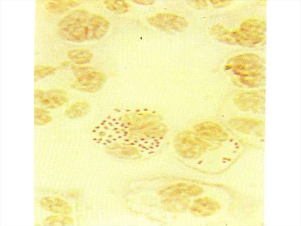
Causative agent - Neisseria meningitidis- Small gram-negative diplococci;
- typical location in a pair of coffee beans;
- do not form spores, the capsule is nonconstant;
- nonmotile;
- aerobic, obligate intracellular microbe;
- grows on artificial nutrient media (blood agar);
- for capsular AG allocate nine serogroups (A, B, C, D, X, Y, Z, W135
and E);
- releases endotoxin;
- produces hyaluronidase and neuraminidase;
- secretes IgA-proteases that cleave the IgA molecule that protects
the bacteria from Ig;
- nonstable in environment;
- sensitive to drying and cooling, to penicillin, tetracycline,
erythromycin and disinfectants
21.
22.
23.
PATHOGENESIS - disease develops in 3 stages:- PENETRATION - the local forms of the diseases are
developed as nasopharyngitis or pneumonia or
carrier-state;
- BACTERIEMIA - infiltration N.m. in a blood, following the
lysis of bacteria and endotoximia - disease proceeds as
acute sepsis with hemorrhagic rush and hemorrhagic sd;
-DISTRIBUTION - into the brain by hematogenous,
lymphogenous way, perineuraly and through ethmoidal
bone;
Serum antibodies and high concentration of IgA on the
mucous of upper RT - play an important role in protection
from N.m.!!!
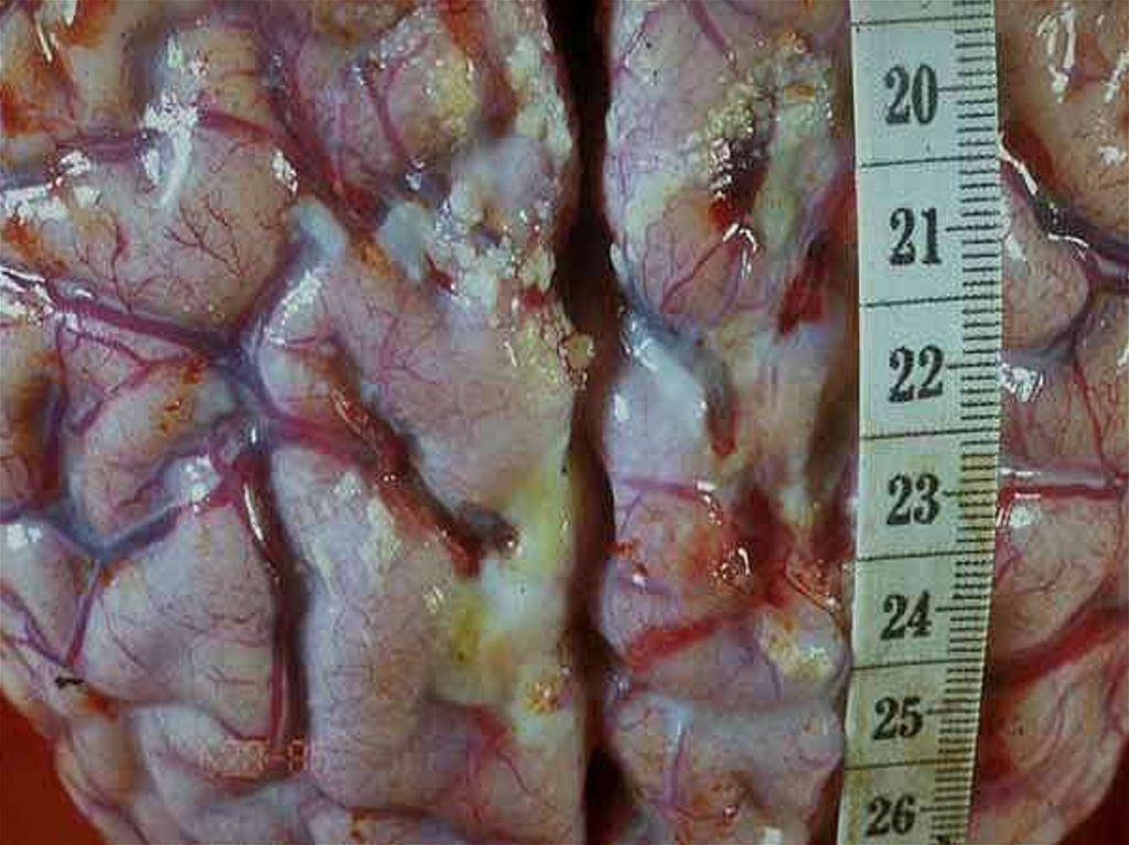
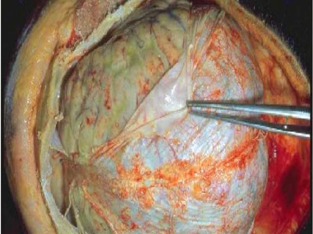
24.
PATHOMORPHOLOGY- the N.m. causes acute inflammatory response in a place
of implantation (statified pavement epithelium).
Endotoximia results of a diffuse vasculitis and DIC (disseminated intravascular coagulopathy)
Vessels are filled by clots of blood with a major contents
of a fibrin and leucocytes, that results in hemorrhages on
the body, but on a skin they are accompanied with
necrosis at the centre of large eruptions.
The cause of a generalisation of the process is not clear.
N.m. “love" well-nourished children and adults »- probably
influence of the genetic factors and inadequate response
of the organism on implantation of the N.m.!!!
25.
CLASSIFICATIONLocalized forms:
- Carriage –without clinical manifestation, can be revealed
by detection of N.m. in culture and smears from a
nasopharynx;
- Nasopharyngitis;
- Meningococcal pneumonia;
Generalized forms:
- Meningococcal sepsis (acute and chronic)
- Meningitis
- Meningoencephalitis
- Mixed (sepsis + meningitis and etc. )
- Infrequent forms of disease: endocarditis, arthritis,
pneumonia, iridocyclitis, otitis etc.)
26.
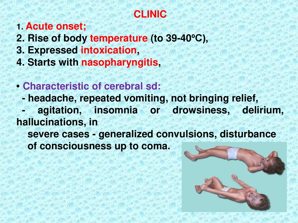
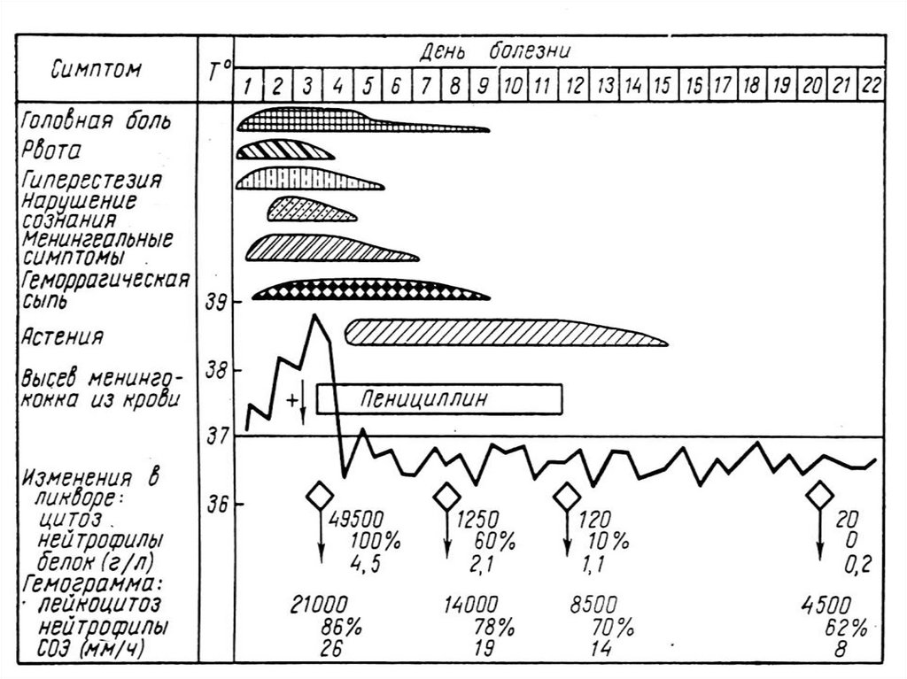
CLINIC1. Acute onset;
2. Rise of body temperature (to 39-40ºC),
3. Expressed intoxication,
4. Starts with nasopharyngitis,
• Characteristic of cerebral sd:
- headache, repeated vomiting, not bringing relief,
- agitation, insomnia or drowsiness, delirium,
hallucinations, in
severe cases - generalized convulsions, disturbance
of consciousness up to coma.
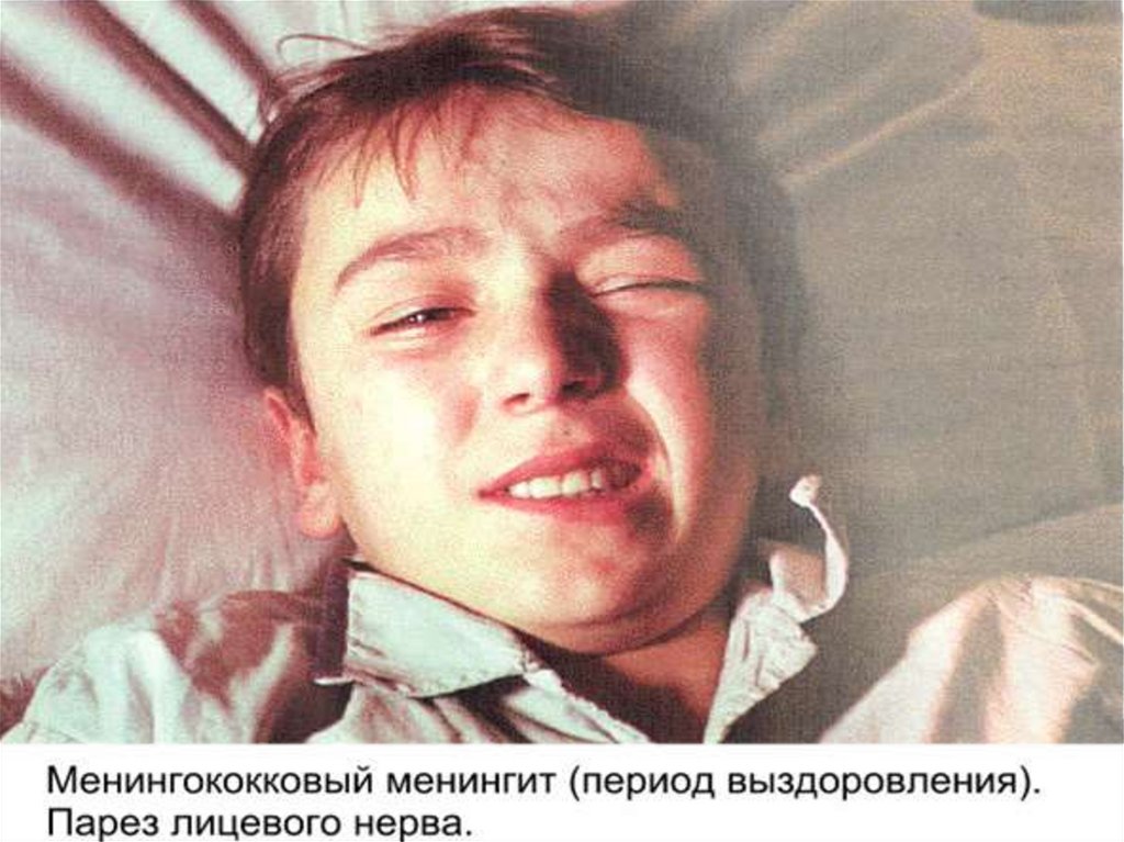
27.
• Meningeal syndrome appears in the first days ofthe disease of different severity:
- often occurs with development of edemaswelling of brain
(the attacks of psychomotor agitation);
-1-1,5% of the patients revealed signs
of encephalitis (paralysis of the facial muscles
paraparesis, paralysis, disorder of coordination);
- affection of cranial nerves;
- visual and auditory hallucinations, euphoria or
depression;
28.
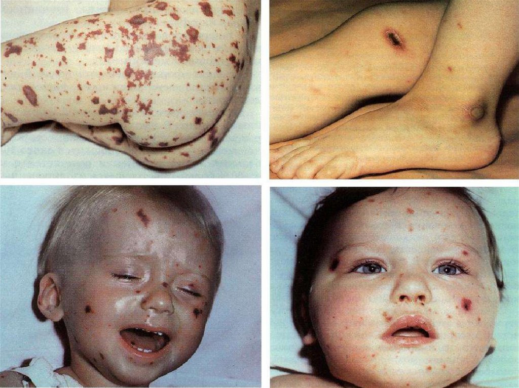
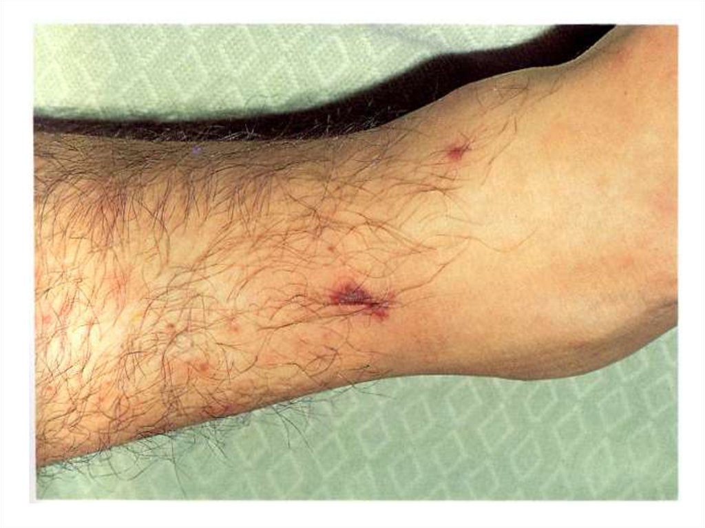
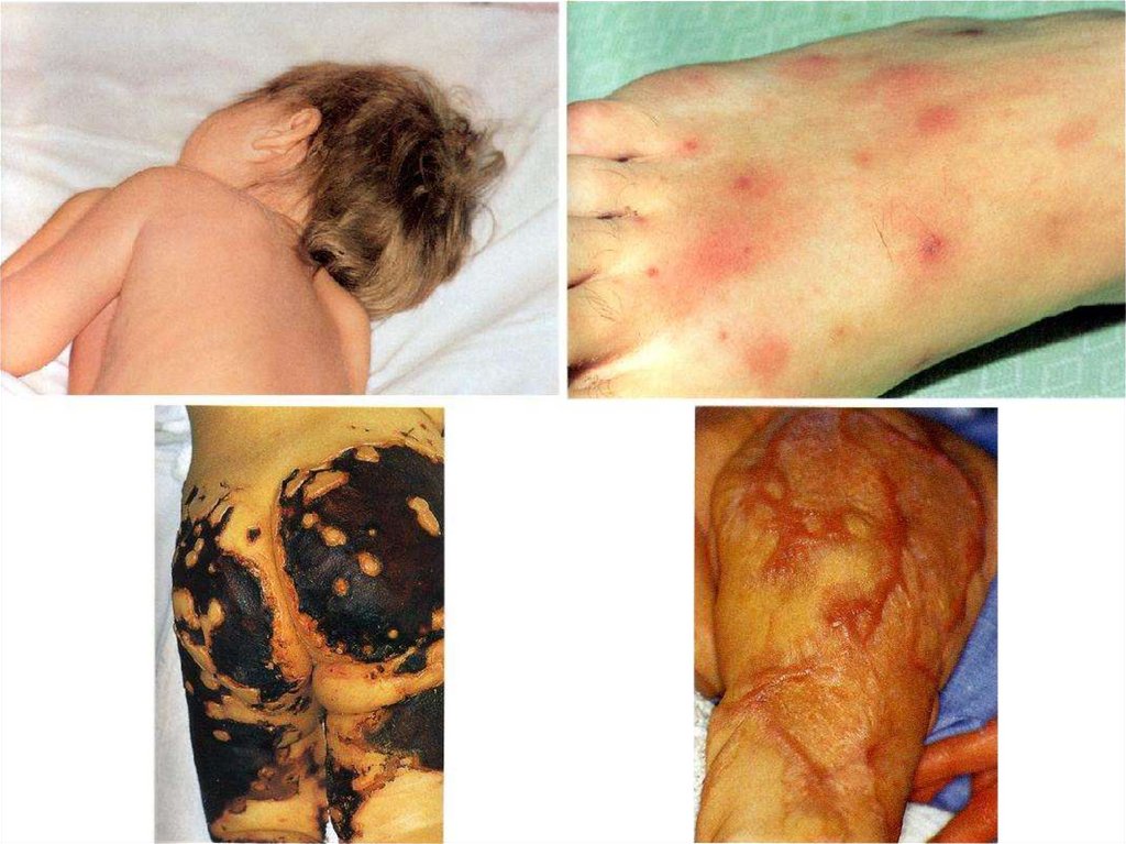
• 30-40%cases of meningitis is combined with
Meningococcemia:
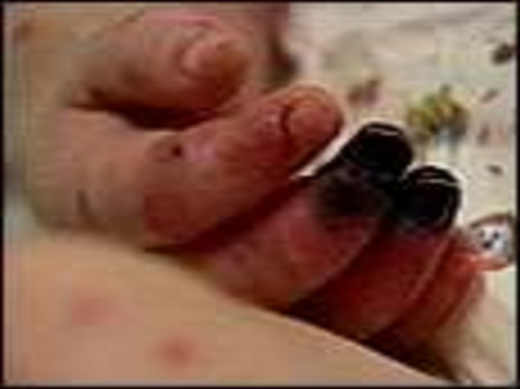
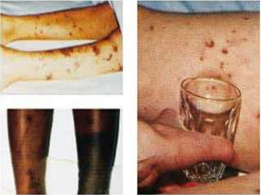
-hemorrhagic rash on the skin with petechial character,
sometimes up to 5-15 cm in diameter with necrosis in
the center, localized on the buttocks, lower limbs,
-infectious-toxic shock,
- serious complication - is the development of
hemorrhages in the adrenal glands (syndrome
Waterhouse – Friderichsen),
- heart (endocarditis, pericarditis, myocarditis) and other
organs.
29.
• CBC: leukocytosis, neutrophilia, shift of the formula tomyelocytes, lymphopenia, increased ESR.
• CSF:
-is cloudy, yellow-colored,
- flows streamly,
- protein is increased to 1g/l,
- count of neutrophils - more than 1,000 cells in 1 ml,
- in the first hours of the disease can be seen only
increased CSF pressure or signs of serous meningitis,
• Microscopy of CSF, thick drops of blood or discharging
from the nasopharynx can be used to detect gramnegative diplococci, located intracellular.
30.
• Bacteriological examination of mucus from thenasopharynx, blood, cerebrospinal fluid is leading in the
diagnosis of meningococcal disease.
Material for bac. exam. should be taking before
etiotropic therapy;
• Serology (RIHA) carried out with diagnostics of
meningococci of group A and C (conditionally
diagnostic titer of antibodies in adults 1:40-1:80).
In severe meningococcal infection and infectious-toxic
shock antibodies are detected in low titers, and in some
cases not detected at all.
31.
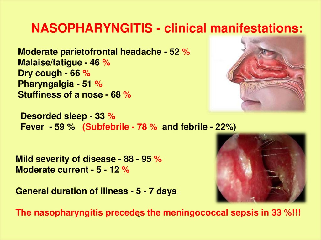
NASOPHARYNGITIS - clinical manifestations:Moderate parietofrontal headache - 52 %
Malaise/fatigue - 46 %
Dry cough - 66 %
Pharyngalgia - 51 %
Stuffiness of a nose - 68 %
Desorded sleep - 33 %
Fever - 59 % (Subfebrile - 78 % and febrile - 22%)
Mild severity of disease - 88 - 95 %
Moderate current - 5 - 12 %
General duration of illness - 5 - 7 days
The nasopharyngitis precedes
- the meningococcal sepsis in 33 %!!!
32.
MENINGOCOCCAL SEPSIS:- acute beginning with chill and fast rise of intermittent
temperature up to 38 - 40ºC
- expressed intoxication (headache, thirst, weakness,
paleness and dryness of a skin)
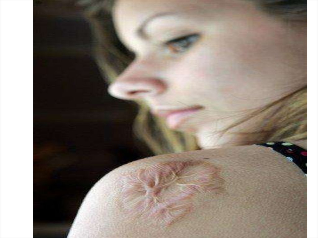
- appearance of hemorrhagic syndrome: hemorrhagic rush
with necrosis on the skin of buttocks, legs, trunk, arms,
eyelids (in 2-4-6 hours from the onset of disease!!!)
- enanthema in a transitive folds of conjuctiva, hemorrhages
on sclera
- the hemorrhages in joints - on 5 - 13 % are more often
than in fingers and legs
- common manifestations of a hemorrhagic syndrome nasal, uterine, internal bleeding
33.
34.
35.
36.
37.
38.
39.
40.
41.
42.
43.
44.
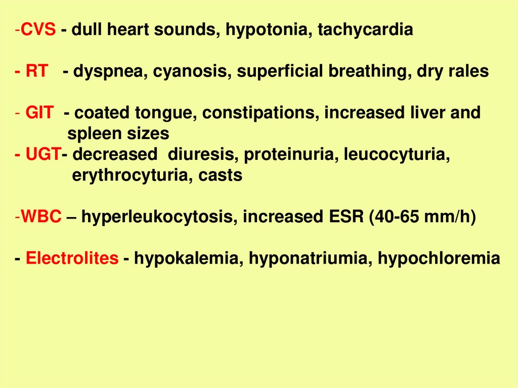
-CVS - dull heart sounds, hypotonia, tachycardia- RT - dyspnea, cyanosis, superficial breathing, dry rales
- GIT - coated tongue, constipations, increased liver and
spleen sizes
- UGT- decreased diuresis, proteinuria, leucocyturia,
erythrocyturia, casts
-WBC – hyperleukocytosis, increased ESR (40-65 mm/h)
- Electrolites - hypokalemia, hyponatriumia, hypochloremia
45.
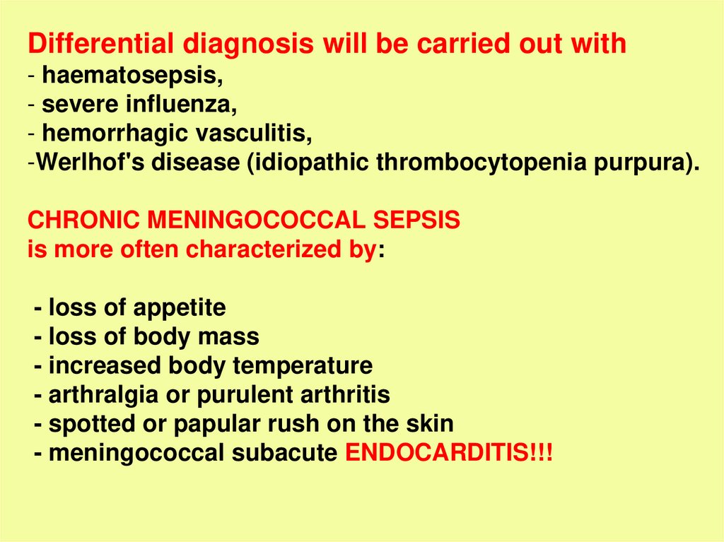
Differential diagnosis will be carried out with- haematosepsis,
- severe influenza,
- hemorrhagic vasculitis,
-Werlhof's disease (idiopathic thrombocytopenia purpura).
CHRONIC MENINGOCOCCAL SEPSIS
is more often characterized by:
- loss of appetite
- loss of body mass
- increased body temperature
- arthralgia or purulent arthritis
- spotted or papular rush on the skin
- meningococcal subacute ENDOCARDITIS!!!
46.
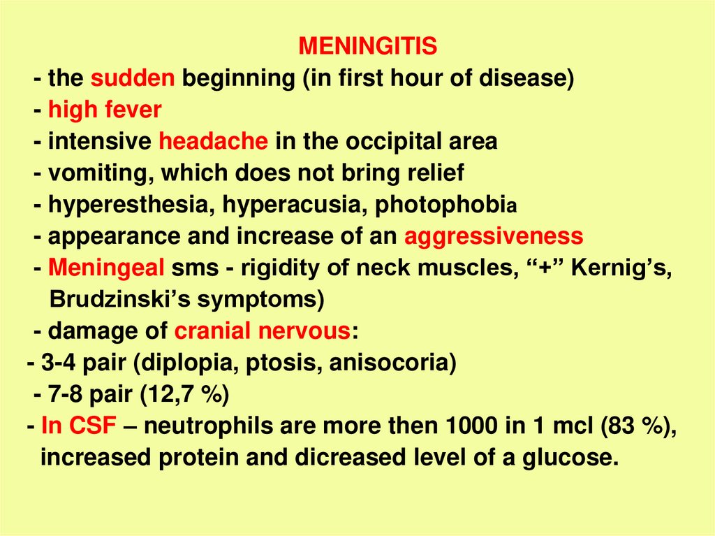
MENINGITIS- the sudden beginning (in first hour of disease)
- high fever
- intensive headache in the occipital area
- vomiting, which does not bring relief
- hyperesthesia, hyperacusia, photophobia
- appearance and increase of an aggressiveness
- Meningeal sms - rigidity of neck muscles, “+” Kernig’s,
Brudzinski’s symptoms)
- damage of cranial nervous:
- 3-4 pair (diplopia, ptosis, anisocoria)
- 7-8 pair (12,7 %)
- In CSF – neutrophils are more then 1000 in 1 mcl (83 %),
increased protein and dicreased level of a glucose.
47.
48.
49.
50.
51.
52.
53.
54.
55.
56.
57.
58.
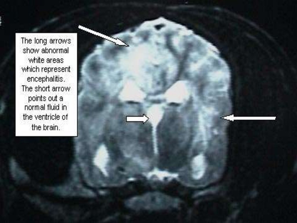
MENINGOCOCCAL MENINGOENCEPHALITIS-more often appears due to diffuse damage of a brain with
desorded consciousness and focal changes:
- Aphasia - 3 %
- Psychosensorial disorders - 1 %
- Cramps, mono- and hemiparesis - 3 %
- Oculomotor disorders - 27 %
COMPLICATIONS:
- Acute renal unsufficiency
- Dural and subdural exudates
- Viral or bacterial superinfections
- Activation of simple herpes in 38 %
- Pneumonia - 6 - 22 %, otites, cystopyelitis 4,1 %
59.
60.
61.
LABORATORY DIAGNOSIS:Microscopy of smear from a mucous of nasopharynx,
CSF and thick drop of a blood (detection of intracellular
gramme (-) diplococci)
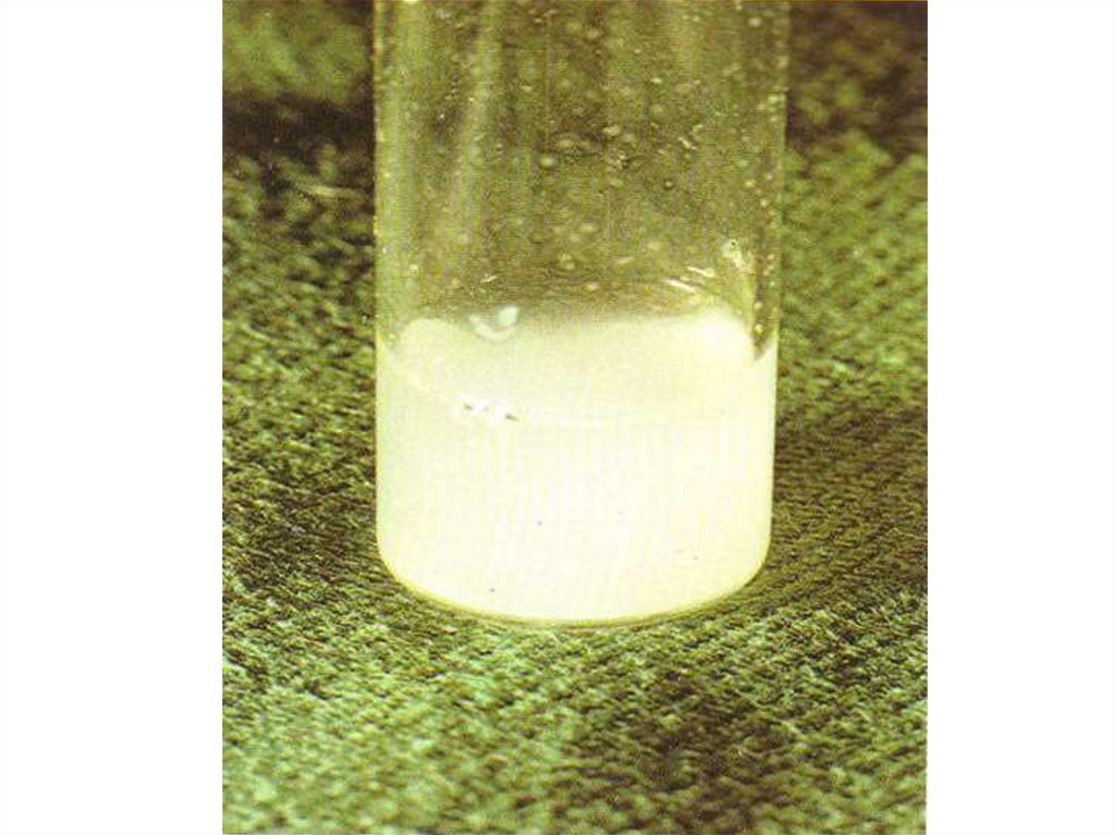
Biochemical and morphological investigation of CSF
(cytosis, contents of protein and glucose, detection of a film
at the bottom test tubes)
Common analysis of a blood and urine
Immunological investigation - RHA (diagnostic titer - 1:160)
and RIHA (1:200), ELISA
62.
63.
64.
Examination of cerebrospinal fluid- The tube №1 (1 ml) – erythrocytes, leucocytes, cells ratio
- The tube №2 (1 ml) – protein, glucose, chlorides
- The tube №3 (2 ml) – growing of bacteria (a blood or
chocolate agar)
- The tube №4 (1 ml) - after centrifugation and sedimentation
coloring by Gram and with Indian ink
- The tube №5 (1 – 2 ml (ml) - is saved and used for
detection of other causative agents
65.
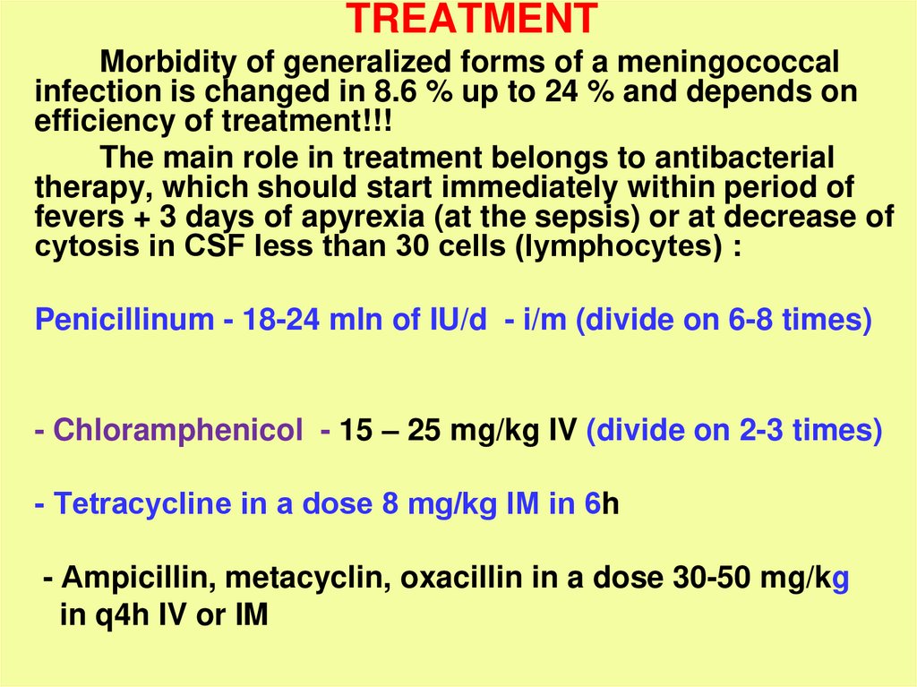
TREATMENTMorbidity of generalized forms of a meningococcal
infection is changed in 8.6 % up to 24 % and depends on
efficiency of treatment!!!
The main role in treatment belongs to antibacterial
therapy, which should start immediately within period of
fevers + 3 days of apyrexia (at the sepsis) or at decrease of
cytosis in СSF less than 30 cells (lymphocytes) :
Penicillinum - 18-24 mln of IU/d - i/m (divide on 6-8 times)
- Chloramphenicol - 15 – 25 mg/kg IV (divide on 2-3 times)
- Tetracycline in a dose 8 mg/kg IМ in 6h
- Аmpicillin, metacyclin, oxacillin in a dose 30-50 mg/kg
in q4h IV or IM
66.
- Cefatoxim 1g. IV or IM in q 12h- Ceftriaxon 1 - 2 g. IV or IМ in q8-12h
- Byceptol 980 мg PO, IV in q12h
- Sulfamonomethoxinum 4 g the first day, then 2 g. PO in
q12h
The nasopharyngitis is treated 5-7 days with
erythromycin, аzytromicin or sulfanilamidums
Pathogenetic therapy:
- dehydrational therapy (at edema of brain)
- desintoxication therapy and glucocorticoids at ТIS
- metabolites
-treatment of a hemorrhagic syndrome
Symptomatic therapy
67.
Pneumococcal meningitis• Anamnesis –
Source - patient with various forms
of
the
pneumococcal
infection
and
carriers
of
pneumococcus.
Route of transmission — airborne, but exogenous
and endogenous route possible.
18-51% of purulent meningitis in adults is
pneumococcal etiology,
1/3 of cases are secondary and develop on the
background of otitis, sinusitis, pneumonia, sepsis.
100% of recurrent meningitis with pneumococcal
liquorrhea.
Pathogen – Streptococcus pneumoniae, stained gram
positive.
68.
• Onset is acute.- Rapid rising of body temperature to 39-40ºC;
- Progressing of general intoxication and cerebral
symptoms (headache, vomiting, loss of consciousness,
repeated seizures).
• Meningeal syndrome appears early and expressed, but
sometimes incomplete, accompanied by signs of edemaswelling of the brain.
• Quick involvement of brain substance in the pathological
process,
accompanied
by
the
development
of
meningoencephalitis, appearance of mono - and
hemiparesis, oculomotor disorders, hyperkinesis, ataxia.
On the 3-4 day of the disease can develop coma status with the
symptoms of brain stem dislocation.
69.
• Course of the disease varies from malignantforms with lethal outcome to a proloned and
recurrent.
• With the development of pneumococcal
sepsis may be affection of other organs:
hemorrhagic
rash
on
the
skin
(like
meningococcemia), pneumonia, endo - and
pericarditis, arthritis.
• In 50% of patients residual effects remain,
may be seen epilepsy, paresis, paralysis.
70.
• Cerebrospinal fluid is turbid, of yellow-greycolor with metallic shade.
Cell count up to 30000 in 1 mkl, protein - 2-5 g/l.
Pressure of CSF is increased moderately due
to accumulation of pus in the subarachnoid
space and block of CSF pathways.
• CBC - expressed inflammatory changes.
• Bacteriological examination of blood, CSF
detects the Streptococcus pneumoniae.
71.
SEROUS MENINGITIS- is a group of CNS diseases with the similar
clinical
manifestations
and
morphology,
characterized by the serous inflammation of the
meninges, mostly of viral, rarely bacterial,
fungal or parasitic etiology,
accompanied by acute development of
hydrocephalic syndrome and nonexpressed
meningeal syndrome.
Depending on the mechanism of CNS demerge
serous meningitis are divided into primary and
secondary.
72.
The etiology of primary serous meningitis is mostoften of viral origin, the clinic is characterized only by
meningeal syndrome
(Acute lymphocytic choriomeningitis, mosquito and tick-borne
encephalitis, the meningeal form of poliomyelitis).
Secondary serous meningitis develop on the
background of main disease, usually of viral etiology
(enterovirus infection, respiratory viral infections, chickenpox,
measles, rubella, epidemic. mumps, HIV infection, herpesvirus
infection, cytomegalovirus, rabies, arbovirus infection).
Less serous meningitis occurs by bacterial infections
(typhoid, tuberculosis, brucellosis, leptospirosis, syphilis, Lyme
disease, yersiniosis), parasitic (malaria, toxoplasmosis,
amebiasis)
or
fungal
infections
(cryptococcosis,
coccidioidomycosis, aspergillosis).
73.
Pathogenesis1. Penetration of the pathogens in the blood
2. Activation of biologically active substances, disturbed homeostasis,
increases the permeability of cellular and vascular membranes,
including the HEB
3. Causative agents (viruses, bacteria and their toxins) penetrate into the
CNF
4. Hyperproduction of CSF due to irritation of the vascular plexus of the
ventricles (lower protein level in the cerebrospinal fluid) or serous
inflammation of the meninges (increased protein and lymphocytes in
the exudate)
Formation of hydrocephalic-hypertensive syndrome is the main link of
pathogenesis of serous meningitis and determines the clinical picture
and severity of the disease.
74.
Clinical manifestations in serous meningitis are formed from the samesyndromes of purulent meningitis, but they have some peculiarities:
- General intoxication syndrome. Its symptoms depend on the
character and properties of the pathogen, but common is the presence
of fever (38-39ºC), moderate intoxication (headache, dizziness,
lethargy, apathy or agitation, loss of appetite), pale skin, dull tones of
the heart, sometimes dysfunction of the gastrointestinal tract;
- Meningeal syndrome may be absent in asymptomatic or mild forms of
meningitis with inflammatory changes of the CSF.
Often reveals incomplete or dissociation of meningeal syndrome (loss
of some symptoms). In most cases, the severity of meningeal
syndrome corresponds to the value of intracranial hypertension.
The most constant symptoms are rigidity of occipital muscles, upper
Brudzinsky-symptom, visual and tactile hyperesthesia;
75.
-Hydrocephalic-hypertensive syndrome manifested byheadache, vomiting, hyperreflexia, seizures, and
pyramidal signs;
- CSF - increased pressure, colorless, transparent or
opalescent, lymphocytic pleocytosis (100-2000/µl), protein
level is normal or slightly increased, glucose is normal (in
tuberculous meningitis is reduced).
In the first days of the disease in CSF can be up to 30-50%
of neutrophils.
76.
Enteroviral meningitisIs caused by ECHO and Coxacky virus of different
serotypes;
Infection is highly contagious;
Spring-summer seasonality, often outbreaks;
70-80% of population - children of 5-9 years;
Source of infection is a sick man and a virus carrier;
Mechanism of transmission is aerogenic (airborne),
but posible fecal-oral transmission of the pathogen.
77.
1. Incubation - 2 - 8 days;2. Acute onset with expressed intoxication syndrome
(wave-like fever in 2-7 days, headache in frontotemporal region,
pain in the eyeballs, dizziness, repeated,
uncontrollable vomiting, lethargy, weakness);
sometimes
3. Encephalitic sd - may be convulsions or delirium,
loss of consciousness;
4. Hyperemia of the face with pale nosal triangle,
injection of sclera;
5. Sometimes combined with other forms of enterovirus
infection – myalgia, herpangina, polymorphic spotted,
papular or hemorrhagic rash.
78.
6. Hypertensive-hydrocephalic syndrome - in the firstdays of the disease (headache, vomiting, hyperreflexia,
pyramidal signs);
7. Meningeal syndrome appears on the 2-3-d day ,
moderate, short-term, characterized by the fever and
inflammatory changes in CSF;
8. CSF is transparent, colorless, pressure is increased,
moderate lymphocytic pleocytosis (30 to 800 cells),
protein content is normal or reduced, the level of glucose
and chlorides are not changed;
9. CBC - leukopenia, nonexpressed neutrophilosis, with
insignificant shift of the formula to the left, moderate
increasing of ESR, at the beginning of the second week –
eosinophilia.
79.
10. Confirmation of the diagnosis is based on isolation ofthe virus from CSF, blood, stool, nasopharyngeal swabs;
11. Serologic investigation reveals increase of AB-titer in
four times in paired serums (taken with an interval in 1012 days);
12. Serouse meningitis is usually benign disease of
moderate severity;
13. Feature of enteroviral meningitis is a tendency to
recurrence: usually on the 20-28-th day of normal
temperature the state is getting worse again (fever,
increases pleocytosis in CSF).
Such recurrences may repeat, but relapsing forms are
always severe.