


 Медицина
Похожие презентации:


Origin, differential diagnosis and thrapy of jaundices in neonates
1. Origin, differential diagnosis and thrapy of jaundices in neonates
Assistant professor of hospital pediatricsdepartment
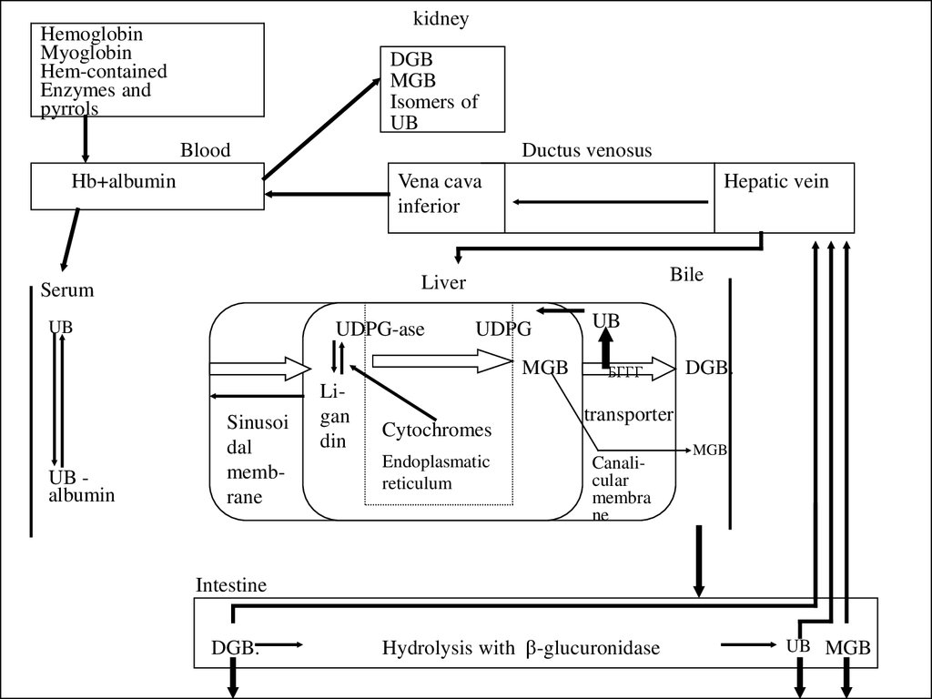
2.
kidneyHemoglobin
Myoglobin
Hem-contained
Enzymes and
pyrrols
DGB
MGB
Isomers of
UB
Blood
Ductus venosus
Hb+albumin
Vena cava
inferior
Hepatic vein
Bile
Liver
Serum
UB
UDPG-ase
UDPG
MGB
UB albumin
Sinusoi
dal
membrane
Ligan
din
Cytochromes
Endoplasmatic
reticulum
UB
БГГГ
DGB.
transporter
Canalicular
membra
ne
MGB
Intestine
DGB.
Hydrolysis with b-glucuronidase
UB MGB
3.
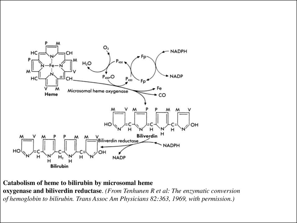
Catabolism of heme to bilirubin by microsomal hemeoxygenase and biliverdin reductase. (From Tenhunen R et al: The enzymatic conversion
of hemoglobin to bilirubin. Trans Assoc Am Physicians 82:363, 1969, with permission.)
4.
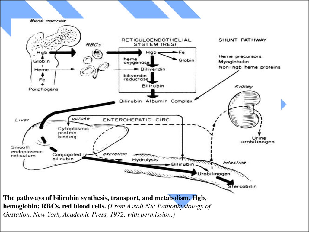
The pathways of bilirubin synthesis, transport, and metabolism. Hgb,hemoglobin; RBCs, red blood cells. (From Assali NS: Pathophysiology of
Gestation. New York, Academic Press, 1972, with permission.)
5.
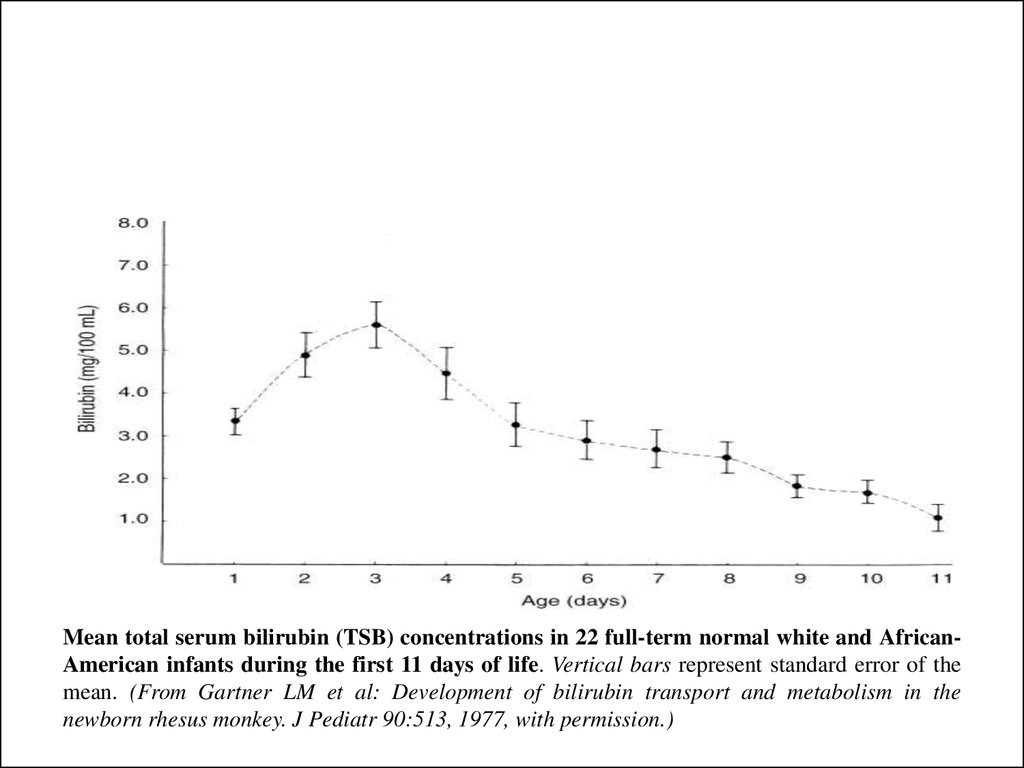
Mean total serum bilirubin (TSB) concentrations in 22 full-term normal white and AfricanAmerican infants during the first 11 days of life. Vertical bars represent standard error of themean. (From Gartner LM et al: Development of bilirubin transport and metabolism in the
newborn rhesus monkey. J Pediatr 90:513, 1977, with permission.)
6.
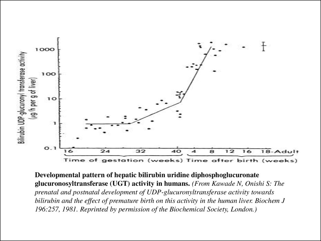
Developmental pattern of hepatic bilirubin uridine diphosphoglucuronateglucuronosyltransferase (UGT) activity in humans. (From Kawade N, Onishi S: The
prenatal and postnatal development of UDP-glucuronyltransferase activity towards
bilirubin and the effect of premature birth on this activity in the human liver. Biochem J
196:257, 1981. Reprinted by permission of the Biochemical Society, London.)
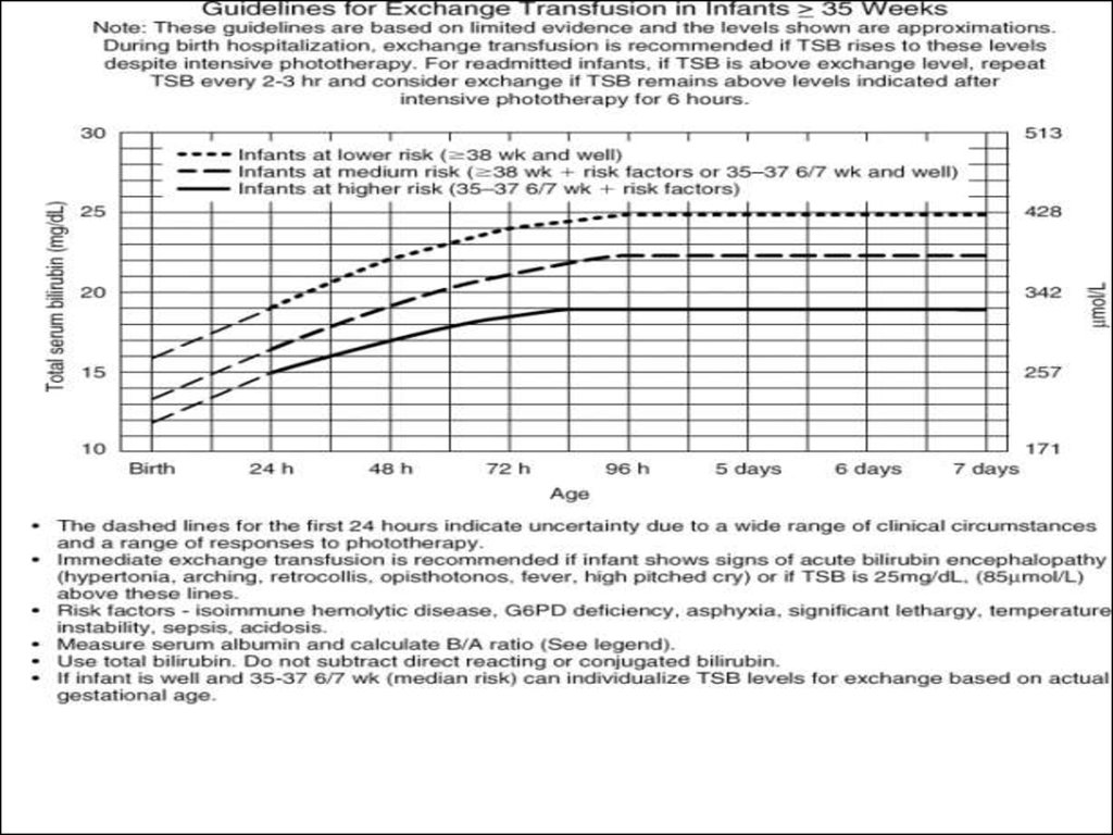
7.
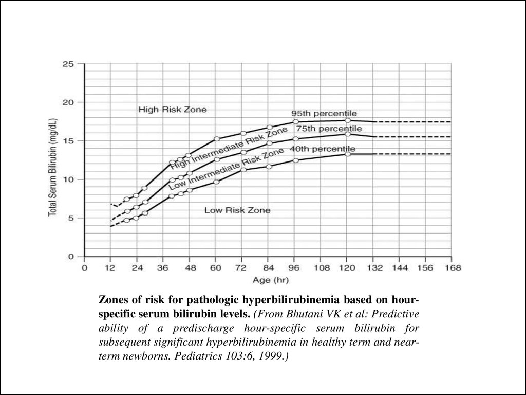
Zones of risk for pathologic hyperbilirubinemia based on hourspecific serum bilirubin levels. (From Bhutani VK et al: Predictiveability of a predischarge hour-specific serum bilirubin for
subsequent significant hyperbilirubinemia in healthy term and nearterm newborns. Pediatrics 103:6, 1999.)
8.
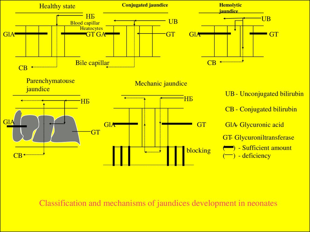
Conjugated jaundiceHealthy state
НБ
UB
UB
Blood capillar
Heatocytes
GlA
Hemolytic
jaundice
GT GA
GT
GlA
Bile capillar
CB
Parenchymatouse
jaundice
GT
CB
Mechanic jaundice
UB - Unconjugated bilirubin
НБ
НБ
CB - Conjugated bilirubin
GlA
GlA
GT
GT
CB
GlA- Glycuronic acid
GT- Glycuroniltransferase
blocking
(
(
) - Sufficient amount
) - deficiency
Classification and mechanisms of jaundices development in neonates
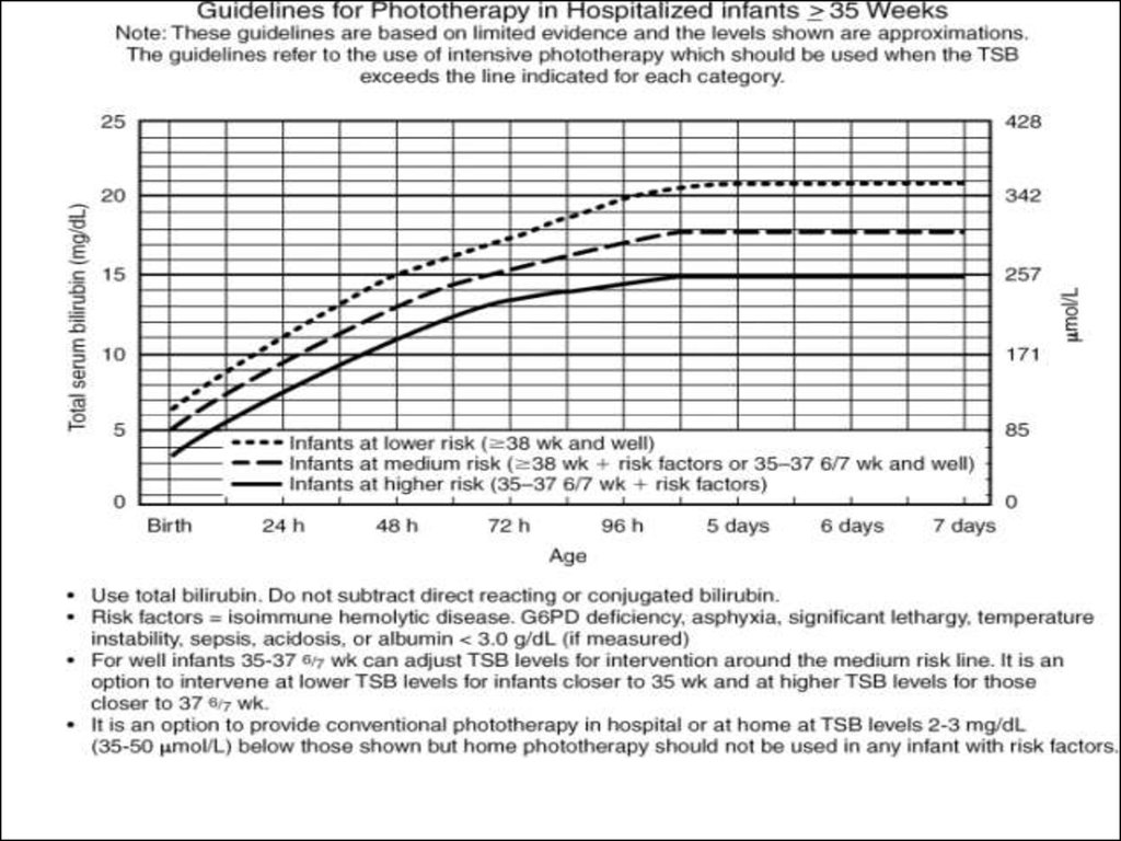
9.
Hour-specific bilirubin nomogram with the predictive ability of thepredischarge bilirubin value for subsequent severe hyperbilirubinemia,
>95th percentile tract. Reproduced with permission from Bhutani VK,
Johnson LH. Jaundice technologies; prediction of hyperbilirubinemia in
term and near term newborns. J Perinatol 2001;21:576
10. Clinical and serologic differences of hemolytic disease among ABO and Rh sensibilisation
1. a - и b –agglutinins normally exists in blood serum of mother and capable topenetrate fetus. Rh antibodies normally are absent both in mother and fetus.
2. Anti-A and Anti-B being full agglutinins as other antibodies could penetrate
placenta whereas full Rh antibodies couldn’t penetrate it.
3. Fetus tissues in “extractors”( people who reveals A and B substances not only in
blood but in humors as well) and in “non-extractors” contains both A and B
substances which is usually neutralizes anti-A and anti-B antibodies. Rh –
antibodies doesn’t neutralizes by the tissue antibodies therefore their infiltration of
Rh positive fetus causes hemolysis. This very characteristic differential feature of
ABO antibodies leads to hemolytic disease development without previous
sesibilisation as mother blood already consists of a and b agglutinins.
11. The basic principles of change blood transfusion.
1.The tip of correctly fixed umbilical vein catheter must be placedinto vena cava being situated between the diaphragm and left
atrium.
2. The length of umbilical vein catheter from it end to label at the
level of umbilical ring is equal to the distance from brachium to the
belly-button – 5 cm; the procedure initiates with removing of 30 -40
ml of blood( 20 ml in preterms).
3. The total amount of injected blood must be 50 ml more than
removed; operation must carried slowly at 3-4 ml per minute
alternating with injecting and rejecting of 20 ml blood (10 ml in
preterms) with total duration no less than 2 hour; every 100 ml of
entering blood need to administrate 1 ml of 10 % calcium gloconas
solution.
4. In the blood serum before change transfusion and just after the
bilirubin level must be detected.
12.
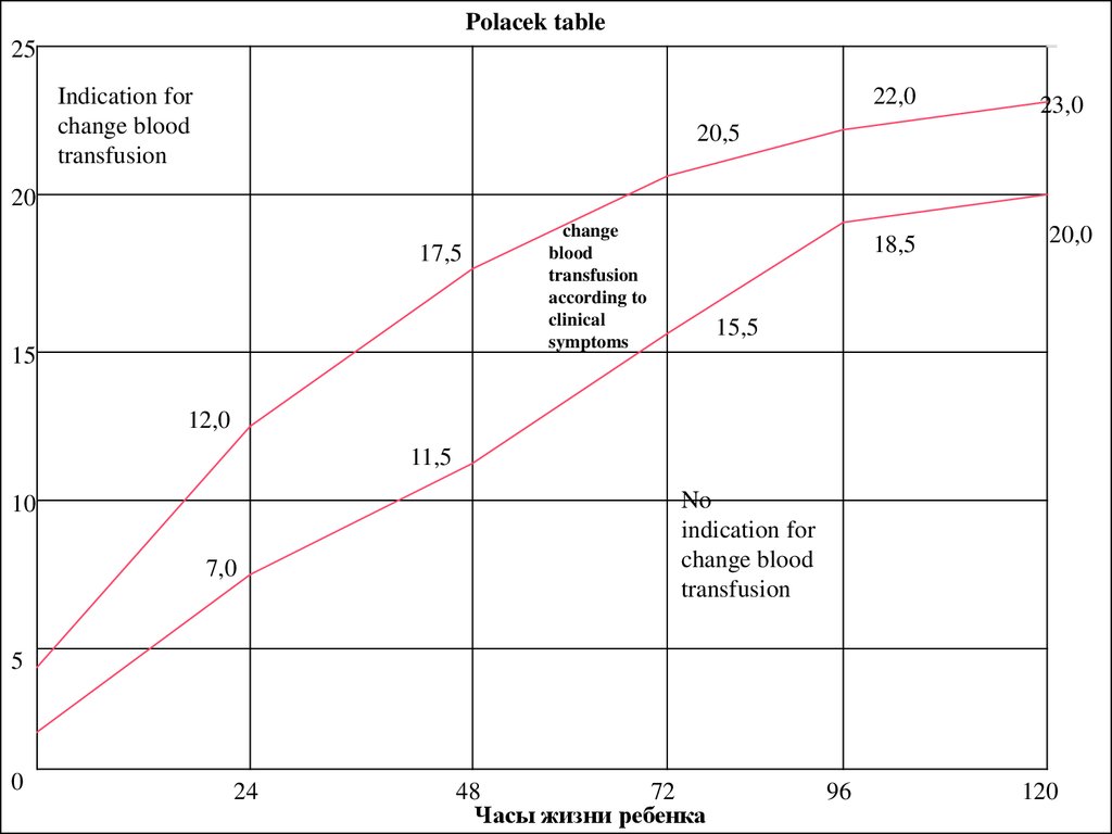
Polacek table25
Indication for
change blood
transfusion
22,0
23,0
20,5
20
17,5
15
change
blood
transfusion
according to
clinical
symptoms
18,5
20,0
15,5
12,0
11,5
10
7,0
No
indication for
change blood
transfusion
5
0
24
48
72
Часы жизни ребенка
96
120