


.")


 Биология
Похожие презентации:


")


")
Physiological bases of hemo dynamic
1. Physiological bases of hemodynamic.
2. Kinds of blood movements
3.
4. Formulas of hemodynamic
r * PQ
,
8 * l
4
8 l
R
.
r
5.
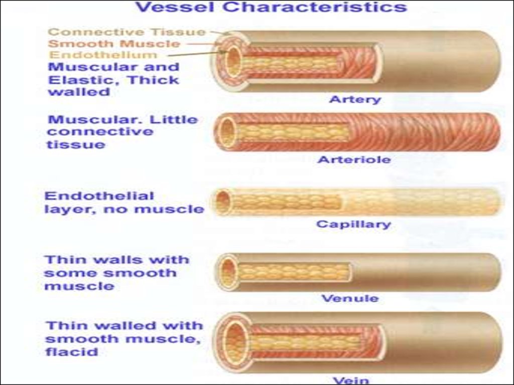
6. Vessel Structure
Structure/function relationships
change as one
moves through
the
cardiovascular
tree
Tunic thickness
and composition
of the three
layers are
variable
7.
8.
9.
10.
11. Functional types of vessels
Amortization orcompensatory
vessels – arteries
Volume vessels or
veins
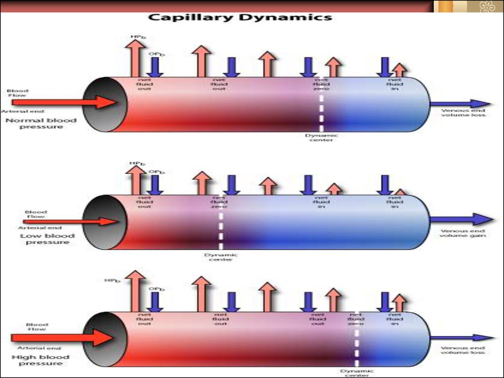
Exchanged vessels
or Capillary
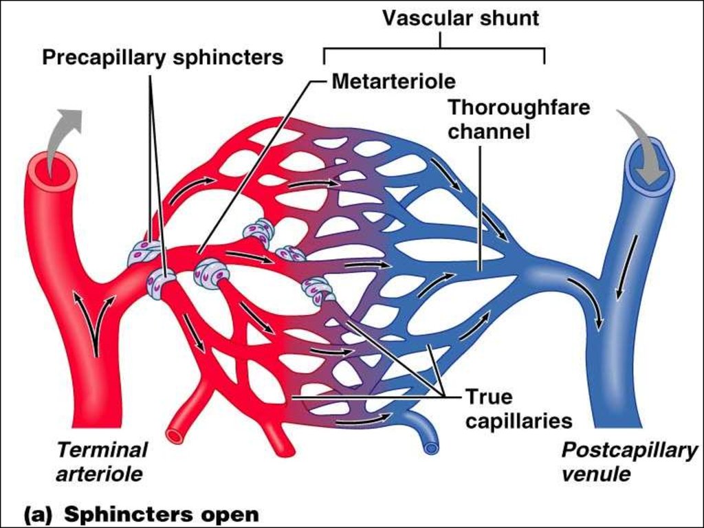
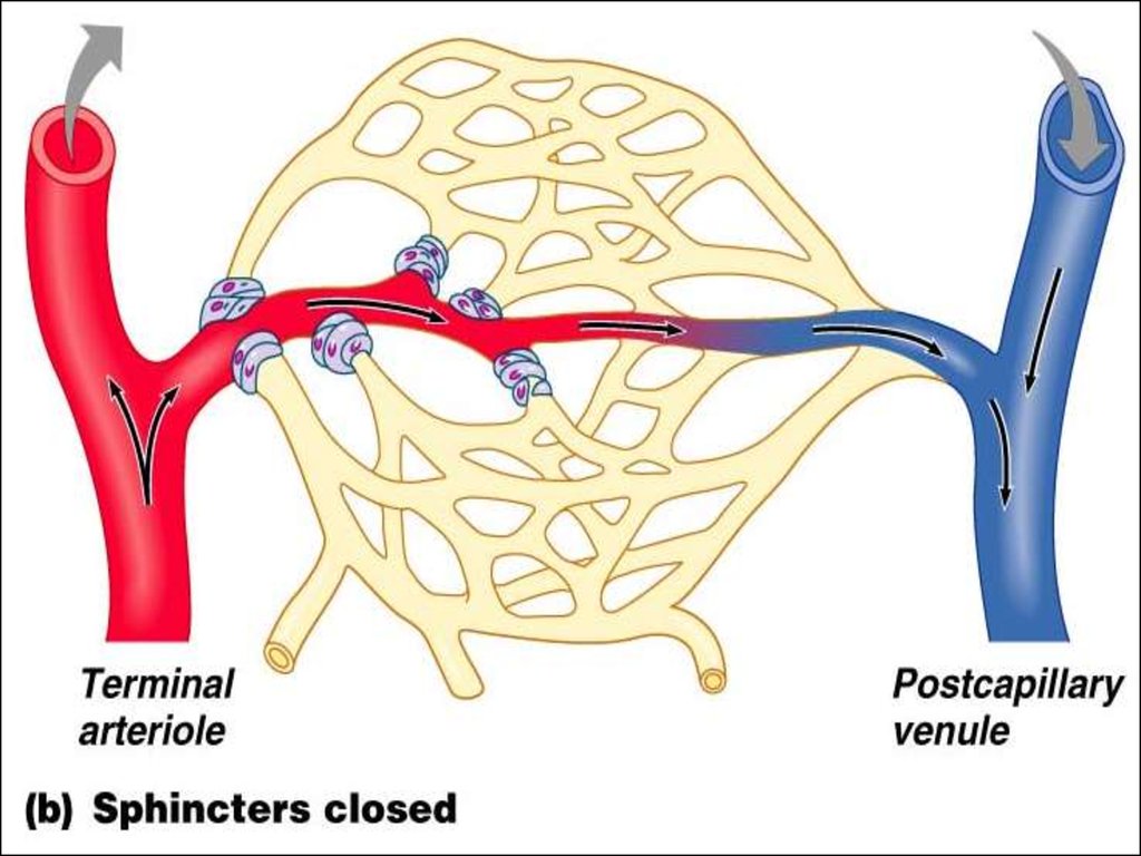
12. Functional types of vessels
Resistive vessels or arterioles, smallestarteries; lead to capillary beds
Sphincters
Shunts
Arterial anastomoses provide alternate
pathways (collateral channels) for blood to
reach a given body region. If one branch is
blocked, the collateral channel can supply
the area with adequate blood supply
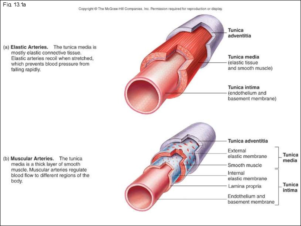
13.
Fig. 13.1a14.
15.
16.
17. Arterial pressure
Determine the influences of factors:1. cardiac – systolic volume, speed of blood ejection
from the ventricles, heart beat;
2. vascular – elasticity of compensatory arteries,
tone of resistive vessels, volume of volume vessels;
3. blood – volume of blood, viscosity, hydrostatic
pressure of blood.
18.
19. Vasomotor control: Sympathetic Innervation of Blood Vessels
Sympathetic nerve fibersinnervate all vessels except
capillaries and precapillary
sphincters (precapillary
sphincters follow local control)
Innervation of small arteries
and arterioles allow
sympathetic nerves to increase
vascular resistance.
Figure 18-2; Guyton and Hall
Large veins and the heart are
also sympathetically innervated.
19
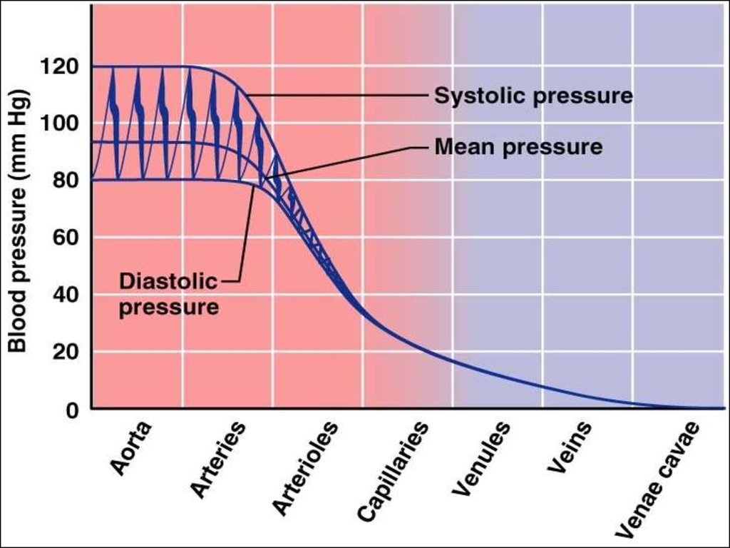
20. Kinds of arterial pressure
1. Systolic or maximal2. Side or absolute systolic
3. Stroke (hemodynamic)
4. Diastolic or minimal
5. Pulse
6. Result – P Pd Pc Pd ,
3
де Р – middle-dynamic pressure; Pd – diastolic
pressure; Pc – systolic pressure.
Ideal pressure:
Systolic = 102 + (0,6 · age) mm Hg
Diastolic = 63 + (0,4 · age) mm Hg
21.
Systolic pressure – pressure exerted onarterial walls during ventricular contraction
Diastolic pressure – lowest level of
arterial pressure during a ventricular cycle
Pulse pressure – the difference between
systolic and diastolic pressure
Mean arterial pressure (MAP) – pressure
that propels the blood to the tissues
MAP = diastolic pressure + 1/3 pulse
pressure
22.
Classification of hypertension (1999)Index
Level of arterial pressure
Systolic, mm
Hg
Diastolic, mm
Hg
Оptimal AP
< 120
< 80
Normal AP
< 130
<85
Higher-normal АP
130-139
85-89
Hypertension І degree
Measure hypertension
140-159
90-99
140-149
90-94
Hypertension ІI degree
160-179
100-109
Hypertension of IIІ degree
>180
>110
Isolated systolic hypertension
Measure hypertension
>140
<90
140-149
<90
23. Classification of hypertension (NHLBI, 2003).
IndexNormal AP
Prehypertension
Hypertension І
degree
Hypertension ІІ
degree
Level of arterial pressure
Systolic, mm Hg
Diastolic, mm Hg
< 120
120-139
140-159
< 80
or 80-89
or 90-99
>160
or >100
24. Apparatuses
25.
RECOMMENDED BLOOD PRESSUREMEASUREMENT TECHNIQUE
2.
2.
••The
Thecuff
cuffmust
mustbe
belevel
levelwith
withheart.
heart.
••IfIfarm
circumfe
rence
e
xceeds
arm circumfe rence e xceeds 33
33cm,
cm,
aalarge
cuff
mu
st
b
e
used.
large cuff mu st b e used.
••Pla
Place
cestethoscope
stethoscopediaphr
diaphragm
agmover
over
brachia
l
artery.
brachia l artery.
1.
1.
••The
Thepatient
patientshould
should
be
relaxed
be relaxedand
andthe
the
arm
mu
st
b
e
arm mu st b e
supported.
supported.
••Ensure
Ensureno
notight
tight
clothing
co
nstricts
clothing co nstricts
the
thearm.
arm.
3.
3.
Stethoscope
Mercury
machine
••The
Thecol
column
umnofof
mercury
mercurymust
mustbe
be
vertical
.
vertical .
••Infla
Inflateteto
toocclude
occludethe
the
pulse.
pulse. Deflate
Deflateatat22to
to
33mm/s.
Me
asu
re
mm/s. Me asu re
systolic
systolic(first
(firstsound)
sound)
and
dia
stolic
and dia stolic
(disapp
(disappearance)
earance)toto
nea
nearest
rest 22mm
mmHg.
Hg.
3
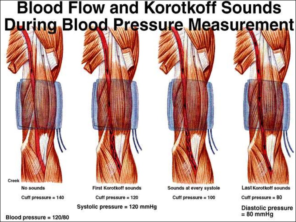
26. Korotkov Sounds caused by vibration collapse of the arterial wall??
Korotkoff IV is a betterindication of diastolic
pressure according to
theory
However Korotkoff V is
the commonly
recommended
measuring point except
in pregnant patients
because
It is associated with
less inter-observer
variations
It is easier to detect by
most observers
27.
28. Sphygmogram
Anacrota -аCatacrota b
Incisura (i)
Addition wave с or
secondary increase
29.
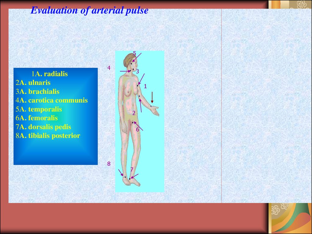
Evaluation of arterial pulse5
1А. radialis
2A. ulnaris
3A. brachialis
4A. carotica communis
5А. temporalis
6A. femoralis
7A. dorsalis pedis
8A. tibialis posterior
4
3
1
2
6
8
7