


")
")


")


")
")

")


")
")


")


 Биология
Похожие презентации:


")


Processes of Respiration
1.
Chapter 23 - Part 2Lecture Outline
See separate PowerPoint slides for all figures and tables preinserted into PowerPoint without notes.
Copyright © McGraw-Hill Education. Permission required for reproduction or display.
1
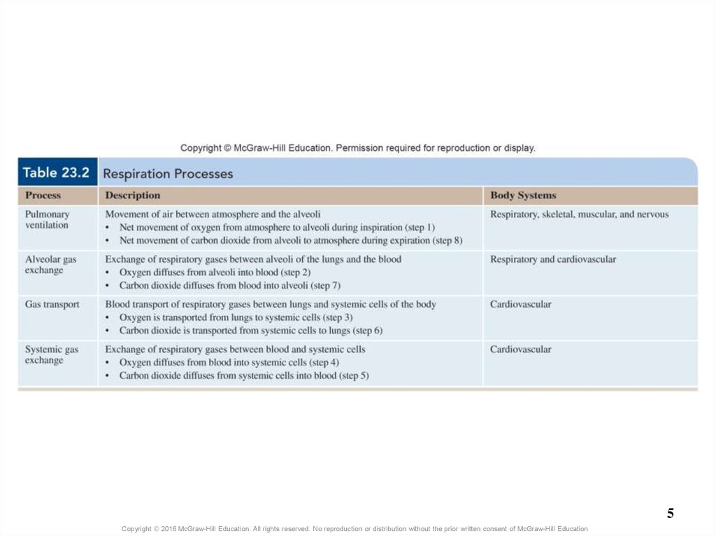
2. Processes of Respiration
• Respiration (exchange of gases between atmosphere andbody’s cells) involves four processes
– Pulmonary ventilation: movement of gases between
atmosphere and alveoli
– Alveolar gas exchange (external respiration): exchange of
gases between alveoli and blood
– Gas transport: transport of gases in blood between lungs and
systemic cells
– Systemic gas exchange (internal respiration): exchange of
respiratory gases between the blood and the systemic cells
2
Copyright © 2016 McGraw-Hill Education. All rights reserved. No reproduction or distribution without the prior written consent of McGraw-Hill Education
3. Processes of Respiration
• Net movement of respiratory gases1)
2)
3)
4)
5)
6)
7)
8)
Air containing O2 is inhaled into alveoli during inspiration
O2 diffuses from alveoli into pulmonary capillaries
Blood from lungs transports O2 to systemic cells
O2 diffuses from systemic capillaries into systemic cells
CO2 diffuses from systemic cells into systemic capillaries
CO2 is transported in blood from systemic cells to lungs
CO2 diffuses from pulmonary capillaries into alveoli
Air containing CO2 is exhaled from alveoli into the
atmosphere
3
Copyright © 2016 McGraw-Hill Education. All rights reserved. No reproduction or distribution without the prior written consent of McGraw-Hill Education
4. Overview of Respiration
Figure 23.184
Copyright © 2016 McGraw-Hill Education. All rights reserved. No reproduction or distribution without the prior written consent of McGraw-Hill Education
5.
5Copyright © 2016 McGraw-Hill Education. All rights reserved. No reproduction or distribution without the prior written consent of McGraw-Hill Education
6. 23.5 Respiration: Pulmonary Ventilation
LearningObjectives:
1.
Give an overview of the process
of pulmonary ventilation.
2.
Explain how pressure gradients
are established and result in
pulmonary ventilation.
3.
State the relationship between
pressure and volume as
described by Boyle’s law.
4.
Distinguish between quiet and
forced breathing.
5.
Describe the anatomic structures
involved in regulating breathing.
Copyright © 2016 McGraw-Hill Education. All rights reserved. No reproduction or distribution without the prior written consent of McGraw-Hill Education
6
7. 23.5 Respiration: Pulmonary Ventilation (continued)
6.7.
Learning
Objectives:
8.
9.
10.
Explain the physiologic events
associated with controlling quiet
breathing.
Explain the different reflexes
that alter breathing rate and
depth.
Distinguish between nervous
system control of structures of
the respiratory system and
nervous system control of
structures involved in breathing.
Define airflow.
Explain how pressure gradients
and resistance determine airflow.
Copyright © 2016 McGraw-Hill Education. All rights reserved. No reproduction or distribution without the prior written consent of McGraw-Hill Education
7
8. 23.5 Respiration: Pulmonary Ventilation (continued)
11.12.
Learning
Objectives:
13.
14.
15.
Distinguish between pulmonary
ventilation and alveolar ventilation,
and discuss the significance of
each.
Explain the relationship between
anatomic dead space and
physiologic dead space.
Define the four different respiratory
volume measurements.
Explain the four respiratory
capacities that are calculated from
the volume measurements.
Give the meaning of forced
expiratory volume (FEV) and
maximum voluntary ventilation
(MVV).
Copyright © 2016 McGraw-Hill Education. All rights reserved. No reproduction or distribution without the prior written consent of McGraw-Hill Education
8
9. 23.5a Introduction to Pulmonary Ventilation
• Pulmonary ventilation (breathing): air movement– Consists of two cyclic phases
o Inspiration brings air into the lungs (inhalation)
o Expiration forces air out of the lungs (exhalation)
–
–
–
–
–
Quiet, rhythmic breathing occurs at rest
Forced, vigorous breathing accompanies exercise
Autonomic nuclei in brainstem regulate breathing activity
Skeletal muscles contract and relax changing thorax volume
Volume changes result in changes in pressure gradient
between lungs and atmosphere
– Air moves down its pressure gradient
o Air enters lung during inspiration; exits during expiration
9
Copyright © 2016 McGraw-Hill Education. All rights reserved. No reproduction or distribution without the prior written consent of McGraw-Hill Education
10. 23.5b Mechanics of Breathing
• Involve several integrated aspects–
–
–
–
–
Specific actions of skeletal muscles of breathing
Dimensional changes within the thoracic cavity
Pressure changes resulting from volume changes
Pressure gradients
Volumes and pressures associated with breathing
10
Copyright © 2016 McGraw-Hill Education. All rights reserved. No reproduction or distribution without the prior written consent of McGraw-Hill Education
11. 23.5b Mechanics of Breathing
Skeletal muscles of breathing• Muscles of quiet breathing
– Diaphragm and external intercostals contract for inspiration
o Diaphragm flattens when it contracts; external intercostals elevate ribs
o These muscles relax for expiration
• Muscles of forced inspiration
– Sternocleidomastoid, scalenes, pectoralis minor, and
serratus posterior superior, contract for deep inspiration
o All are located superiorly in thorax
o Move rib cage superiorly, laterally, and anteriorly, increasing volume
– Erector spinae located along length of vertebral column
o Contracts to help lift rib cage
11
Copyright © 2016 McGraw-Hill Education. All rights reserved. No reproduction or distribution without the prior written consent of McGraw-Hill Education
12. Diaphragm
12Copyright © 2016 McGraw-Hill Education. All rights reserved. No reproduction or distribution without the prior written consent of McGraw-Hill Education
13. Diaphragm
13Copyright © 2016 McGraw-Hill Education. All rights reserved. No reproduction or distribution without the prior written consent of McGraw-Hill Education
14. Phrenic Nerve—Diaphragm
14Copyright © 2016 McGraw-Hill Education. All rights reserved. No reproduction or distribution without the prior written consent of McGraw-Hill Education
15. External Intercostals
15Copyright © 2016 McGraw-Hill Education. All rights reserved. No reproduction or distribution without the prior written consent of McGraw-Hill Education
16. External Intercostals
OriginsInsertions
Copyright © 2016 McGraw-Hill Education. All rights reserved. No reproduction or distribution without the prior written consent of McGraw-Hill Education
17. Sternocleidomastoid
17Copyright © 2016 McGraw-Hill Education. All rights reserved. No reproduction or distribution without the prior written consent of McGraw-Hill Education
18. Sternocleidomastoid
insertionorigin
Copyright © 2016 McGraw-Hill Education. All rights reserved. No reproduction or distribution without the prior written consent of McGraw-Hill Education
18
19. Scalenes
22-19Copyright © 2016 McGraw-Hill Education. All rights reserved. No reproduction or distribution without the prior written consent of McGraw-Hill Education
19
20. Pectoralis Minor
20Copyright © 2016 McGraw-Hill Education. All rights reserved. No reproduction or distribution without the prior written consent of McGraw-Hill Education
21. Pectoralis Minor
OriginsInsertion
21
Copyright © 2016 McGraw-Hill Education. All rights reserved. No reproduction or distribution without the prior written consent of McGraw-Hill Education
22. Serratus Posterior Superior
serrate = scalloped or zigzagCopyright © 2016 McGraw-Hill Education. All rights reserved. No reproduction or distribution without the prior written consent of McGraw-Hill Education
22
23. Erector Spinae
insertionorigin
23
Copyright © 2016 McGraw-Hill Education. All rights reserved. No reproduction or distribution without the prior written consent of McGraw-Hill Education
24. Iliocostalis of Erector Spinae
24Copyright © 2016 McGraw-Hill Education. All rights reserved. No reproduction or distribution without the prior written consent of McGraw-Hill Education
25. Longissimus of Erector Spinae
originCopyright © 2016 McGraw-Hill Education. All rights reserved. No reproduction or distribution without the prior written consent of McGraw-Hill Education
insertion
26. Spinalis of Erector Spinae
originCopyright © 2016 McGraw-Hill Education. All rights reserved. No reproduction or distribution without the prior written consent of McGraw-Hill Education
insertion
27. 23.5b Mechanics of Breathing
Skeletal muscles of breathing (continued)• Muscles of forced expiration
– Include internal intercostals, abdominal muscles,
transversus thoracis, and serratus posterior inferior
– Contract during a hard expiration, e.g., coughing
– Either pull the rib cage inferiorly, medially, posteriorly, or
compress abdominal contents
– Collectively termed accessory muscles of breathing when
paired with the muscles of forced inspiration
27
Copyright © 2016 McGraw-Hill Education. All rights reserved. No reproduction or distribution without the prior written consent of McGraw-Hill Education
28. Internal Intercostals
28Copyright © 2016 McGraw-Hill Education. All rights reserved. No reproduction or distribution without the prior written consent of McGraw-Hill Education
29. Lateral Abdominal Muscles
External AbdominalOblique
Internal Abdominal
Oblique
Transverse Abdominis
29
Copyright © 2016 McGraw-Hill Education. All rights reserved. No reproduction or distribution without the prior written consent of McGraw-Hill Education
30. Internal Abdominal Oblique and Transversus Abdominis
30Copyright © 2016 McGraw-Hill Education. All rights reserved. No reproduction or distribution without the prior written consent of McGraw-Hill Education
31. Rectus Abdominis
Tendinousintersections
Linea alba
Insertions
31
Copyright © 2016 McGraw-Hill Education. All rights reserved. No reproduction or distribution without the prior written consent of McGraw-Hill Education
32. Rectus Abdominis
32Copyright © 2016 McGraw-Hill Education. All rights reserved. No reproduction or distribution without the prior written consent of McGraw-Hill Education
33. Serratus Posterior Inferior
serrate = scalloped or zigzagCopyright © 2016 McGraw-Hill Education. All rights reserved. No reproduction or distribution without the prior written consent of McGraw-Hill Education
33
34. Skeletal Muscles of Breathing
Figure 23.19-top34
Copyright © 2016 McGraw-Hill Education. All rights reserved. No reproduction or distribution without the prior written consent of McGraw-Hill Education
35. 23.5b Mechanics of Breathing
Volume changes in the thoracic cavity• Thoracic volume changes vertically, laterally, and
anterior-posteriorly
• Vertical changes result from diaphragm movement
– Flattens (by moving inferiorly) when contracted
– When relaxed, returns to original position, vertical
dimensions decrease
– For relaxed breathing: only small movements required
– For forced expiration: abdominal muscle contraction causes
larger movement of diaphragm superiorly
35
Copyright © 2016 McGraw-Hill Education. All rights reserved. No reproduction or distribution without the prior written consent of McGraw-Hill Education
36. Thoracic Cavity Dimensional Changes Associated with Breathing
Figure 23.2036
Copyright © 2016 McGraw-Hill Education. All rights reserved. No reproduction or distribution without the prior written consent of McGraw-Hill Education
37. 23.5b Mechanics of Breathing
Volume changes in the thoracic cavity (continued)• Lateral dimension changes
– Rib cage elevation widens thoracic cavity in inspiration
– Rib cage depression narrows thoracic cavity in expiration
– Changes due to activity (or relaxation) of all breathing
muscles except diaphragm
• Anterior-posterior dimension changes
– Inferior part of sternum moves anteriorly in inspiration;
posteriorly in expiration
– According to activity level of all breathing muscles except
diaphragm
37
Copyright © 2016 McGraw-Hill Education. All rights reserved. No reproduction or distribution without the prior written consent of McGraw-Hill Education
38. Figure 23.21a
23.5b Mechanics of Breathing• Boyle’s gas law:
Relationship of volume
and pressure
– At constant temperature,
pressure (P) of a gas
decreases if volume (V) of
the container increases, and
vice versa
– P1 and V1 represent initial
conditions and P2 and V2 the
changed conditions
– P 1 V1 = P 2 V 2
– Inverse relationship between
gas pressure and volume
Figure 23.21a
Copyright © 2016 McGraw-Hill Education. All rights reserved. No reproduction or distribution without the prior written consent of McGraw-Hill Education
38
39. 23.5b Mechanics of Breathing
• An air pressure gradient exists when force per unit areais greater in one place than another
– If the two places are interconnected, air flows from high to
low pressure until pressure is equal
Figure 23.21b
39
Copyright © 2016 McGraw-Hill Education. All rights reserved. No reproduction or distribution without the prior written consent of McGraw-Hill Education
40. 23.5b Mechanics of Breathing
Volumes and pressures associated with breathing• Atmospheric pressure: pressure of air in environment
– Changes with altitude
o Increased altitude = “thinner air” = lower pressure
o Sea level value is 760 mm Hg = 14.7 lbs per square inch = 1 atm
– Unchanged in process of breathing
• Alveolar volume: collective volume of alveoli
• Intrapulmonary pressure: pressure in alveoli
– Fluctuates with breathing
o May be higher, lower, or equal to atmospheric pressure
o Is equal to atmospheric pressure at end of inspiration and expiration
40
Copyright © 2016 McGraw-Hill Education. All rights reserved. No reproduction or distribution without the prior written consent of McGraw-Hill Education
41. 23.5b Mechanics of Breathing
Volumes and pressures associated with breathing (cont’d.)• Intrapleural pressure: pressure in pleural cavity
– Fluctuates with breathing
– Is lower than intrapulmonary pressure (keeps lungs inflated)
o About 4 mm Hg lower than intrapulmonary pressure between breaths
• Volume changes create pressure changes and air flows
down its pressure gradient
– During inspiration: thoracic volume increases, thoracic
pressure decreases, so air flows in
– During expiration: thoracic volume decreases, thoracic
pressure increases, so air flows out
41
Copyright © 2016 McGraw-Hill Education. All rights reserved. No reproduction or distribution without the prior written consent of McGraw-Hill Education
42. Pressures Associated with Breathing
42Figure 23.21c
Copyright © 2016 McGraw-Hill Education. All rights reserved. No reproduction or distribution without the prior written consent of McGraw-Hill Education
43. 23.5b Mechanics of Breathing
Quiet breathing: Inspiration1) Intrapulmonary pressure and atmospheric pressure are initially
equal (760 mg Hg)
– Intrapleural pressure is 4 mm Hg lower
2) Diaphragm and external intercostals contract increasing
thoracic volume
– Diaphragm movement accounts for 2/3 of volume change; external
intercostal movement accounts for 1/3
– Intrapleural volume increases, so intrapleural pressure decreases
– Lungs pulled by pleurae, so lung volume increases and
intrapulmonary pressure decreases
– Because intrapulmonary pressure is less than atmospheric pressure,
air flows in until these pressures are equal
o Typically 0.5 L flows in as tidal volume
43
Copyright © 2016 McGraw-Hill Education. All rights reserved. No reproduction or distribution without the prior written consent of McGraw-Hill Education
44. 23.5b Mechanics of Breathing
Quiet breathing: Expiration3) Initially, intrapulmonary pressure equals atmospheric pressure
– Intrapleural pressure is about 6 mm Hg lower
4) Diaphragm and external intercostals relax decreasing thoracic
volume
– Pleural cavity volume decreases, so intrapleural pressure increases
– Elastic recoil pulls lungs inward, so alveolar volume decreases and
intrapulmonary pressure increases
– Since intrapulmonary pressure is greater than atmospheric pressure,
air flows out until these pressures are equal
o About 0.5 L of air leaves the lung
44
Copyright © 2016 McGraw-Hill Education. All rights reserved. No reproduction or distribution without the prior written consent of McGraw-Hill Education
45. Volume and Pressure Changes During Quiet Breathing
Figure 23.22aCopyright © 2016 McGraw-Hill Education. All rights reserved. No reproduction or distribution without the prior written consent of McGraw-Hill Education
46. Volume and Pressure Changes During Quiet Breathing
Figure 23.22b-c46
Copyright © 2016 McGraw-Hill Education. All rights reserved. No reproduction or distribution without the prior written consent of McGraw-Hill Education
47.
47Copyright © 2016 McGraw-Hill Education. All rights reserved. No reproduction or distribution without the prior written consent of McGraw-Hill Education
48. 23.5b Mechanics of Breathing
• Forced breathing– Involves steps similar to quiet breathing
– Requires contraction of additional muscles
– Causes greater changes in thoracic cavity volume and
intrapulmonary pressure
– More air moves into and out of lungs
– Significant chest volume changes are apparent
48
Copyright © 2016 McGraw-Hill Education. All rights reserved. No reproduction or distribution without the prior written consent of McGraw-Hill Education
49. 23.5c Nervous Control of Breathing
• Autonomic nuclei within the brain coordinate breathing– Respiratory center of the brainstem
o Medullary respiratory center contains two groups
– Ventral respiratory group (VRG) in anterior medulla
– Dorsal respiratory group (DRG) in posterior medulla
o Pontine respiratory center in pons also known as pneumotaxic center
• Brainstem neurons influence respiratory muscles
– VRG neurons synapse with lower motor neurons of skeletal
muscles in spinal cord
– Lower motor neuron axons project to respiratory muscles
o Axons innervating diaphragm travel in phrenic nerves
o Axons innervating intercostal muscles travel in intercostal nerves
49
Copyright © 2016 McGraw-Hill Education. All rights reserved. No reproduction or distribution without the prior written consent of McGraw-Hill Education
50. Neural Control of Breathing
Medullaoblongata
Pons
50
Copyright © 2016 McGraw-Hill Education. All rights reserved. No reproduction or distribution without the prior written consent of McGraw-Hill Education
51. 23.5c Nervous Control of Breathing
• Chemoreceptors monitor changes in concentrations ofH+, PCO2 and PO2
– Central chemoreceptors in medulla monitor pH of CSF
o CSF pH changes are caused by changes in blood PCO2
– CO2 diffuses from blood to CSF where carbonic anhydrase is
– Carbonic anhydrase builds carbonic acid from CO2 and water
– Peripheral chemoreceptors are in aortic and carotid bodies
o Stimulated by changes in H+ or respiratory gases in blood
– Respond to H+ produced independently of CO2
» E.g., H+ from ketoacidosis (from fatty acid metabolism)
o Carotid chemoreceptors send signals to respiratory center via
glossopharyngeal nerve
o Aortic chemoreceptors send signals to respiratory center via vagus nerve
51
Copyright © 2016 McGraw-Hill Education. All rights reserved. No reproduction or distribution without the prior written consent of McGraw-Hill Education
52. 23.5c Nervous Control of Breathing
• Other receptors also influence respiration– Proprioceptors of muscles and joints are stimulated by
body movements
– Baroreceptors in pleurae and bronchioles respond to stretch
– Irritant receptors in air passageways stimulated by
particulate matter
52
Copyright © 2016 McGraw-Hill Education. All rights reserved. No reproduction or distribution without the prior written consent of McGraw-Hill Education
53. Respiratory Center
Figure 23.2353
Copyright © 2016 McGraw-Hill Education. All rights reserved. No reproduction or distribution without the prior written consent of McGraw-Hill Education
54. 23.5c Nervous Control of Breathing
• Physiology of quiet breathing– Inspiration begins when VRG inspiratory neurons fire
spontaneously
– Signals are sent from VRG to nerve pathways exciting skeletal
muscles for about 2 seconds
o Diaphragm and external intercostals contract causing air to flow in
– Quiet expiration occurs when VRG is inhibited
o Signals from inspiratory neurons are relayed to VRG expiratory neurons
o Expiratory neurons send inhibitory signals back (negative feedback)
– Signals no longer sent to inspiratory muscles (for about 3 sec)
o Diaphragm and external intercostals relax causing air to flow out
54
Copyright © 2016 McGraw-Hill Education. All rights reserved. No reproduction or distribution without the prior written consent of McGraw-Hill Education
55. 23.5c Nervous Control of Breathing
• Physiology of quiet breathing (continued)– Respiration rate for normal, quiet breathing is eupnea
o Average of 12–15 breaths per minute
– Pontine respiratory center facilitates smooth transitions
between inspiration and expiration
o Sends signals to medullary respiratory center
o Damage to pons causes erratic breathing
55
Copyright © 2016 McGraw-Hill Education. All rights reserved. No reproduction or distribution without the prior written consent of McGraw-Hill Education
56. Clinical View: Apnea
• Apnea = absence of breathing– Can occur voluntarily
o Swallowing or holding your breath
– Can be drug-induced
– Can result from neurological disease or trauma
• Sleep apnea = temporary cessation of breathing
during sleep
56
Copyright © 2016 McGraw-Hill Education. All rights reserved. No reproduction or distribution without the prior written consent of McGraw-Hill Education
57. 23.5c Nervous Control of Breathing
• Reflexes that alter breathing rate and depth– Chemoreceptors alter breathing by sending signals to DRG,
which are then relayed to VRG
o VRG triggers changes in rhythm and force of breathing
– Rate changes by altering amount of time in inspiration and expiration
– Depth changes by stimulation of accessory muscles
– Ventilation increases in response to
o Central chemoreceptors detecting increase in H+ concentration of CSF
o Peripheral chemoreceptors detecting increase in blood H+ or PCO2
– Increased ventilation expels more CO2 returning conditions to
normal
– Ventilation decreases if chemoreceptors detect decreases in
H+ or PCO2
57
Copyright © 2016 McGraw-Hill Education. All rights reserved. No reproduction or distribution without the prior written consent of McGraw-Hill Education
58. 23.5c Nervous Control of Breathing
• Reflexes that alter breathing rate and depth (cont’d.)– Blood PCO2 is most important stimulus affecting breathing
o Raising blood PCO2 by 5 mm Hg causes doubling of breathing rate
o CO2 fluctuations influence sensitive central chemoreceptors
– CO2 combines with water to form carbonic acid in CSF
– CSF lacks protein buffers and so its pH change triggers reflexes
o Blood PO2 is not a sensitive regulator of breathing
– Arterial oxygen must decrease from 95 to 60 mm Hg to have major
effect independent of PCO2
– When PO2 drops it causes peripheral chemoreceptors to be more
sensitive to blood PCO2
58
Copyright © 2016 McGraw-Hill Education. All rights reserved. No reproduction or distribution without the prior written consent of McGraw-Hill Education
59. Clinical View: Hypoxic Drive
• Normally the most important stimulus affecting breathing rateand depth is blood PCO2
• Hypoxic drive = PO2 levels become stimulus for breathing
– Occurs in some respiratory disorders such as emphysema with
decreased ability to exhale carbon dioxide
– Carbon dioxide levels in the blood remain elevated for a long period
– Chemoreceptors become less sensitive to PCO2
– By default, decreased PO2 stimulates them
– Administering oxygen can elevate PO2 and interfere with the person’s
ability to breathe on his own
59
Copyright © 2016 McGraw-Hill Education. All rights reserved. No reproduction or distribution without the prior written consent of McGraw-Hill Education
60. 23.5c Nervous Control of Breathing
• Reflexes that alter breathing rate and depth (cont’d.)– Altering breathing through other receptors
o Joint and muscle proprioceptors are stimulated by body movement
– Signal respiratory center to increase breathing depth
o Baroreceptors within visceral pleura and bronchiole smooth muscle
– Send signals to respiratory center when overstretched
– Initiate inhalation reflex (Hering-Breuer reflex) to shut off inspiration
and protect against overinflation
o Irritant receptors initiate sneezing and coughing
– Exaggerated intake of breath followed by closure of larynx
– Contraction of abdominal muscles
– Abrupt opening of vocal cords and explosive blast of exhaled air
60
Copyright © 2016 McGraw-Hill Education. All rights reserved. No reproduction or distribution without the prior written consent of McGraw-Hill Education
61. 23.5c Nervous Control of Breathing
• Reflexes that alter breathing rate and depth (cont’d.)– Action of higher brain centers
o Hypothalamus increases breathing rate if body is warm
– Works through respiratory center
o Limbic system alters breathing rate in response to emotions
– Works through respiratory center
o Frontal lobe of cerebral cortex controls voluntary changes in
breathing patterns
– Bypasses respiratory center stimulating lower motor neurons directly
61
Copyright © 2016 McGraw-Hill Education. All rights reserved. No reproduction or distribution without the prior written consent of McGraw-Hill Education
62. 23.5c Nervous Control of Breathing
Nervous control of respiratory system structures andbreathing structures
• Respiratory system includes smooth muscles and glands
– Innervated by axons of lower motor neurons of autonomic nervous system
– Controlled by autonomic brainstem nuclei
• Breathing muscles are skeletal muscles
– Innervated by lower motor neurons of somatic nervous system
– Controlled by brainstem autonomic nuclei, cerebral cortex, and somatic
nervous system
• Thus, there are both reflexive and conscious controls of breathing
62
Copyright © 2016 McGraw-Hill Education. All rights reserved. No reproduction or distribution without the prior written consent of McGraw-Hill Education
63. 23.5d Airflow, Pressure Gradients, and Resistance
• Airflow: amount of air moving in and out of lungswith each breath
– Depends on
1) The pressure gradient established between atmospheric pressure
and intrapulmonary pressure
2) The resistance that occurs due to conditions within the airways,
lungs, and chest wall
63
Copyright © 2016 McGraw-Hill Education. All rights reserved. No reproduction or distribution without the prior written consent of McGraw-Hill Education
64. 23.5d Airflow, Pressure Gradients, and Resistance
• F = ∆P/R– F = flow
– ∆P = difference in pressure between atmosphere and
intrapulmonary pressure = pressure gradient = Patm – Palv
– R = resistance
– Flow directly related to pressure gradient and inversely
related to resistance
– If pressure gradient increases, airflow to lungs increases
– If resistance increases, airflow lessens
64
Copyright © 2016 McGraw-Hill Education. All rights reserved. No reproduction or distribution without the prior written consent of McGraw-Hill Education
65. 23.5d Airflow, Pressure Gradients, and Resistance
• Pressure gradient– Can be changed by altering volume of thoracic cavity
o Small volume changes of quiet respiration allow 500 mL of air to enter
o If accessory muscles of inspiration are used, volume increases more
– Airflow increases due to greater pressure gradient
• Resistance: greater difficulty moving air
– May be altered by
1) Change in elasticity of chest wall and lungs
2) Change in bronchiole diameter (size of air passageway)
3) Collapse of alveoli
65
Copyright © 2016 McGraw-Hill Education. All rights reserved. No reproduction or distribution without the prior written consent of McGraw-Hill Education
66. 23.5d Airflow, Pressure Gradients, and Resistance
• Resistance (continued)– Decreases in chest wall elasticity increase resistance
o Chest wall elasticity decreases with aging and disease
– Vertebral malformations (scoliosis) can decrease elasticity
– Arthritis in thoracic cage
– Replacement of elastic tissue with scar tissue (pulmonary fibrosis)
– Bronchiole diameter varies inversely with resistance
o Bronchoconstriction or occlusion increase resistance
– Constriction caused by parasympathetic activity, histamine, or cold
– Occlusion by excess mucus or inflammation
o Bronchodilation decreases resistance
– Caused by sympathetic stimulation, epinephrine
66
Copyright © 2016 McGraw-Hill Education. All rights reserved. No reproduction or distribution without the prior written consent of McGraw-Hill Education
67. 23.5d Airflow, Pressure Gradients, and Resistance
• Resistance (continued)– Collapsed alveoli increase resistance
o Can occur if alveolar type II cells are not producing surfactant (high
surface tension of alveoli is not overcome)
– An important factor for premature infants
– Alveoli collapse with expiration increasing resistance
– Condition referred to as acute respiratory distress syndrome (ARDS)
• Compliance
– Ease with which lungs and chest wall expand
– Determined by surface tension and elasticity of chest and lung
– The easier the lung expands, the greater the compliance
67
Copyright © 2016 McGraw-Hill Education. All rights reserved. No reproduction or distribution without the prior written consent of McGraw-Hill Education
68. 23.5d Airflow, Pressure Gradients, and Resistance
• Several conditions can increase resistance to airflow–
–
–
–
Decreases in size of bronchiole lumen (asthma)
Decrease in compliance (pulmonary fibrosis)
The result is a need for more forceful inspirations
More forceful inspirations of respiratory disorders require
high amount of energy
o Can cause four-fold to six-fold increase in energy need
– From 5% to 25% of body’s total energy expenditure
o Individuals with these conditions can become exhausted
68
Copyright © 2016 McGraw-Hill Education. All rights reserved. No reproduction or distribution without the prior written consent of McGraw-Hill Education
69. 23.5e Pulmonary and Alveolar Ventilation
• Pulmonary ventilation– Process of moving air into and out of the lungs
– Amount of air moved between atmosphere and alveoli in
1 minute
• Tidal volume = amount of air per breath
• Respiration rate = number of breaths per minute
• Tidal volume × Respiration rate = Pulmonary
ventilation
500 mL × 12 breaths/min = 6 L/ minute (typical amount)
69
Copyright © 2016 McGraw-Hill Education. All rights reserved. No reproduction or distribution without the prior written consent of McGraw-Hill Education
70. 23.5e Pulmonary and Alveolar Ventilation
• Anatomic dead space: conducting zone space– No exchange of respiratory gases here
– About 150 mL
• Alveolar ventilation
– Amount of air reaching alveoli per minute
– (Tidal volume – anatomic dead space) × Respiration rate =
Alveolar ventilation
(500 mL – 150 mL) × 12 = 4.2 L/min
– Deep breathing maximizes alveolar ventilation
70
Copyright © 2016 McGraw-Hill Education. All rights reserved. No reproduction or distribution without the prior written consent of McGraw-Hill Education
71. 23.5e Pulmonary and Alveolar Ventilation
• Physiologic dead space– Normal anatomic dead space + any loss of alveoli
– Some disorders decrease number of alveoli participating in
gas exchange
o Due to damage to alveoli or changes in respiratory membrane
(e.g., pneumonia)
71
Copyright © 2016 McGraw-Hill Education. All rights reserved. No reproduction or distribution without the prior written consent of McGraw-Hill Education
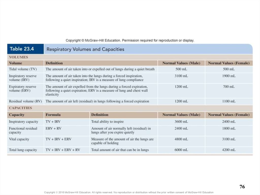
72. 23.5f Volume and Capacity
• Spirometer measures respiratory volume– Can be used to assess respiratory health
o Standard values are available (e.g., for people of different ages)
– Four volumes measured by spirometry
o Tidal volume: amount of air inhaled or exhaled per breath during
quiet breathing
o Inspiratory reserve volume (IRV): amount of air that can be
forcibly inhaled beyond the tidal volume
– Measure of compliance
o Expiratory reserve volume (ERV): amount that can be forcibly
exhaled beyond tidal volume
– Measure of elasticity
o Residual volume: amount of air left in the lungs after the most
forceful expiration
72
Copyright © 2016 McGraw-Hill Education. All rights reserved. No reproduction or distribution without the prior written consent of McGraw-Hill Education
73. 23.5f Volume and Capacity
• Four capacities calculated from respiratory volumes– Inspiratory capacity (IC)
o Tidal volume + inspiratory reserve volume
– Functional residual capacity (FRC)
o Expiratory reserve volume + residual volume
o Volume left in the lungs after a quiet expiration
– Vital capacity
o Tidal volume + inspiratory and expiratory reserve volumes
o Total amount of air a person can exchange through forced breathing
– Total lung capacity (TLC)
o Sum of all volumes, including residual volume
o Maximum volume of air that the lungs can hold
73
Copyright © 2016 McGraw-Hill Education. All rights reserved. No reproduction or distribution without the prior written consent of McGraw-Hill Education
74. 23.5f Volume and Capacity
• Additional respiratory measurements—rates of airmovement
– Forced expiratory volume (FEV)
o Percent of vital capacity that can be expelled in a set period of time
o FEV1 = percentage expelled in one second
o 75–85% of vital capacity in a healthy person
– Less in emphysema patients and others with poor expiration
– Maximum voluntary ventilation (MVV)
o Greatest amount of air that can be taken in and then expelled from the
lungs in 1 minute
o Breathing as quickly and as deeply as possible
o Can be as high as 30 L/min (compared to 6 L/min at rest)
o All respiratory disorders impair this
74
Copyright © 2016 McGraw-Hill Education. All rights reserved. No reproduction or distribution without the prior written consent of McGraw-Hill Education
75. Respiratory Volumes and Capacities
Figure 23.2475
Copyright © 2016 McGraw-Hill Education. All rights reserved. No reproduction or distribution without the prior written consent of McGraw-Hill Education
76.
76Copyright © 2016 McGraw-Hill Education. All rights reserved. No reproduction or distribution without the prior written consent of McGraw-Hill Education
77. What did you learn?
What is Boyle’s law and how
does it relate to respiration?
Which muscles are involved in
quiet respiration, and what
nerves control them?
To what chemical signal is the
body most sensitive with
regard to respiratory control?
What parts of the brain control
respiration
What is vital capacity?
Copyright © 2016 McGraw-Hill Education. All rights reserved. No reproduction or distribution without the prior written consent of McGraw-Hill Education
77
78. 23.6 Respiration: Alveolar and Systemic Gas Exchange
LearningObjectives:
Define partial pressure and the
movement of gases relative to
a partial pressure gradient.
2. Describe the partial pressures
that are relevant to gas
exchange.
3. Explain the laws that govern
gas solubility.
4. Describe alveolar gas
exchange and the partial
pressure gradients responsible.
1.
Copyright © 2016 McGraw-Hill Education. All rights reserved. No reproduction or distribution without the prior written consent of McGraw-Hill Education
78
79. 23.6 Respiration: Alveolar and Systemic Gas Exchange (continued)
LearningObjectives:
5.
6.
7.
8.
Name the two anatomic features
of the respiratory membrane that
contribute to efficient alveolar
gas exchange.
Explain ventilation-perfusion
coupling and how it maximizes
alveolar gas exchange.
Explain the partial pressure
gradients between the systemic
cells and the blood in capillaries.
Differentiate between alveolar
and systemic gas exchange.
Copyright © 2016 McGraw-Hill Education. All rights reserved. No reproduction or distribution without the prior written consent of McGraw-Hill Education
79
80. 23.6a Chemical Principles of Gas Exchange
Partial pressure and Dalton’s law• Partial pressure: pressure exerted by each gas within
a mixture of gases, measured in mm Hg
– Written with P followed by gas symbol (i.e., PO2 )
– Each gas moves independently down its partial pressure gradient during
gas exchange
• Atmospheric pressure = 760 mm Hg at sea level
– Total pressure all gases collectively exert in the environment
– Includes N2, O2, CO2, H2O, and other minor gases
80
Copyright © 2016 McGraw-Hill Education. All rights reserved. No reproduction or distribution without the prior written consent of McGraw-Hill Education
81. 23.6a Chemical Principles of Gas Exchange
Partial pressure and Dalton’s law (continued)• Total pressure × % of gas = Partial pressure of that gas
– Nitrogen is 78.6% of the gas in air
– 760 mm HG × 78.6% = 597 mm Hg = partial pressure of
nitrogen
– Partial pressures added together equal the total atmospheric
pressure
• Dalton’s law
– The total pressure in a mixture of gases is equal to the sum
of the individual partial pressures
81
Copyright © 2016 McGraw-Hill Education. All rights reserved. No reproduction or distribution without the prior written consent of McGraw-Hill Education
82. 23.6a Chemical Principles of Gas Exchange
Partial pressure gradients• Gradient exists when partial pressure for a gas is higher in one
region of the respiratory system than another
• Gas moves from region of higher partial pressure to region of
lower partial pressure until pressures become equal
• Both types of gas exchange depend on gradients
– Alveolar gas exchange: between blood in pulmonary capillaries and
alveoli
– Systemic gas exchange: between blood in systemic capillaries and
systemic cells
82
Copyright © 2016 McGraw-Hill Education. All rights reserved. No reproduction or distribution without the prior written consent of McGraw-Hill Education
83. 23.6a Chemical Principles of Gas Exchange
Relevant partial pressures in the body• Reasons partial pressures in alveoli differ from atmospheric
partial pressures
– Air from environment mixes with air remaining in anatomic
dead space
– Oxygen diffuses out of alveoli into the blood; carbon dioxide diffuses
from blood into alveoli
– More water vapor is present in alveoli than in atmosphere
• Within alveoli, the…
– percentage and partial pressure of O2 are lower than in atmosphere
– percentage and partial pressure of CO2 are higher than in atmosphere
– partial pressures of respiratory gases normally stay constant
83
Copyright © 2016 McGraw-Hill Education. All rights reserved. No reproduction or distribution without the prior written consent of McGraw-Hill Education
84. 23.6a Chemical Principles of Gas Exchange
Relevant partial pressures in the body (continued)• In systemic cells, partial pressures of gases reflect cellular
respiration (use of O2, production of CO2)
– The percentage of O2 lower and CO2 higher than in alveoli
– Under resting, normal conditions the partial pressures remain constant
• In circulating blood, gas partial pressures are not constant
– O2 enters blood in pulmonary capillaries; CO2 leaves
– O2 leaves blood in systemic capillaries; CO2 enters
84
Copyright © 2016 McGraw-Hill Education. All rights reserved. No reproduction or distribution without the prior written consent of McGraw-Hill Education
85. Alveolar and Systemic Gas Exchange
Figure 23.2585
Copyright © 2016 McGraw-Hill Education. All rights reserved. No reproduction or distribution without the prior written consent of McGraw-Hill Education
86. 23.6a Chemical Principles of Gas Exchange
Gas solubility and Henry’s law• Henry’s law: at a given temperature, the solubility of a gas in
liquid is dependent upon the
– Partial pressure of the gas in the air
– Solubility coefficient of the gas in the liquid
• Partial pressure: driving force moving gas into liquid
– Determined by total pressure and percentage of gas in the mixture
– E.g., CO2 is forced into soft drinks under high pressure
• Solubility coefficient: volume of gas that dissolves in a
specified volume of liquid at a given temperature and pressure
– A constant that depends upon interactions between molecules of the gas
and liquid
86
Copyright © 2016 McGraw-Hill Education. All rights reserved. No reproduction or distribution without the prior written consent of McGraw-Hill Education
87. 23.6a Chemical Principles of Gas Exchange
Gas solubility and Henry’s law (continued)• Gases vary in their solubility in water
– Carbon dioxide about 24 times as soluble as oxygen
– Nitrogen about half as soluble as oxygen
o It does not normally dissolve in blood in significant amounts
– Gases with low solubility require larger pressure gradients
to “push” the gas into the liquid
87
Copyright © 2016 McGraw-Hill Education. All rights reserved. No reproduction or distribution without the prior written consent of McGraw-Hill Education
88. Clinical View: Decompression Sickness and Hyperbaric Oxygen Chambers
• Decompression sickness (the bends)– Occurs when a diver is submerged in water beyond a certain
depth and returns too quickly to the surface
– Nitrogen forced into the blood due to the higher pressure
– Dissolved nitrogen bubbles out of solution while still in blood
and tissues
– Treated with hyperbaric oxygen chambers
o Partial pressure gradient for oxygen increased
o Additional oxygen can dissolve in blood plasma
o Can be used for other disorders such as carbon monoxide poisoning
88
Copyright © 2016 McGraw-Hill Education. All rights reserved. No reproduction or distribution without the prior written consent of McGraw-Hill Education
89. 23.6b Alveolar Gas Exchange (External Respiration)
• Oxygen– PO2 in alveoli is 104 mm Hg
– PO2 of blood entering pulmonary capillaries is 40 mm Hg
– Oxygen diffuses across respiratory membrane from alveoli
into the capillaries
– Continues until blood PO2 is equal to that of alveoli
– Levels in alveoli remain constant as fresh air continuously
enters
89
Copyright © 2016 McGraw-Hill Education. All rights reserved. No reproduction or distribution without the prior written consent of McGraw-Hill Education
90. 23.6b Alveolar Gas Exchange (External Respiration)
• Carbon dioxide– PCO2 in alveoli 40 mm Hg
– PCO2 in blood of
pulmonary capillaries
45 mm Hg
– Carbon dioxide diffuses
from blood to alveoli
– Continues until blood
levels equal alveoli levels
– Levels in alveoli remain
constant
Figure 23.25b
90
Copyright © 2016 McGraw-Hill Education. All rights reserved. No reproduction or distribution without the prior written consent of McGraw-Hill Education
91. Clinical View: Emphysema
• Emphysema causes–
–
–
–
–
Irreversible loss of pulmonary gas exchange surface area
Inflammation of air passageways distal to terminal bronchioles
Widespread destruction of pulmonary elastic connective tissue
Dilation and decreased total number of alveoli
Inability to expire effectively
• Emphysema is caused by
– In most cases, smoking
– Rarely from an alpha-1 antitrypsin deficiency
91
Copyright © 2016 McGraw-Hill Education. All rights reserved. No reproduction or distribution without the prior written consent of McGraw-Hill Education
92. 23.6b Alveolar Gas Exchange (External Respiration)
Efficiency of gas exchange at respiratory membrane• Anatomical features of membrane contributing to efficiency
– Large surface area (70 square meters)
– Minimal thickness (0.5 micrometers)
• Physiologic adjustments: ventilation-perfusion coupling
– Ability of bronchioles to regulate airflow and arterioles to regulate blood
flow
o Ventilation changes by bronchodilation or bronchoconstriction
– E.g., dilation in response to increased PCO2 in air in bronchiole
o Perfusion changes by pulmonary arteriole dilation or constriction
– E.g., dilation in response to either decreased PCO2 or increased PO2 in blood
92
Copyright © 2016 McGraw-Hill Education. All rights reserved. No reproduction or distribution without the prior written consent of McGraw-Hill Education
93. Ventilation-Perfusion Coupling
Figure 23.2693
Copyright © 2016 McGraw-Hill Education. All rights reserved. No reproduction or distribution without the prior written consent of McGraw-Hill Education
94. Clinical View: Respiratory Diseases and Efficiency of Alveolar Gas Exchange
• Certain diseases decrease the efficiency of oxygen andcarbon dioxide exchange
– Due to decreased number of alveoli (e.g., lung cancer)
– Due to thickened respiratory membrane (e.g., congestive
heart failure)
– Due to changes in ventilation-perfusion coupling (e.g.,
asthma or pulmonary embolism)
• Diseases result in decreased blood PO2 and increased
blood CO2
94
Copyright © 2016 McGraw-Hill Education. All rights reserved. No reproduction or distribution without the prior written consent of McGraw-Hill Education
95. 23.6c Systemic Gas Exchange (Internal Respiration)
• Oxygen diffuses out of systemic capillaries to entersystemic cells
– Partial pressure gradient drives the process
o PO2 in systemic cells 40 mm Hg
o PO2 in systemic capillaries is 95 mm Hg
– Continues until blood PO2 is 40 mm Hg
– Systemic cell PO2 stays fairly constant
o Oxygen delivered at same rate it is used unless engaging in strenuous
activity
95
Copyright © 2016 McGraw-Hill Education. All rights reserved. No reproduction or distribution without the prior written consent of McGraw-Hill Education
96. 23.6c Systemic Gas Exchange (Internal Respiration)
• Carbon dioxide– Diffuses from systemic
cells to blood
– Partial pressure gradient
driving process
o PCO2 in systemic cells
45mm Hg
o PCO2 in systemic
capillaries 40 mm Hg
– Diffusion continuing until
blood PCO2 is 45 mm Hg
Figure 23.25c
Copyright © 2016 McGraw-Hill Education. All rights reserved. No reproduction or distribution without the prior written consent of McGraw-Hill Education
97.
Integration of Gas Exchange• Alveolar gas exchange decreases blood PCO2, whereas systemic gas
exchange increases it
• Alveolar gas exchange increases blood PO2, whereas systemic gas
exchange decreases it
̶
̶
As blood leaves pulmonary capillaries PO2 is 104 mm Hg
Mingling with deoxygenated blood from bronchial veins (in pulmonary veins)
results in a PO2 of 95 mm Hg in left heart
97
Copyright © 2016 McGraw-Hill Education. All rights reserved. No reproduction or distribution without the prior written consent of McGraw-Hill Education
98. What did you learn?
What is a partial pressure?
How does Henry’s law relate
to human respiration?
What are the partial pressure
gradients for the respiratory
gases in the alveoli?
What anatomical features of
the respiratory membrane
foster gas exchange?
Copyright © 2016 McGraw-Hill Education. All rights reserved. No reproduction or distribution without the prior written consent of McGraw-Hill Education
98
99. 23.7 Respiration: Gas Transport
LearningObjectives:
1.
2.
3.
4.
5.
Explain why hemoglobin is
essential to oxygen transport.
Describe the three ways carbon
dioxide is transported in the
blood.
Explain the conversion of CO2 to
and from HCO3– within
erythrocytes.
Name the three substances
carried by hemoglobin.
Explain the significance of the
oxygen-hemoglobin saturation
curve for both alveolar and
systemic gas exchange.
Copyright © 2016 McGraw-Hill Education. All rights reserved. No reproduction or distribution without the prior written consent of McGraw-Hill Education
99
100. 23.7a Oxygen Transport
• Blood’s ability to transport oxygen depends on– Solubility coefficient of oxygen
o This is very low, and so very little oxygen dissolves in plasma
– Presence of hemoglobin
o The iron of hemoglobin attaches oxygen
o About 98% of O2 in blood is bound to hemoglobin
– HbO2 is oxyhemoglobin (with oxygen bound)
– HHb is deoxyhemoglobin (without bound oxygen)
100
Copyright © 2016 McGraw-Hill Education. All rights reserved. No reproduction or distribution without the prior written consent of McGraw-Hill Education
101. Erythrocytes
101Copyright © 2016 McGraw-Hill Education. All rights reserved. No reproduction or distribution without the prior written consent of McGraw-Hill Education
102. Clinical View: Measuring Blood Oxygen Levels with a Pulse Oximeter
• Noninvasive and indirect way to measure oxygen̶
Applied to finger or earlobe
• Measure hemoglobin saturation by determining the
ratio of oxyhemoglobin to deoxyhemoglobin
• Normal reading hemoglobin saturation >95%
102
Copyright © 2016 McGraw-Hill Education. All rights reserved. No reproduction or distribution without the prior written consent of McGraw-Hill Education
103. 23.7b Carbon Dioxide Transport
• Carbon dioxide has three means of transport– As CO2 dissolved in plasma (7%)
– As CO2 attached to amine group of globin portion of
hemoglobin (23%)
o HbCO2 is carbaminohemoglobin
– As bicarbonate dissolved in plasma (70%)
o CO2 diffuses into erythrocytes and combines with water to form
bicarbonate and hydrogen ion
o Bicarbonate diffuses into plasma
o CO2 is regenerated when blood moves through pulmonary capillaries
and the process is reversed
103
Copyright © 2016 McGraw-Hill Education. All rights reserved. No reproduction or distribution without the prior written consent of McGraw-Hill Education
104. Conversion of CO2 to Bicarbonate (Figure 23.27)
Copyright © 2016 McGraw-Hill Education. All rights reserved. No reproduction or distribution without the prior written consent of McGraw-Hill Education105. 23.7c Hemoglobin as a Transport Molecule
• Hemoglobin transports– Oxygen attached to iron
– Carbon dioxide bound to globin part
– Hydrogen ions bound to globin part
• Binding of one substance causes a change in shape of
the hemoglobin molecule
– Influences the ability of hemoglobin to bind or release the
other two substances
105
Copyright © 2016 McGraw-Hill Education. All rights reserved. No reproduction or distribution without the prior written consent of McGraw-Hill Education
106. 23.7c Hemoglobin as a Transport Molecule
Oxygen-hemoglobin saturation curve• Each hemoglobin can bind up to four O2 molecules
– One on each iron atom in the hemoglobin molecule
• Percent O2 saturation of hemoglobin is crucial
– It is the amount of oxygen bound to available hemoglobin
– Saturation increases as PO2 increases
o Cooperative binding effect: each O2 that binds causes a change in
hemoglobin making it easier for next O2 to bind
– Graphed in the oxygen-hemoglobin saturation curve
o S-shaped, nonlinear relationship
106
Copyright © 2016 McGraw-Hill Education. All rights reserved. No reproduction or distribution without the prior written consent of McGraw-Hill Education
107. Oxygen-Hemoglobin Saturation Curve
Figure 23.28107
Copyright © 2016 McGraw-Hill Education. All rights reserved. No reproduction or distribution without the prior written consent of McGraw-Hill Education
108. 23.7c Hemoglobin as a Transport Molecule
Oxygen-hemoglobin saturation curve (continued)– Large changes in saturation occur with small increases of PO2
at lower partial pressures (i.e., curve is initially steep)
– At PO2 higher than 60 mm Hg only small changes in
saturation occur
o About 90% saturation at 60 mm Hg
– Hemoglobin saturation is about 98% at pulmonary capillaries
as PO2 is 104 mm Hg
– Saturation can only reach 100% at pressures above 1 atm
(e.g., in hyperbaric oxygen chambers)
108
Copyright © 2016 McGraw-Hill Education. All rights reserved. No reproduction or distribution without the prior written consent of McGraw-Hill Education
109. 23.7c Hemoglobin as a Transport Molecule
Oxygen-hemoglobin saturation curve (continued)• Can use graph to determine saturation at a given PO2
– At 5000 ft, alveolar PO2 is 81 mm Hg
o Corresponds to a hemoglobin saturation of 95%
– At 17,000 ft, alveolar PO2 is 40 mm Hg
o Corresponds to a hemoglobin saturation of 75%
• Altitude sickness
– Adverse physiologic effects from a decrease in alveolar PO2
and low oxygen saturation
– Includes symptoms of headache, nausea, pulmonary edema,
and cerebral edema
109
Copyright © 2016 McGraw-Hill Education. All rights reserved. No reproduction or distribution without the prior written consent of McGraw-Hill Education
110. 23.7c Hemoglobin as a Transport Molecule
Oxygen-hemoglobin saturation curve (continued)• Some (not all) oxygen released from hemoglobin at systemic
capillaries
– 98% saturation as it leaves the lungs (at sea level)
– About 75% saturation after passing systemic cells at rest
o Only 20–25% of transported oxygen is released
– Oxygen reserve: O2 remaining bound to hemoglobin after passing
through systemic circulation
o Provides a means for additional oxygen to be delivered under increased
metabolic demands (e.g., exercise)
o Vigorous exercise produces a significant drop in saturation
– Blood leaving capillaries in active muscles only about 35% saturated
110
Copyright © 2016 McGraw-Hill Education. All rights reserved. No reproduction or distribution without the prior written consent of McGraw-Hill Education
111. 23.7c Hemoglobin as a Transport Molecule
Other variables that influence oxygen release fromhemoglobin during systemic exchange
• Temperature
– Elevated temperature diminishes hemoglobin’s hold on
oxygen
• H+ binding to hemoglobin
– Hydrogen ion binds to hemoglobin and causes a
conformational change
– This causes decreased affinity for O2 and oxygen release
o Called the Bohr effect
111
Copyright © 2016 McGraw-Hill Education. All rights reserved. No reproduction or distribution without the prior written consent of McGraw-Hill Education
112. 23.7c Hemoglobin as a Transport Molecule
Other variables that influence oxygen release fromhemoglobin during systemic exchange (continued)
• Presence of 2,3-BPG: a molecule in erythrocytes
– Molecule binds hemoglobin, causing release of additional oxygen
– Certain hormones stimulate erythrocytes to produce 2,3-BPG
o Thyroid, epinephrine, growth hormone, and testosterone
• CO2 binding to hemoglobin
– Binding causes release of more oxygen from hemoglobin
• Haldane effect
– Release of oxygen causes a conformational change in hemoglobin
– Conformational change increases the amount of carbon dioxide that can
bind
112
Copyright © 2016 McGraw-Hill Education. All rights reserved. No reproduction or distribution without the prior written consent of McGraw-Hill Education
113. 23.7c Hemoglobin as a Transport Molecule
Other variables that influence oxygen release fromhemoglobin during systemic exchange (continued)
• Shifts to the saturation curve
– Some variables decrease oxygen affinity for hemoglobin
o Known as a shift right
o E.g., increased temperature, increase in hydrogen ion
– Other variables increase oxygen affinity to hemoglobin
o Known as a shift left
o E.g., decreased temperature, decrease in hydrogen ion
113
Copyright © 2016 McGraw-Hill Education. All rights reserved. No reproduction or distribution without the prior written consent of McGraw-Hill Education
114. Hemoglobin and Oxygen Release
Figure 23.29114
Copyright © 2016 McGraw-Hill Education. All rights reserved. No reproduction or distribution without the prior written consent of McGraw-Hill Education
115. Summary of Respiration
Figure 23.30a115
Copyright © 2016 McGraw-Hill Education. All rights reserved. No reproduction or distribution without the prior written consent of McGraw-Hill Education
116. Summary of Respiration
Figure 23.30b116
Copyright © 2016 McGraw-Hill Education. All rights reserved. No reproduction or distribution without the prior written consent of McGraw-Hill Education
117. Clinical View: Fetal Hemoglobin and Physiologic Jaundice
• Unborn babies have a different type of hemoglobinmolecule
– Fetal hemoglobin has a greater affinity for binding oxygen
than adult hemoglobin
– This ensures a net movement of oxygen from the blood of
the mother to the blood of the fetus
– Infants may have physiologic jaundice as fetal hemoglobin
breaks down
o Yellowish tinge to skin due to elevated levels of bilirubin
117
Copyright © 2016 McGraw-Hill Education. All rights reserved. No reproduction or distribution without the prior written consent of McGraw-Hill Education
118. What did you learn?
Why is so little O2 dissolved
in plasma?
How is most CO2 transported
in blood?
What does increased
temperature do to
hemoglobin’s hold on
oxygen?
What sort of shift to the
saturation curve is caused by
factors that decrease the
affinity between O2 and Hb?
Copyright © 2016 McGraw-Hill Education. All rights reserved. No reproduction or distribution without the prior written consent of McGraw-Hill Education
118
119. 23.8 Breathing Rate and Homeostasis
LearningObjectives:
1.
Explain how hyperventilation
and hypoventilation influence
the chemical composition of
blood.
2.
Describe how breathing rate
and depth effects venous return
of blood and lymph.
3.
Explain the changes in
breathing that accompany
exercise.
Copyright © 2016 McGraw-Hill Education. All rights reserved. No reproduction or distribution without the prior written consent of McGraw-Hill Education
119
120. 23.8a Effects of Hyperventilation and Hypoventilation on Cardiovascular Function
• Hyperventilation: breathing rate or depth above body’sdemand
– Caused by anxiety, ascending to high altitude, or voluntarily
– PO2 rises and PCO2 fall in the air of alveoli
– Additional oxygen does not enter blood because hemoglobin
is already 98% saturated
– There is greater loss of CO2 from blood, called hypocapnia
120
Copyright © 2016 McGraw-Hill Education. All rights reserved. No reproduction or distribution without the prior written consent of McGraw-Hill Education
121. 23.8a Effects of Hyperventilation and Hypoventilation on Cardiovascular Function
• Hyperventilation (continued)– Low blood CO2 causes vasoconstriction
o Brain vessel constriction can decrease oxygen delivery to the brain
– May decrease blood hydrogen ion concentration
o If buffers cannot compensate, result is respiratory alkalosis
– Hyperventilation may cause
o Feeling faint or dizzy, numbness, tingling, cramps, and tetany
o If prolonged, disorientation, loss of consciousness, coma, possible
death
121
Copyright © 2016 McGraw-Hill Education. All rights reserved. No reproduction or distribution without the prior written consent of McGraw-Hill Education
122. 23.8a Effects of Hyperventilation and Hypoventilation on Cardiovascular Function
• Hypoventilation: breathing too slow (bradypnea) ortoo shallow (hypopnea)
– Causes include: airway obstruction, pneumonia, brainstem
injury, other respiratory conditions
– O2 levels down, CO2 levels up in alveoli
– Blood PO2 decreases (hypoxemia); and can lead to low
oxygen in tissues (hypoxia)
– Blood PCO2 increases (hypercapnia)
122
Copyright © 2016 McGraw-Hill Education. All rights reserved. No reproduction or distribution without the prior written consent of McGraw-Hill Education
123. 23.8a Effects of Hyperventilation and Hypoventilation on Cardiovascular Function
• Hypoventilation (continued)– May result in inadequate oxygen delivery
– May result in increased hydrogen ion concentration due to
high blood PCO2
o Might result in respiratory acidosis
– May cause
o Lethargy, sleepiness, headache, polycythemia, cyanotic tissues
o If prolonged, convulsions, loss of consciousness, death
123
Copyright © 2016 McGraw-Hill Education. All rights reserved. No reproduction or distribution without the prior written consent of McGraw-Hill Education
124. 23.8b Breathing and Exercise
• While exercising, breathing shows hyperpnea to meetincreased tissue needs
– Breathing depth increases while rate remains the same
• Blood PO2 and Blood PCO2 remain relatively constant
– Increased cellular respiration compensated for by deeper
breathing, increased cardiac output, greater blood flow
– The respiratory center is stimulated from one or more causes
o Proprioceptive sensory signals in response to movement
o Corrollary motor output from cerebral cortex relayed to respiratory
center
o Conscious anticipation of exercise
124
Copyright © 2016 McGraw-Hill Education. All rights reserved. No reproduction or distribution without the prior written consent of McGraw-Hill Education
125. What did you learn?
If hyperventilation is
excessive, what change occurs
in blood pH?
What distinguishes
hyperventilation from
hyperpnea?
What happens to blood levels
of oxygen and carbon dioxide
during exercise?
Copyright © 2016 McGraw-Hill Education. All rights reserved. No reproduction or distribution without the prior written consent of McGraw-Hill Education
125