


")
")


")
. Two mechanisms are believed to cause inflammation at the sites of deposition:")


")
 Медицина
Похожие презентации:
")


Pathology of immune system
1.
ZAPOROZHZHIAN STATE MEDICAL UNIVERSITYThe department of pathological anatomy and forensic
medicine with basis of law
Pathology of
immune system
Lecture on pathological anatomy for the
3-rd year students
2.
Functions of immune system1. To provide defense of organism from:
- any infection,
- cells-mutants,
- tumor cells,
- transplanted cells,
- any substances which are recognized
by IS as foreign.
2. Permanent control by lymphocytes of
AG composition of own cells in
accordance to the HLA type 1 or 2
3.
Organs of IS:Central organs:
bone marrow and thymus
There is a permanent rhythmic new
formation of immune cells not
depending on immune (AG)
stimulation of organism
Peripheral organs:
lymphatic nodules, spleen, mucosaassociated lymphoid tissue
Additional reproduction and AG-related
differentiation of immune cells takes place
there in reply on the AG-stimulation
4. Bone marrow
1. Active (red) bone marrow consists oflymphopoetic cells, fatty tissue, vessels
and fibroblasts. At adults it is situated in
a breastbone, bodies of vertebrae, ribs
and pelvic bones.
Children
have active marrow in the
tubular bones, but after children
became grown up it change into yellow.
2. Yellow bone marrow can transform into
red if it is necessary.
After 70 years old the atrophy of myeloid
tissue is seen, it is consists of fatty tissue
and fibrocytes.
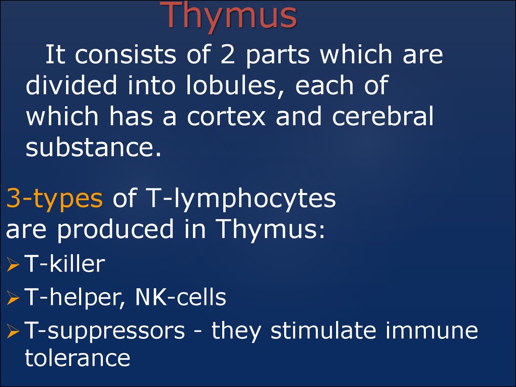
5.
ThymusIt consists of 2 parts which are
divided into lobules, each of
which has a cortex and cerebral
substance.
3-types of Т-lymphocytes
are produced in Thymus:
Т-killer
Т-helper, NK-cells
T-suppressors - they stimulate immune
tolerance
6.
LYMPHATIC NODULESControl and AG recognition is provided in a lymph:
Size of lymphatic nodules: 3-30мм.
Lymphatic nodules
consists of: - cortex
- medullar substances
In cortex there are lymphatic follicles
(primary and secondary – with the center of
reproduction
–B-cells are formed there (B-zone)
In cerebral substances there are
veins and lymphatic sinuses.
7.
SPLEENControl and the AGrecognition of blood.
Every artery is
surrounded by lymphatic
follicles. Red pulp is a
potential active
mesenchymal tissue (there are B-lymphocytes)
Mass =150-180 gr.
A spleen can deposit up to the 2/3 of vein blood
volume.
In pathology it is an organ of extra-medullar
blood-formation.
8.
MALTMucosa-associated lymphoid tissue includes the
following structures:
— Lymphatic pharyngeal ring with the
pharyngeal, lingual, and palatine tonsils;
— Gut-associated lymphoid tissue (the follicles
of the duodenum, appendix, colon);
— Bronchi-associated lymphoid tissue
(in the peribronchial fascial sheath);
— Exocrine glands (salivary glands and pancreas);
— Mammary glands.
9.
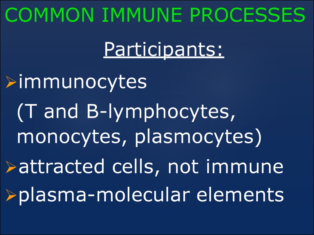
COMMON IMMUNE PROCESSESParticipants:
immunocytes
(Т and B-lymphocytes,
monocytes, plasmocytes)
attracted cells, not immune
plasma-molecular elements
10.
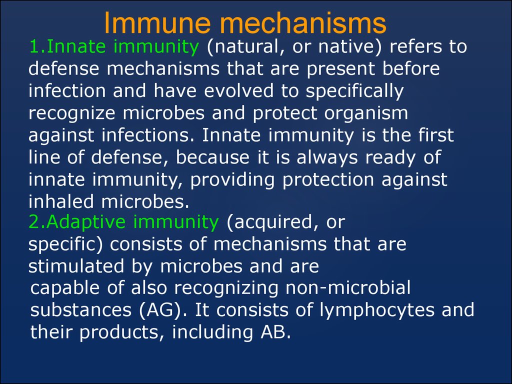
Immune mechanisms1.Innate immunity (natural, or native) refers to
defense mechanisms that are present before
infection and have evolved to specifically
recognize microbes and protect organism
against infections. Innate immunity is the first
line of defense, because it is always ready of
innate immunity, providing protection against
inhaled microbes.
2.Adaptive immunity (acquired, or
specific) consists of mechanisms that are
stimulated by microbes and are
capable of also recognizing non-microbial
substances (AG). It consists of lymphocytes and
their products, including AB.
11.
Adaptive immunityThere are two main types:
1.cell-mediated (or cellular) immunity, which is
responsible for defense against intracellular
microbes, it is mediated by T (thymus-derived)
lymphocytes
Performed by: - T-killer
- NK-cells
- macrophages
- labrocytes
- leucocytes (basophiles, neutrophils)
The result is formation of infiltrates and
granulomas, displays of immune cell killing
At final stage – phagocytosis and destruction of
intracellular bacteria and viruses, fungi, tumor
and transplanted cells
12.
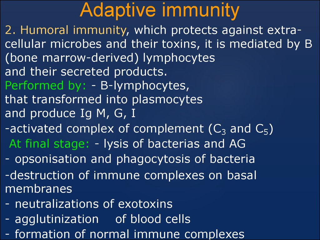
Adaptive immunity2. Humoral immunity, which protects against extracellular microbes and their toxins, it is mediated by B
(bone marrow-derived) lymphocytes
and their secreted products.
Performed by: - B-lymphocytes,
that transformed into plasmocytes
and produce Ig M, G, I
-activated complex of complement (C3 and C5)
At final stage: - lysis of bacterias and AG
- opsonisation and phagocytosis of bacteria
-destruction of immune complexes on basal
membranes
- neutralizations of exotoxins
- agglutinization of blood cells
- formation of normal immune complexes
13.
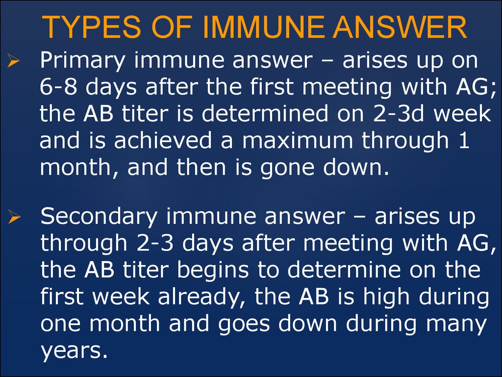
TYPES OF IMMUNE ANSWERPrimary immune answer – arises up on
6-8 days after the first meeting with AG;
the AB titer is determined on 2-3d week
and is achieved a maximum through 1
month, and then is gone down.
Secondary immune answer – arises up
through 2-3 days after meeting with AG,
the AB titer begins to determine on the
first week already, the AB is high during
one month and goes down during many
years.
14. Pathology of the immune system
Reactions of hypersensitivenessImmunodeficiency syndromes
Autoimmune diseases
Amyloidosis
Tumors of the lymphatic system
15.
Reaction of hypersensitivityThe reaction of hypersensitivity is the
individual reaction on the repeated
reception of AG, which is exceeds the
measure of biological expedience on the
express and effect, because it is completed
by destruction of the cells and violation of
function.
These reactions are unusual on the
methods of answer. The basis of these
reactions is a normal immune answer
which is perverted because of the unusual
reception or promoted reception of AG.
16. Types of reactions:
Immediate hypersensitivity –anaphylactic type (Type I),
Antibody-mediated disorders (Type II
hypersensitivity),
Immune complex-mediated disorders
(type III hypersensitivity ),
Cell-mediated immune disorders -
slow type (type IV hypersensitivity ).
17. Immediate hypersensitivity (Type I)
It is a rapidly developingimmunologic reaction occurring
within minutes after the
connection of an AG with AB
bound to mast cells in individuals
previously sensitized to the
antigen
18. Immediate hypersensitivity (type I)
Immune mechanism is:Production of IgE antibody ―immediate release
of vasoactive amines and other mediators from
mast cells; recruitment of inflammatory cells
(late-phase reaction)
Pathologic lesions:
Vascular dilation, edema,
Smooth muscle
contraction,
Mucus production,
Inflammation
19.
Immediate hypersensitivity (type I) may occur as:1.a systemic disorder - it follows injection of an AG to
which the host has become sensitized
2.a local reaction - the nature of reactions varies
depending on the entering of the AG
Systemic displays:
- spasm of respirator bronchiole (difficulty of
breathing) - acute respiratory insufficiency,
- respiratory D-stress syndrome (RDS),
- system disorders of haemodynamic and rapid
expansion of vessels – it leads to collapse with the
loss of consciousness and decreasing of arterial
pressure,
- gastroenteritis – it is spastically stomachaches, vomiting and diarrhea,
- allergens cause development of anaphylactic shock,
and medicinal allergens – anaphylactic reaction.
20. Local displays
1.localized cutaneous swellings (skin allergy, hives) as hyperemia (anaemia), edema, blisters,neurodermitis,
2.hay fever,
3. nasal and conjunctiva discharge (allergic
rhinitis, conjunctivitis and sinusopathy), can be
seen at:
- inhalation of pollen of plants (polynosis), wool
of animals
- allergic edema of larynx after the appliqué
of medicines that leads to asphyxia.
4. allergic gastroenteritis (food allergy) develops on food allergens, the spasm of smooth
muscle and secretion of liquid in the road
clearance of bowel is seen (diarrhea).
21.
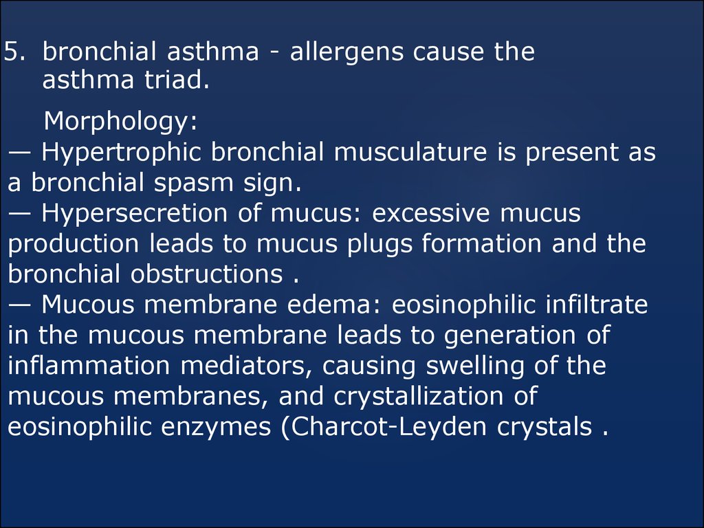
5. bronchial asthma - allergens cause theasthma triad.
Morphology:
— Hypertrophic bronchial musculature is present as
a bronchial spasm sign.
— Hypersecretion of mucus: excessive mucus
production leads to mucus plugs formation and the
bronchial obstructions .
— Mucous membrane edema: eosinophilic infiltrate
in the mucous membrane leads to generation of
inflammation mediators, causing swelling of the
mucous membranes, and crystallization of
eosinophilic enzymes (Charcot-Leyden crystals .
22.
АB-mediated hypersensitivity - type IIIt is mediated by AB directed
toward AG present on cell surfaces
or extra-cellular matrix.
The antigenic determinants may be
intrinsic to the cell membrane or
matrix, or they may take the form
of an exogenous AG, such as a
drug metabolite, that is adsorbed
on a cell surface or matrix.
23.
Mechanisms of AB-mediated reaction:1. Opsonization and Phagocytosis
AB connects with AG at the cellꞌs surface (it is named opsonization).
This process is necessary for recognition the cell-target by
macrophages, with following destruction (phagocytosis).
Clinically it is occur as:
1.transfusion reactions;
2.erythroblastosis fetalis (hemolytic disease of the newborn);
3.autoimmune hemolytic anemia, agranulocytosis, and
thrombocytopenia, in which individuals produce
antibodies to their own blood cells, which are then
destroyed;
4. certain drug reactions, in which antibodies are
produced that react with the drug.
24.
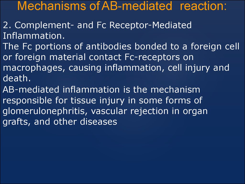
Mechanisms of AB-mediated reaction:2. Complement- and Fc Receptor-Mediated
Inflammation.
The Fc portions of antibodies bonded to a foreign cell
or foreign material contact Fc-receptors on
macrophages, causing inflammation, cell injury and
death.
AB-mediated inflammation is the mechanism
responsible for tissue injury in some forms of
glomerulonephritis, vascular rejection in organ
grafts, and other diseases
25.
Mechanisms of AB-dependent reaction:3. Antibody-Mediated Cellular Dysfunction
AB directed against cell-surface receptors impair or
dysregulate
function without causing cell injury or inflammation.
1. Myasthenia gravis - AB reactive with acetylcholine
receptors in the motor end-plates of skeletal muscles
impair neuromuscular transmission and therefore
cause muscle weakness.
2. In pemphigus vulgaris - AB against desmosomes
disrupt intercellular junctions in epidermis, leading to
the formation of skin vesicles.
3. In Graves disease - AB against the thyroid-stimulating
hormone receptor on thyroid epithelial cells stimulate
the cells, resulting in hyperthyroidism.
26.
Immune complex-mediatedhypersensitivity - type III
Antigen-antibody complexes produce tissue
damage mainly by eliciting inflammation
at the sites of deposition.
27. Immune complex-mediated hypersensitivity – type III
Immune mechanism is:Deposition of antigen-antibody complexes
leads to complement activation
recruitment of leukocytes by complement
products and Fc-receptors
release of
enzymes and other toxic molecules
Pathologic lesions:
Necrotizing vasculitis (fibrinoid necrosis)
Inflammation
28. Types of immune complex-mediated diseases (type III)
Types of immune complexmediateddiseases (type III)
1.generalized, if immune complexes are
formed in the blood circulation and are
deposited in many organs
2. localized in particular organs, such as:
- kidneys (glomerulonephritis),
- joints (arthritis),
- small blood vessels of the skin
if the complexes are formed and
deposited locally.
29.
Phases of immune complex disease1.formation of AG-AB complexes in
the blood circulation
2.deposition of the immune
complexes in various tissues, thus
initiating
3. an inflammatory reaction at the
sites of immune complex deposition
30.
The first phase is initiated by the introduction ofAG, usually a protein, and its interaction with
immune-competent cells, resulting in the
formation of AB.
These
AB
are
secreted into the
blood, where they
react with the AG
still present in the
circulation to form
AG-AB complexes.
In the second phase, the circulating AG-AB
complexes are deposited in various tissues.
31. Once complexes are deposited in the tissues, they initiate an acute inflammatory reaction (third phase). Two mechanisms are believed to cause inflammation at the sites of deposition:
1. activation of the complement cascade,2. activation of neutrophils and macrophages.
Complement
activation promotes
the migration of
polymorphonuclear
leukocytes and
monocytes and
inflammation.
Thrombi are formed
in the vessels,
resulting in local
ischemic injury.
32.
Types of immune complex-mediateddiseases (type III)
During this phase (approximately
10 days after AG administration),
clinical features appear:
- urticaria,
- arthralgias,
- lymph node enlargement,
- proteinuria.
33.
Cell-Mediated hypersensitivity (type IV) - slow typeIt is initiated by AG-activated (sensitized)
T-lymphocytes.
Mechanisms of T cell-mediated (type IV)
reactions
1. The delayed type hypersensitivity
reactions mediated by CD4+ T-cells.
CD4+ T cells (and sometimes CD8+ cells)
respond to tissue AG by secreting cytokines that
stimulate inflammation and activate phagocytes,
leading to tissue injury.
34.
Cell-Mediated hypersensitivity (type IV) - slow type2. T-cell mediated cytolysis. CD8+ cytolytic
T-lymphocytes directly kill tissue cells.
It is the principal pattern of immunologic
response to:
- a variety of intracellular microbiologic
agents, such as Mycobacterium tuberculosis,
- viruses, fungis, protozoa, and simplest.
35. Cell-Mediated hypersensitivity (type IV)
The immune mechanism ofactivating T-lymphocytes leads to:
1)Releasing of cytokines and
macrophage activation;
2)Activation of T-cell-mediated
cytotoxicity
Pathologic lesions:
Perivascular cellular infiltrates;
edema;
cell destruction;
granuloma formation