

 Медицина
Похожие презентации:


")


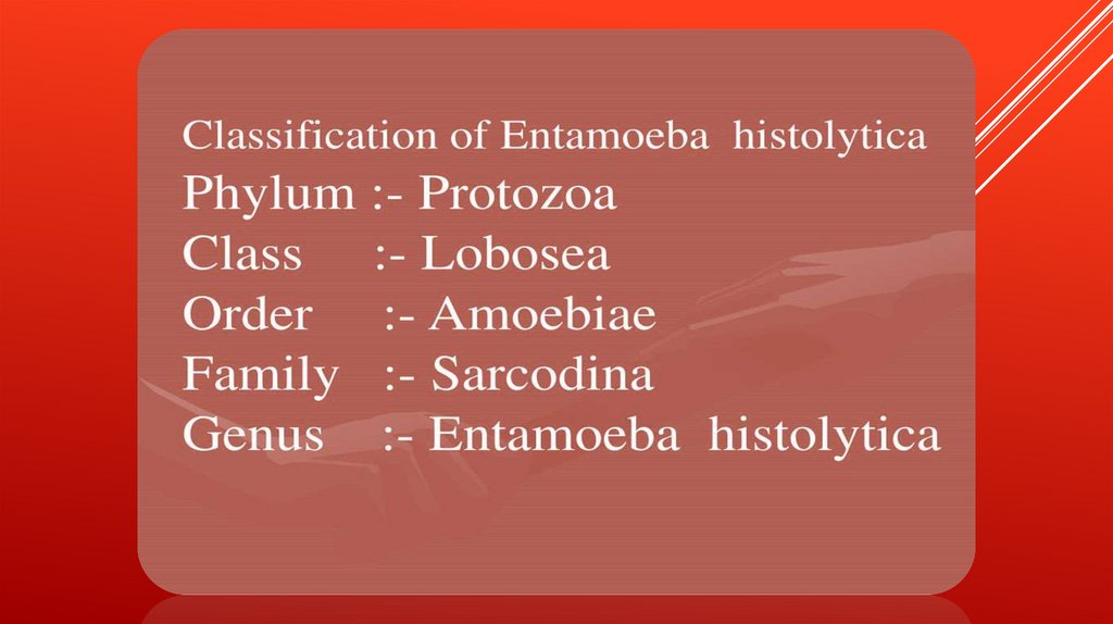
Histolytica
1.
2.
3.
4.
Anaerobic parasitic amoebozoan, part ofthe genus Entamoeba.[1] Predominantly
infecting humans and other primates
causing amoebiasis, E. histolytica is
estimated to infect about 35-50 million
people worldwide.[1] E. histolytica infection is
estimated to kill more than 55,000 people
each year
5.
6.
7.
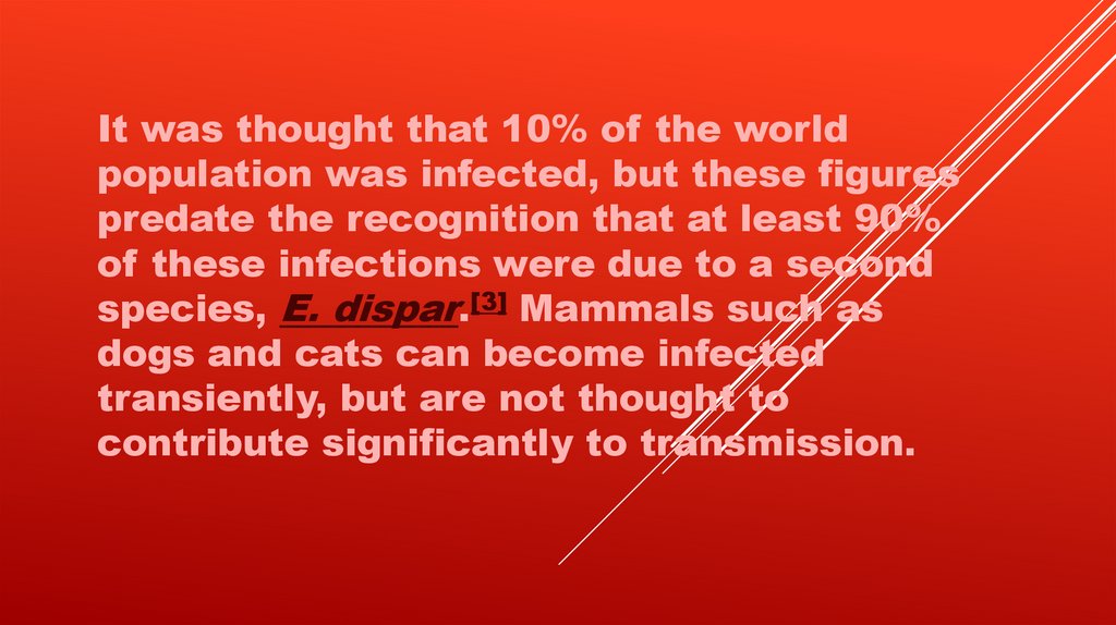
It was thought that 10% of the worldpopulation was infected, but these figures
predate the recognition that at least 90%
of these infections were due to a second
species, E. dispar.[3] Mammals such as
dogs and cats can become infected
transiently, but are not thought to
contribute significantly to transmission.
8.
9.
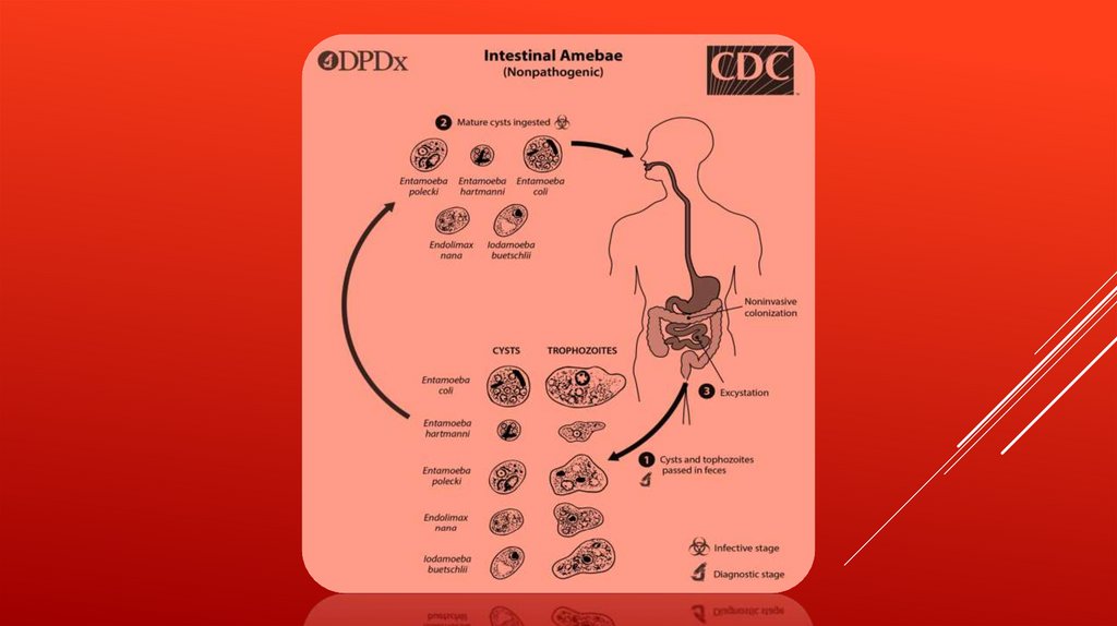
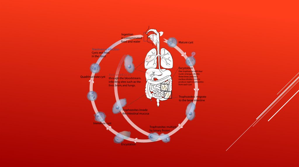
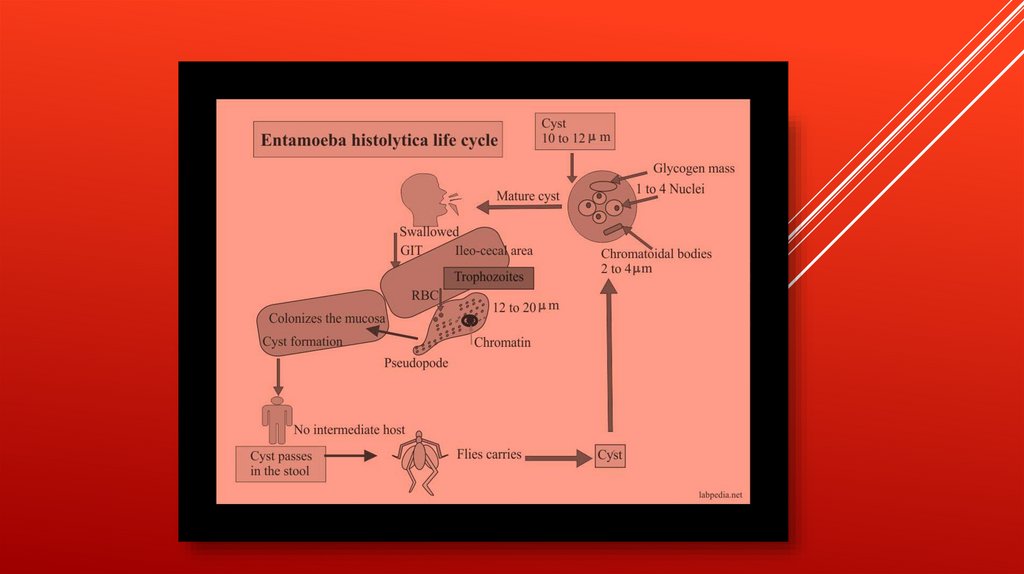
TRANSMISSIONThe active (trophozoite) stage exists only in the host and in
fresh loose feces; cysts survive outside the host in water, in
soils, and on foods, especially under moist conditions on the
latter. The infection can occur when a person puts anything
into their mouth that has touched the feces of a person
who is infected with E. histolytica, swallows something, such
as water or food, that is contaminated with E. histolytica, or
swallows E. histolytica cysts (eggs) picked up from
contaminated surfaces or fingers.
10.
11.
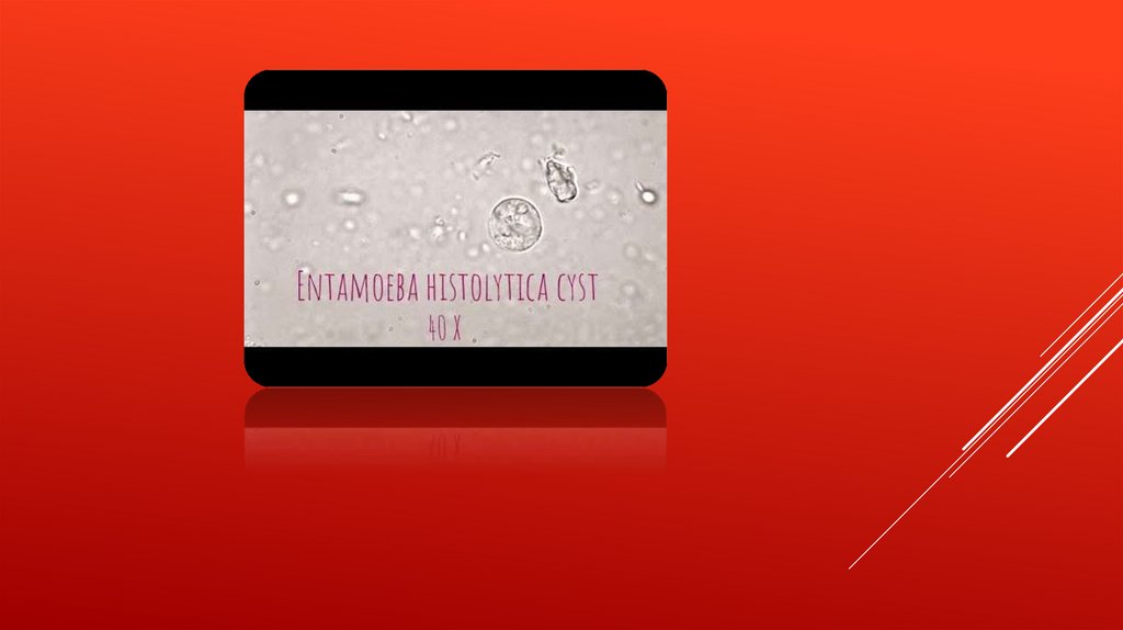
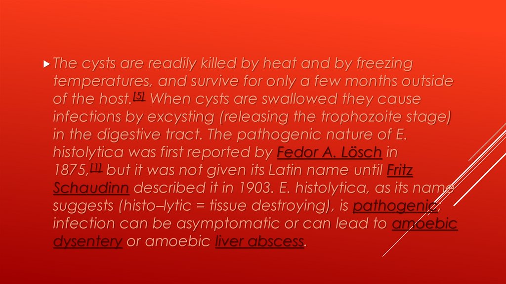
Thecysts are readily killed by heat and by freezing
temperatures, and survive for only a few months outside
of the host.[5] When cysts are swallowed they cause
infections by excysting (releasing the trophozoite stage)
in the digestive tract. The pathogenic nature of E.
histolytica was first reported by Fedor A. Lösch in
1875,[1] but it was not given its Latin name until Fritz
Schaudinn described it in 1903. E. histolytica, as its name
suggests (histo–lytic = tissue destroying), is pathogenic;
infection can be asymptomatic or can lead to amoebic
dysentery or amoebic liver abscess.
12.
13.
14.
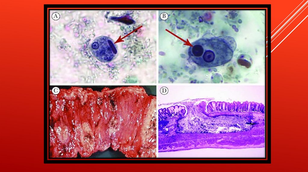
Symptomscan include fulminating dysentery, bloody
diarrhea, weight loss, fatigue, abdominal pain,
and amoeboma. The amoeba can actually 'bore' into
the intestinal wall, causing lesions and intestinal
symptoms, and it may reach the blood stream. From
there, it can reach different vital organs of the human
body, usually the liver, but sometimes the lungs, brain,
spleen, etc. A common outcome of this invasion of
tissues is a liver abscess, which can be fatal if untreated.
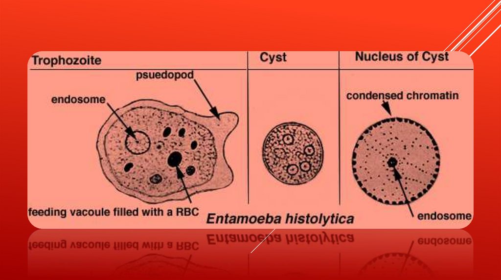
Ingested red blood cells are sometimes seen in the
amoeba cell cytoplasm.
15.
16.
RISK FACTORSPoor sanitary conditions are known to increase the risk of
contracting amebiasis E. histolytica.[8] In the United States,
there is a much higher rate of amebiasis-related mortality in
California and Texas, which might be caused by the
proximity of those states to E. histolytica-endemic areas,
such as Mexico, other parts of Latin America, and Asia.[9] E.
histolytica is also recognized as an emerging sexually
transmissible pathogen, especially in male homosexual
relations, causing outbreaks in non-endemic regions.[10] As
such, high-risk sex behaviour is also a potential source of
infection.[11] Although it is unclear whether there is a causal
link, studies indicate a higher chance of being infected
with E. histolytica if one is also infected with HIV
17.
18.
PATHOGEN INTERACTIONE. histolytica may modulate the virulence of certain human viruses and is itself a
host for its own viruses.
For example, AIDS accentuates the damage and pathogenicity of E.
histolytica.[13] On the other hand, cells infected with HIV are often consumed
by E. histolytica. Infective HIV remains viable within the amoeba, although there
has been no proof of human reinfection from amoeba carrying this virus.[23]
A burst of research on viruses of E. histolytica stems from a series of papers
published by Diamond et al. from 1972 to 1979. In 1972, they hypothesized two
separate polyhedral and filamentous viral strains within E. histolytica that caused
cell lysis. Perhaps the most novel observation was that two kinds of viral strains
existed, and that within one type of amoeba (strain HB-301) the polyhedral
strain had no detrimental effect but led to cell lysis in another (strain HK-9).
Although Mattern et al. attempted to explore the possibility that these protozoal
viruses could function like bacteriophages, they found no significant changes
in Entamoeba histolytica virulence when infected by viruses.
19.
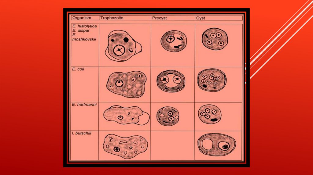
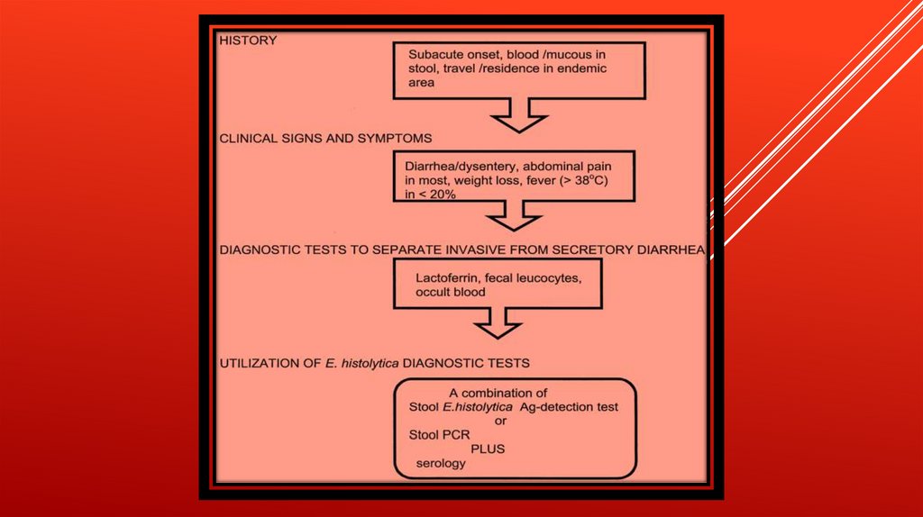
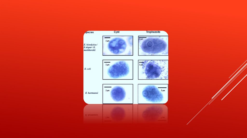
DIAGNOSISDiagnosis
is confirmed by microscopic examination for
trophozoites or cysts in fresh or suitably preserved faecal
specimens, smears of aspirates or scrapings obtained by
proctoscopy, and aspirates of abscesses or other tissue specimen.
A blood test is also available but is only recommended when a
healthcare provider believes the infection may have spread
beyond the intestine (gut) to some other organ of the body, such
as the liver. However, this blood test may not be helpful in
diagnosing current illness because the test can be positive if the
patient has had amebiasis in the past, even if they are not
infected at present.[26] Stool antigen detection and PCR are
available for diagnosis, and are more sensitive and specific than
microscopy.