")


Похожие презентации:


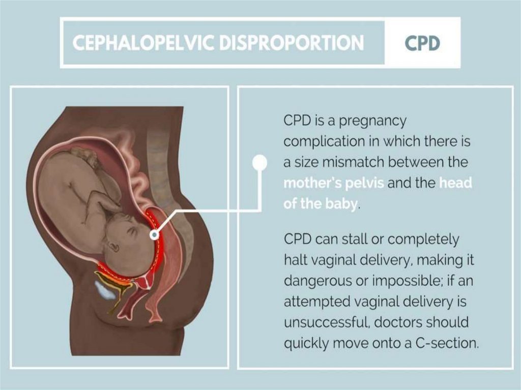
Cephalo pelvic disproportion (Сpd)
1. CEPHALO PELVIC DISPROPORTION (CPD)
Teacher : Kamilova Irina KaharovnaBy: Sulur PerumalSwamy Venkatesh Prabhu
Group : LA1-CO-163(B)
Year : 2020-2021 Course V
Date: 29-09-2020
2.
3. CPD either due to :-
• The baby’s head isproportionally too
large
• the mother’s pelvis is
too small
to easily allow the baby to fit
through the pelvic opening.
4. Causes :-
Causes :1. Large baby due to:• Hereditary factors
• Diabetes
• Postmaturity (still
pregnant after due date
has passed)
• Multiparity (not the
first pregnancy)
2. Abnormal fetal positions
3. contracted pelvis
4. Abnormally shaped pelvis
5. Contracted Pelvis
Contracted
Pelvis
6. Contracted Pelvis
ContractedDefinition:Pelvis
• Anatomical definition: It is a pelvis in
which one or more of its diameters is
reduced below the normal by one or
more centimeters.
• Obstetric definition: It is a pelvis in
which its size & shape is sufficiently
abnormal that interfere with vaginal
delivery of normal size fetus
7. Factors influencing the size and shape
of the pelvis:1. Developmental factor: hereditary or
congenital.
2. Racialfactor.
3. Nutritional factor: malnutrition results in
small pelvis.
4. Sexualfactor: asexcessiveandrogen may
produce android pelvis.
5. Metabolic factor: asrickets andosteomalacia.
6. Trauma, diseases or tumours of thebony
pelvis, legs or spines.
8. Etiology of Contracted Pelvis
Causes in the pelvis• Developmental (congenital):
1.
2.
3.
4.
Smal gynaecoid pelvis (generally contracted pelvis).
Smal androidpelvis.
Smal anthropoid pelvis
Smal platypelloid pelvis (simple flat pelvis)
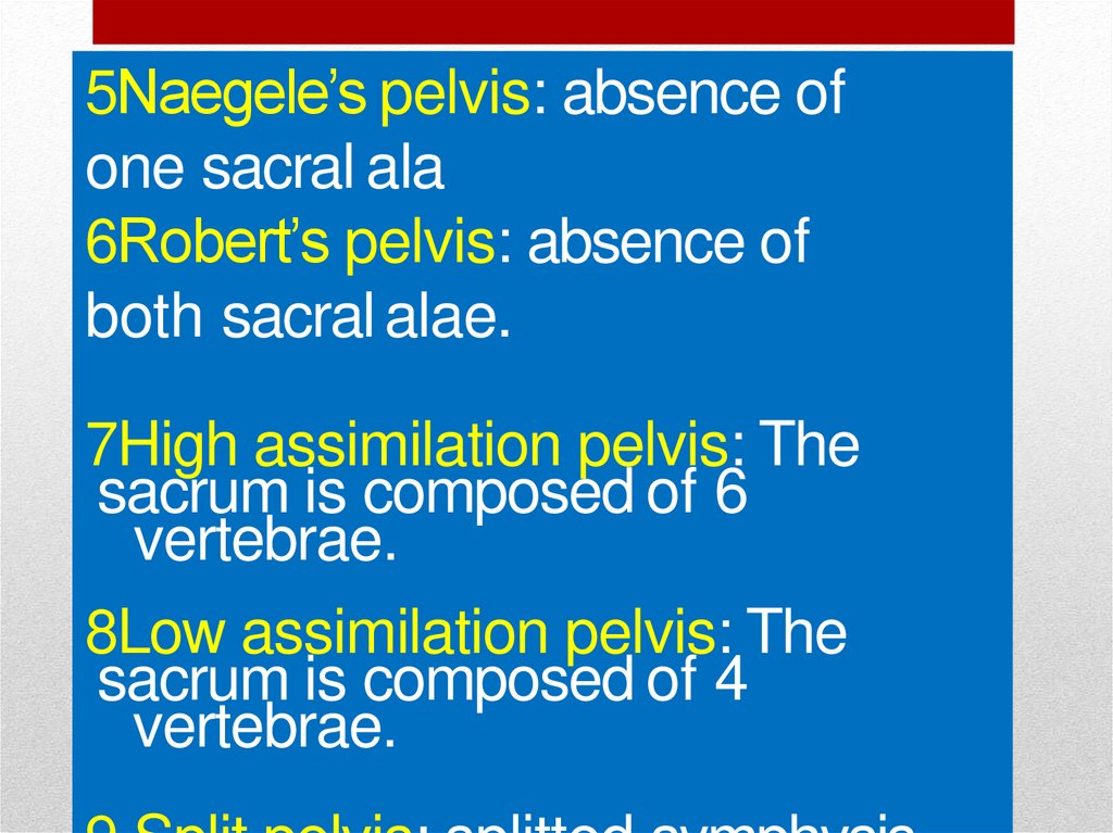
9.
5Naegele’s pelvis: absence ofone sacral ala
6Robert’s pelvis: absence of
both sacral alae.
7High assimilation pelvis: The
sacrum is composed of 6
vertebrae.
8Low assimilation pelvis: The
sacrum is composed of 4
vertebrae.
10. Etiology of ContractedPelvis
• Causes in the pelvis• Metabolic:
- Rickets.
- Osteomalacia (triradiate pelvic brim).
• Traumatic: asfractures.
• Neoplastic: asosteoma.
• Infection : TB
11. Etiology of ContractedPelvis
Causes in the spine• Lumbarkyphosis
• Lumbarscoliosis
• Spondylolisthesis:
The 5th lumbar vertebra with the above vertebral
column is pushed forward while the promontory is
pushed backwards and the tip ofthe sacrum is
pushed forwards leading to outlet contraction.
12. Etiology of Contracted Pelvis
Causes in the lower limbs• Dislocation of one or bothfemurs.
• Atrophy ofone or both lowerlimbs.
N.B.oblique or asymmetric pelvis: one oblique
diameter is obviously shorter than theother.
This can be found in:
• Diseases, fracture or tumours affectingone
side.
13. Pelvis
• History• Rickets: is expected if there is a history of
delayed walking and dentition.
• Traumaor diseases:of the pelvis, spines or
lower limbs.
• Badobstetric history: e.g. prolonged labour
ended by:
difficult forceps
caesarean sectionor
still birth.
14. Pelvis
•Examination• General examination:
Gait: abnormal gait suggesting abnormalities in
the pelvis, spinesor lowerlimbs.
Height: women with lessthan 150 cmheight
usualy have contracted pelvis.
Spinesand lower limbs: may havea diseaseor
lesion.( kyphosis,…)
15. Pelvis
•Examination
•General
examination:
Manifestations of rickets as:
square head
rosary beads in the costalridges.
pigeon chest
Harrison’s sulcus and bowlegs.
Dystocia dystrophia syndrome: the
woman is
*short,obese stocky, subfertile, has android pelvis and
16. Pelvis
Abdominal examination:Nonengagement of the head:
in the last 3-4 weeks in primigravida.
Pendulous abdomen:
in aprimigravida.
Malpresentations:
are morecommon.
17. Pelvis
• Pelvimetry :It is assessment of the pelvic diameters andcapacity
done at 38-39 weeks.It includes:
1. Clinical pelvimetry:
Internal pelvimetry for:
inlet
cavity, and
outlet.
External pelvimetry for:
inlet and
outlet.
18. Diagnosis of Contracted Pelvis
•Pelvimetry :2.Imaging pelvimetry:
X-ray.
Computed tomography (CT).
Magnetic resonance imaging (MRI) .
• N.B. CTand MRI are recent and accurate but
expensive and not always available sothey are
not in commonuse.
19. Internal pelvimetry
is done through vaginalexamination1. The inlet:
a.Palpation of the forepelvis
(pelvicbrim):
The index and middle fingers are moved
along the pelvic brim. Note whether it is
round or angulated, causing the
fingers to dip into a
V- shaped depression behind the
symphysis.
b.Diagonal conjugate:
Try to palpate the sacral promontory to
measure the diagonal conjugate. Normally,
it is
12.5 cm and cannot be reached. If it is felt
the pelvis is considered contracted and the
true conjugate can be calculated by
20. Internal pelvimetry
2.The cavity:a.Height, thickness and inclination of thesymphysis.
b. Shapeand inclination of the sacrum.
c. Side walls: Todetermine whether it is straight,
convergent or divergent starting from the pelvic
brim down to the base of ischial spines in the
direction of the base of the ischial tuberosity.
Then relation between the index and middle
finger of the baseof ischial spines and the thumb
of the other hand on the ischial tuberosity is
detected. If the thumb is medial the side wall is
convergent and if lateral it is divergent.
21. Internal pelvimetry
• 2.Thecavity:• d.Ischial spines:
Whether it is blunt (difficult to identify at
all), prominent (easily felt but not large)or
very prominent (large and encroaching on
the mid- plane).
The ischial spines can be located by
following the sacrospinous ligament to its
lateral end.
22. Internal pelvimetry
2.Thecavity:e.Interspinous diameter: By using the 2
examining fingers, if both spines can be
touched simultaneously, the interspinous
diameter is £ 9.5 cm i.e. inadequate for an
average-sizedbaby.
f. Sacrosciatic notch: If the sacrospinous
ligament is two and half fingers, the
sacrosciatic notch is consideredadequate.
23. Internal pelvimetry
3-The outlet:a. Subpubic angle: Normally, it admits2fingers.
b. Mobility of the coccyx:by pressing firmly on
it while an external hand on it candetermineits
mobility.
c.Anteroposterior diameter of the outlet: from
the tip of the sacrum to the inferior edge of
the symphysis.
24.
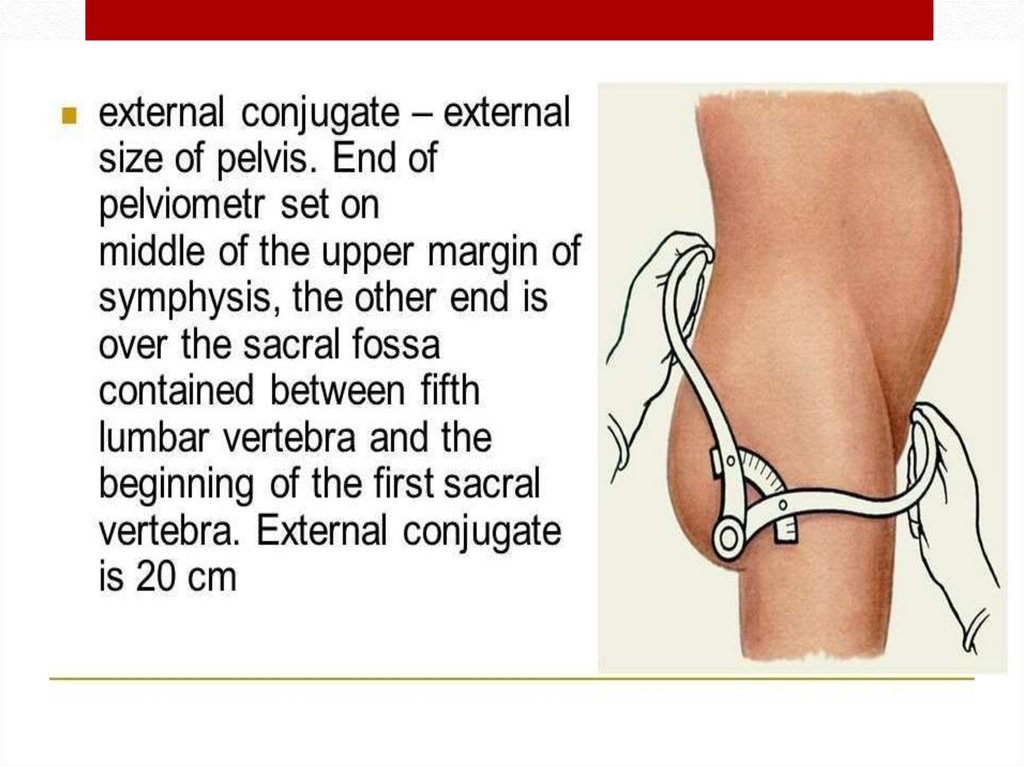
25. External pelvimetry
• Thom’s, Jarcho’s or crossingpelvimeter can be used for
external pelvimetry.
Interspinous diameter
(25cm): between the
anterior superior iliac
spines.
Intercrestal diameter (28
cm): between the most far
points on the outer borders of
the iliaccrests.
External conjugate (20 cm(.
Bituberous diameter
(11cm)
26.
27. Radiological pelvimetry
• Lateral view:Thepatient stands with the X-ray tube on one side
and the film cassetteon the opposite side.
it shows
the anteroposterior diameters of the pelvis,
angle of inclination of the brim, width of
sacrosciatic notch, curvature of the sacrumand
cephalo-pelvic relationship.
• Inlet view: Thepatient sits on the film cassette
and leans backwards so that the plane of the
pelvic brim becomes parallel to thefilm.
• Outlet view: Thepatient sits on the film
cassetteand leans forwards.
28. Cephalometry
• Ultrasonography: is the safeaccurate andeasy method and candetect:
The biparietal diameter(BPD)
The occipito-frontaldiameter.
Thecircumference of the head.
• Radiology (X-ray: isdifficult to interpret.
29. Cephalopelvic disproportion tests
Cephalopelvic disproportiontestsTheseare done to detect contracted inlet if the head
is not engaged in the last 3-4 weeks in a
primigravida.
• (1) Pinard’smethod:
• Thepatient evacuates her bladder andrectum.
• The patient is placed in semi-sitting
position
to bring the foetal axis
perpendiculartothe brim.
• The left hand pushes the head downwards
and backwardsinto the pelvis while the
fingers of the right hand are put on the
symphysis to detect disproportion.
30. Cephalopelvic disproportion tests
Cephalopelvic disproportiontests(2) Muller - Kerr’smethod:
It is more valuable in detection of
the degree of disproportion.
Thepatient evacuates her bladder and
rectum.
Thepatient is placed in the dorsal
position.
Theleft hand pushesthe head into the
pelvis and vaginal examination is done
by the right hand while its thumb is
placed over the symphysisto detect
disproportion.
31. Degrees of Disproportion
1. Minor disproportion:The anterior surface of the head is in line with the
posterior surface of the symphysis.During labour the
head is engageddue to moulding and vaginaldelivery
can beachieved.
2. Moderate disproportion 1st degree
disproportion):The anterior surface of the head is in
line with the anterior surface of the symphysis.Vaginal
delivery may or may not occur.
3. Marked disproportion 2nd degreedisproportion):
Thehead overrides the anterior surface ofthe
symphysis.Vaginaldelivery cannot occur.
32. Degrees of Contracted Pelvis
1.Minor degree: Thetrue conjugate is 9-10 cm.It corresponds to minordisproportion.
2.Moderate degree: Thetrue conjugate is 8-9 cm.
It corresponds to moderatedisproportion.
3.Severedegree: Thetrue conjugate is 6-8 cm.
It corresponds to markeddisproportion.
4.Extreme degree: Thetrue conjugate is lessthan
6 cm. Vaginal delivery is impossible even after
craniotomy asthe bimastoid diameter (7.5 cm) is
not crushed.
33. Management
Contracted pelvis Managementdepends mainly
on the degree of
disproportion
Minor
Moderate
Sever
vaginal delivery
trial labor, if
failed caesarean
section.
caesarean section
34. Trial of Labour
• It is a clinical test for the factors that cannotbe determined before start of labouras:
Efficiency of uterinecontractions.
Moulding of thehead.
Yielding of the pelvis and softtissues.
35. Procedure :
Trial is carried out in a hospital wherefacilities for C.S is available.
Adequate analgesia.
Nothing by mouth.
Avoid premature rupture of membranes
by:
rest in bed,
avoid high enema,
minimise vaginalexaminations.
The patient is left for 2 hours in the 2nd
stage with good uterine contractions
under close supervision to the
mother andfoetus
36. Indications of trial of labour:
1. Young primigravida of goodhealth.
2. Moderate disproportion.
3. Vertexpresentation.
4. No contractedoutlet
5. Average sizedbaby.
6. Vertexpresentation
37. Termination of trial of labour:
Vaginal delivery: either spontaneouslyorby forceps if the head is engaged.
Caesarean section if: failed trial of labour
i.e. the head did not engageor
complications occur during trial as
foetal distress or prolapsed pulsatingcord
beforefull cervical dilatation.
38. Indications of caesarean section in contracted pelvis
1. Moderate disproportion if trial of labouris contraindicated orfailed.
2. Marked disproportion.
3. Extreme disproportion whether the foetus is
living or dead.
4. Contracted outlet.
5. Contracted pelvis with other indicationsas;
I. elderly primigravida,
II. malpresentations, or
III. placenta praevia.
39. Complications
Contracted pelvisComplications
Maternal
During
pregnancy:
↑retroverted
gravid uterus.
Malpresentations.
Pendulous
abdomen
Nonengagement.
Pyelonephritis
due to more
compression of the
ureter.
Fetal
During labour:
Slow cervical
dilatation and
prolonged labour.
PROM and cord
prolapse.
Obstructed labour
and rupture uterus.
Injury to pelvic
joints or nerves from
difficult forceps
delivery.
Postpartum
hemorrhage.
Intracranial
hemorrhage.
Asphyxia.
Fracture skull.
Nerve injuries.
Intra-amniotic
infection
40. Complications of Contracted Pelvis
•Maternal:During pregnancy:
1. Incarcerated retroverted graviduterus.
2. Malpresentations.
3. Pendulous abdomen.
4. Nonengagement.
5. Pyelonephritis especial y in high assimilation
pelvis due to more compression of the ureter.
41. Complications of Contracted Pelvis
During labour:1. Inertia, slow cervical dilatation and
prolonged labour.
2. Premature rupture of membranesand
cord prolapse.
3. Obstructed labour and rupture uterus.
4. Necrotic genito-urinary fistula.
5. Injury to pelvic joints or nerves from
difficult forceps delivery.
6. Postpartum haemorrhage.
42. Complications of Contracted Pelvis
• Foetal:1. Intracranial
haemorrhage.
2. Asphyxia.
3. Fracture skull.
4. Nerve injuries.
5. Intra-amniotic infection.