


")


Похожие презентации:


Trauma + ATLS
1. Trauma + ATLS
2.
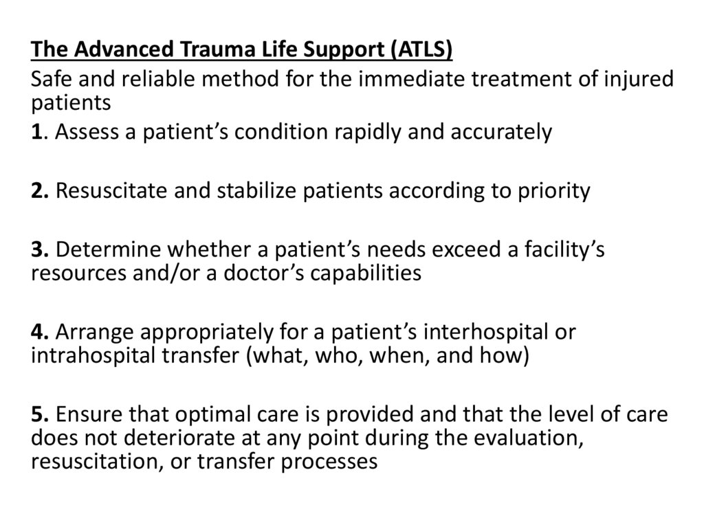
The Advanced Trauma Life Support (ATLS)Safe and reliable method for the immediate treatment of injured
patients
1. Assess a patient’s condition rapidly and accurately
2. Resuscitate and stabilize patients according to priority
3. Determine whether a patient’s needs exceed a facility’s
resources and/or a doctor’s capabilities
4. Arrange appropriately for a patient’s interhospital or
intrahospital transfer (what, who, when, and how)
5. Ensure that optimal care is provided and that the level of care
does not deteriorate at any point during the evaluation,
resuscitation, or transfer processes
3. ABCDE:
Airway with cervical spine protection
Breathing
Circulation, stop the bleeding
Disability or neurologic status
Exposure (undress) and Environment
(temperature control)
4. A and B:
• Speech5.
6.
7.
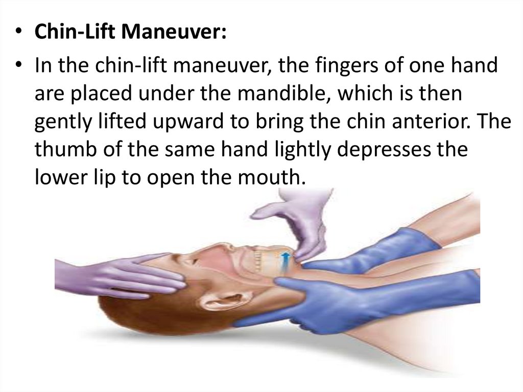
• Chin-Lift Maneuver:• In the chin-lift maneuver, the fingers of one hand
are placed under the mandible, which is then
gently lifted upward to bring the chin anterior. The
thumb of the same hand lightly depresses the
lower lip to open the mouth.
8.
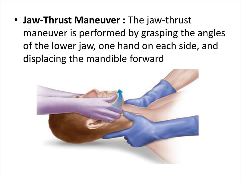
• Jaw-Thrust Maneuver : The jaw-thrustmaneuver is performed by grasping the angles
of the lower jaw, one hand on each side, and
displacing the mandible forward
9.
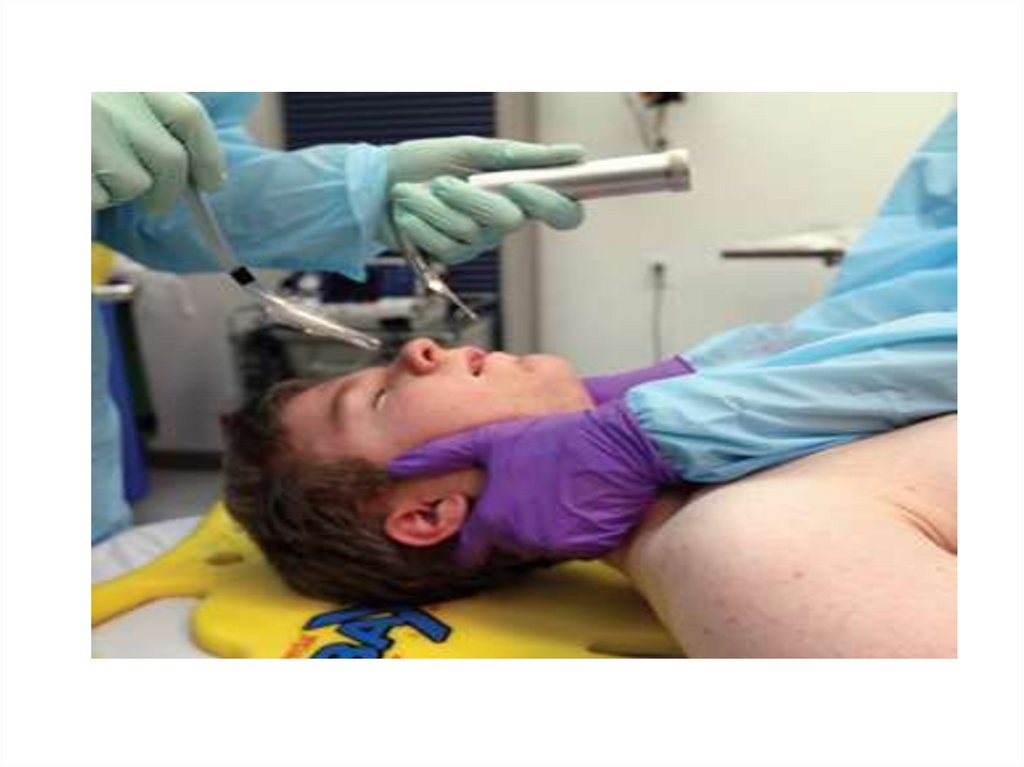
10. Rapid sequence intubation (RSI)
The technique for rapid sequence intubation (RSI) is as follows:• 1. Have a plan in the event of failure that includes the possibility of
performing a surgical airway.
• 2. Ensure that suction and the ability to deliver positive pressure
ventilation are ready.
• 3. Preoxygenate the patient with 100% oxygen.
• 4. Apply pressure over the cricoid cartilage.
• 5. Administer an induction drug (e.g., etomidate, 0.3 mg/kg) or
sedate, according to local practice.
• 6. Administer 1 to 2 mg/kg succinylcholine intravenously (usual
dose is 100 mg).
• 7. After the patient relaxes, intubate the patient
• 8. Inflate the cuff and confirm tube placement by auscultating the
patient’s chest and determining the presence of CO2 in exhaled air.
• 9. Release cricoid pressure.
• 10. Ventilate the patient.
11.
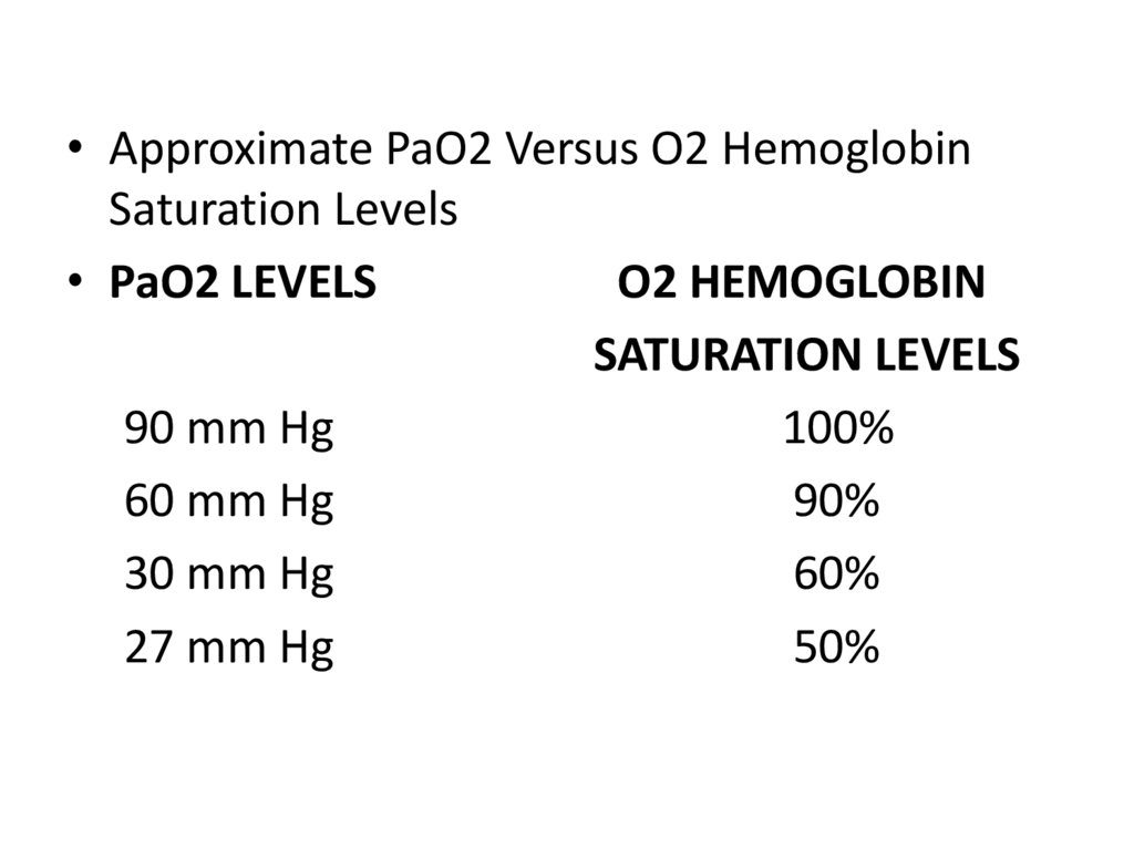
• Approximate PaO2 Versus O2 HemoglobinSaturation Levels
• PaO2 LEVELS
O2 HEMOGLOBIN
SATURATION LEVELS
90 mm Hg
100%
60 mm Hg
90%
30 mm Hg
60%
27 mm Hg
50%
12. Surgical Cricothyroidotomy
13. B: Breathing
• Evaluation:– visualizing chest movement
– auscultating breath sounds
– measuring oxygen
saturation
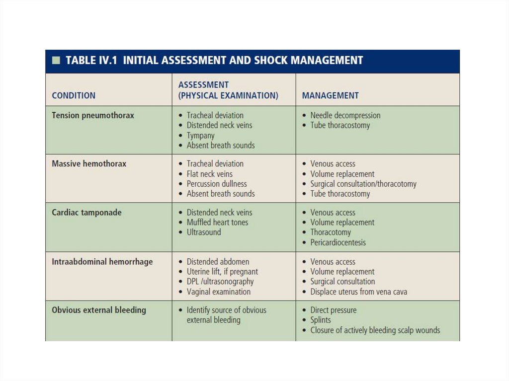
Tension pneumothorax:
- deviation of the tracea,
- unilaterally diminisched /
abscent breathing sound
Large needle or thoracostomy!
What if problematic ?
THINK ON:
• Tension pneumothorax
• Massive hemothorax
• Flail chest with pulmonary
contusion
Massice hemothorax:
Thoracostomy
evacuation of blood
re-expansion of the
lung.
Severe pulmonary Contusion:
aggressive
mechanical ventilation,
often with elevated levels of
expiratory positive end
pressure.
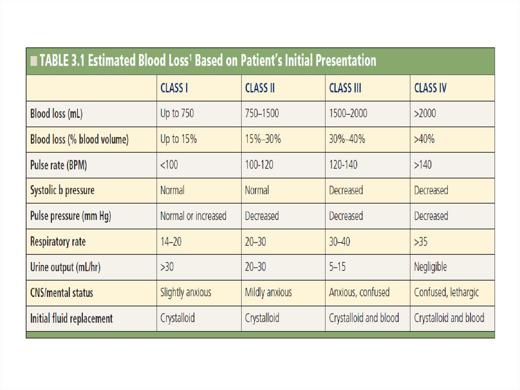
14. C-circulation
15.
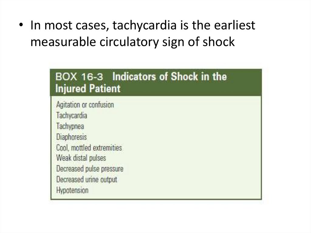
• In most cases, tachycardia is the earliestmeasurable circulatory sign of shock
16.
17.
18.
19. C:
• Venous access• Intraosseous
20. C: Circulation
5 Life-threating bloodloss causes:
external blood loss • 5 Life-threating blood loss
causes:
chest
external blood loss
abdomen,
Retroperitoneum Chest Tube thoracostomy
(pelvic fracture)
Abdomen ustable =
multiple long bone
operation
fractures
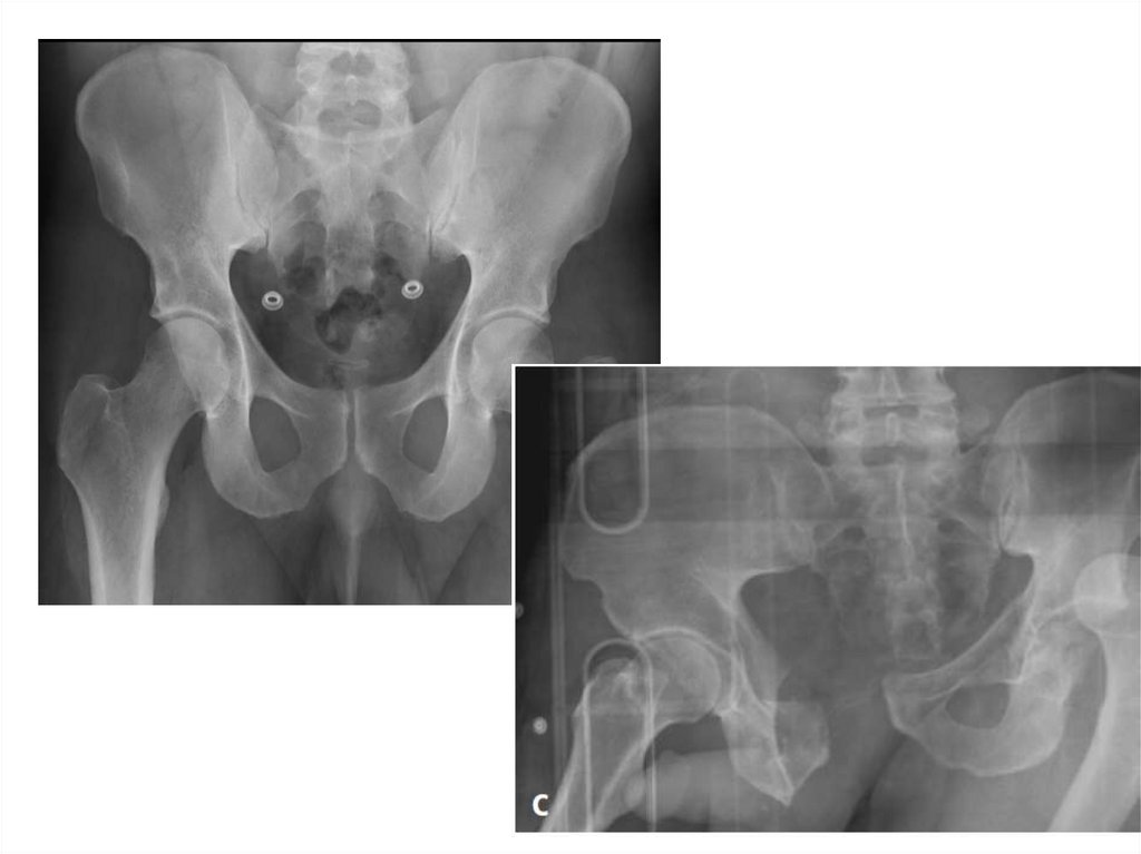
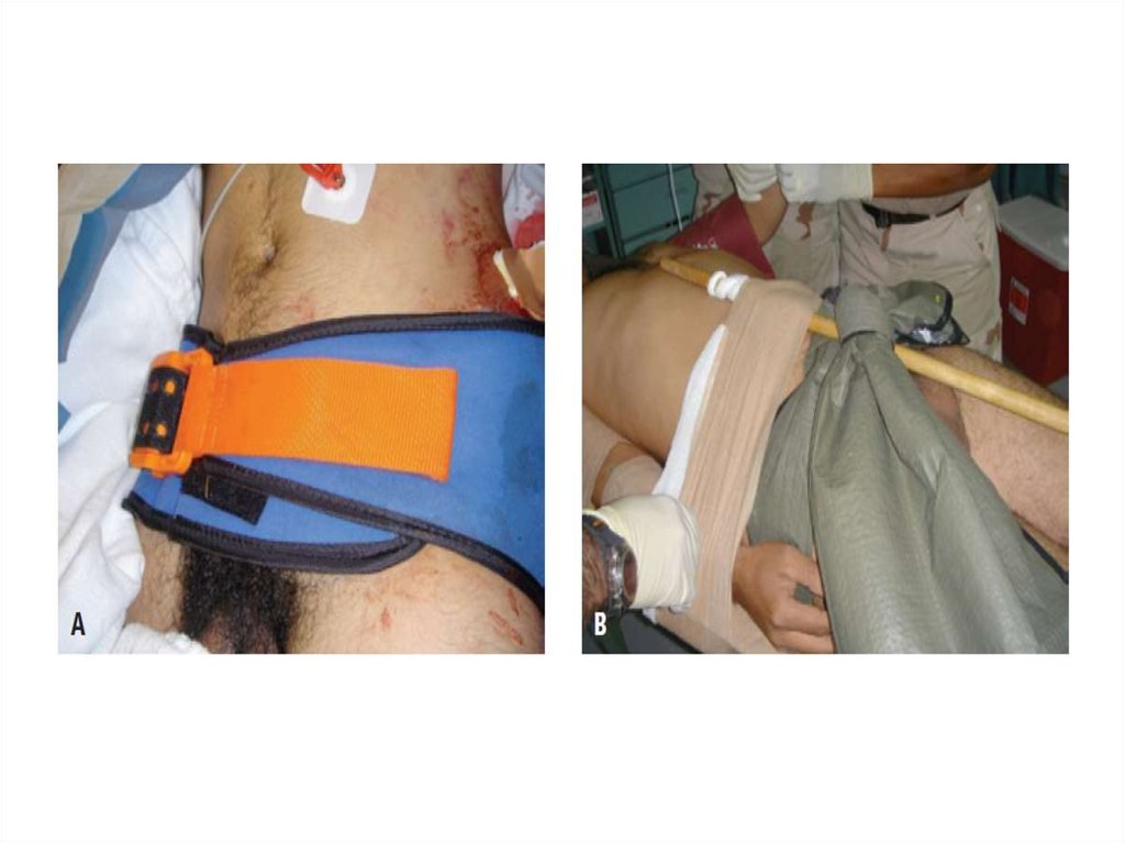
Retroperitoneum (pelvic fracture)
stabilization of the Fx
pelvic volume with a binder
pelvic angiography +/embolization
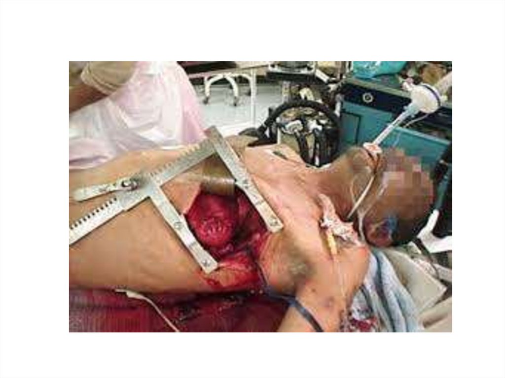
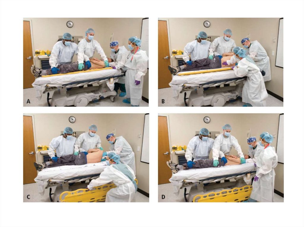
21. Resuscitative Thoracotomy
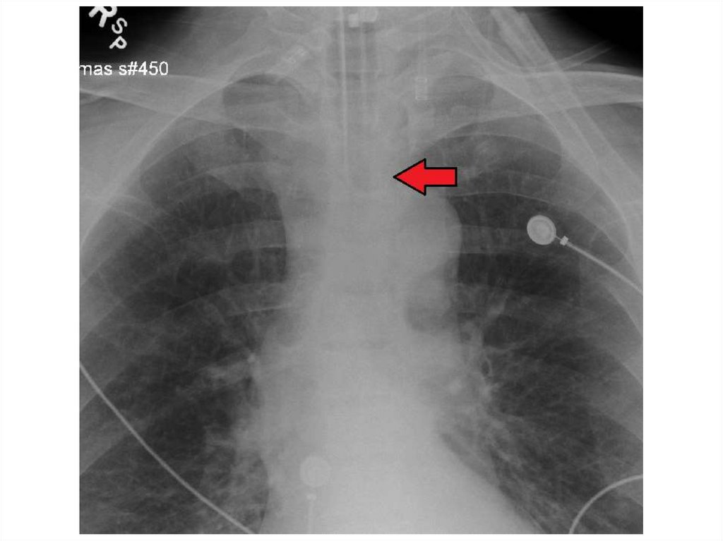
• When? critic injury and Cardiac arrest• For what?
– Opportunity to open pericardium relieves tamponade
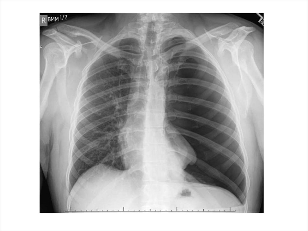
– internal cardiac massage
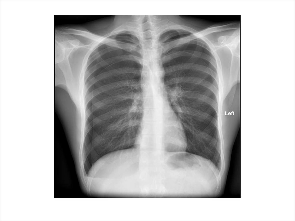
– cross-clamp the distal thoracic aorta (better perfusion for Brain and
heart)
– manage intrathoracic bleeding
who profits?
– The most: penetrating thoracic injury + sign of life on arrival 35%
success rate
22.
23. REBOA resuscitative endovascular balloon occlusion of the aorta
• Obtainig temporaryhemorrhage control in the
agonal patient
• Used in setting of aortic
aneurysms rupture
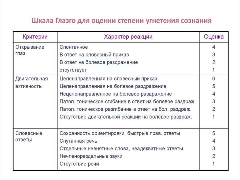
24. D
25.
26. D: Disability
Neurological function evaluation:Neurogenic shock?
Spinal cord injury?
Body temprature?
Keep the patient warm!
27. :E
E:Head to toe
PR
Xray
FAST
NGT
Urine catheter
28.
29.
30.
31.
32.
33.
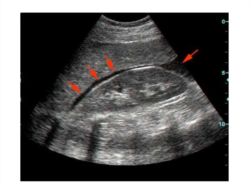
34. FAST
• Focused abdominal sonography in trauma:35.
36.
37. HEAD INJURY
38.
39.
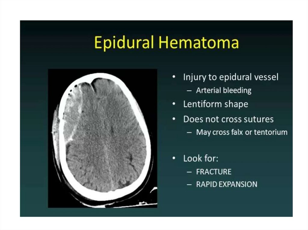
• Epidural hematomas typically result from a lateralfracture of the cranium causing bleeding from the
middle meningeal artery or a nearby vessel.
• Classically, the clinical course consists of an initial loss
of consciousness followed by a lucid interval, during
which time the hematoma is expanding. On reaching a
significant size, the epidural hematoma causes
profound neurologic deterioration.
• Treatment with decompression
• Underlying brain tissue is often not severely injured in
the setting of an epidural hematoma.
40.
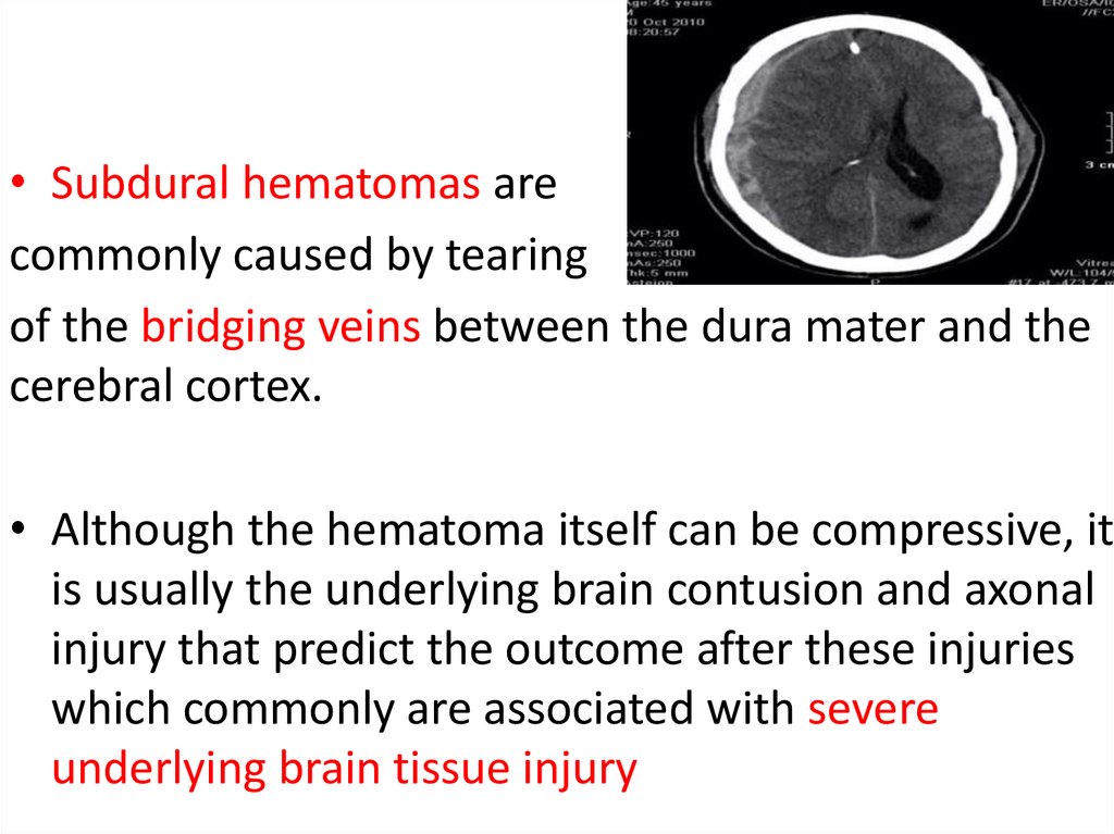
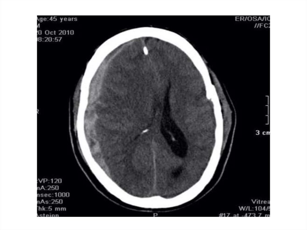
• Subdural hematomas arecommonly caused by tearing
of the bridging veins between the dura mater and the
cerebral cortex.
• Although the hematoma itself can be compressive, it
is usually the underlying brain contusion and axonal
injury that predict the outcome after these injuries
which commonly are associated with severe
underlying brain tissue injury
41.
42.
• Parenchymal contusions of brain tissue result fromthe direct transmission of energy to the cranium
and underlying brain as well as from movement of
the brain within the rigid cranial vault, resulting in
injury on the opposite side
• Secondary brain injury resulting from cerebral
edema is the greatest cause of morbidity after
intraparenchymal contusion.
43. Diffuse axonal injury describes the phenomenon of disruption of the axon from the neuronal body secondary to severe rotational
forces that are believed to create a shearing effect44.
45.
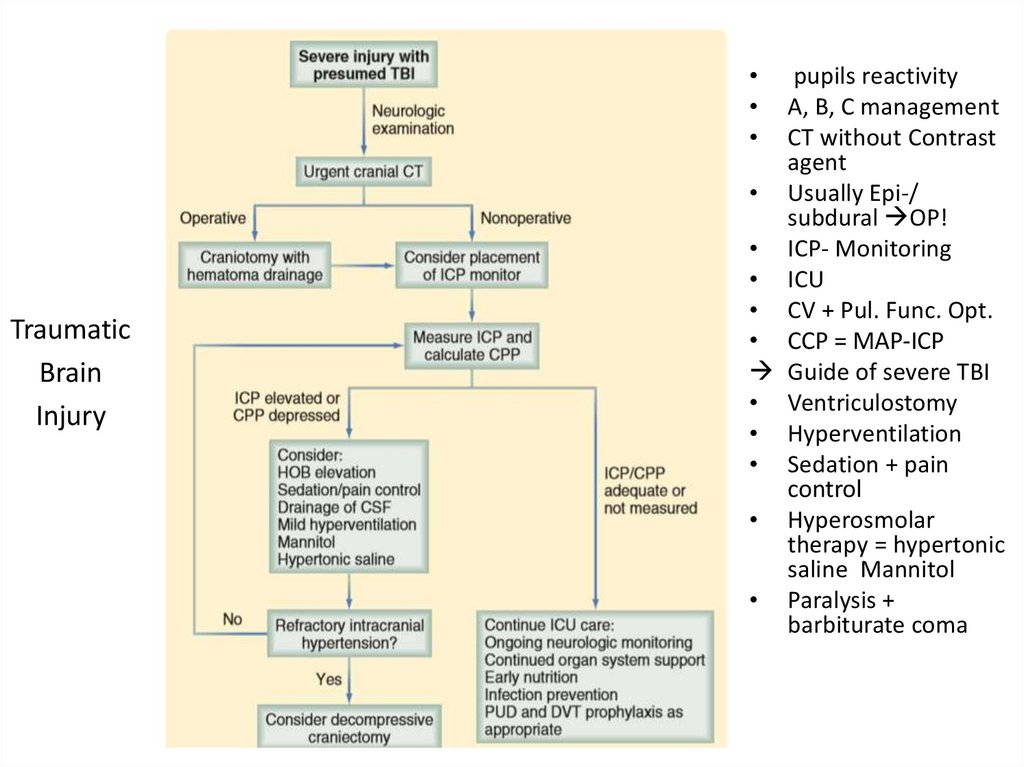
Traumatic
Brain
Injury
pupils reactivity
A, B, C management
CT without Contrast
agent
• Usually Epi-/
subdural OP!
• ICP- Monitoring
• ICU
• CV + Pul. Func. Opt.
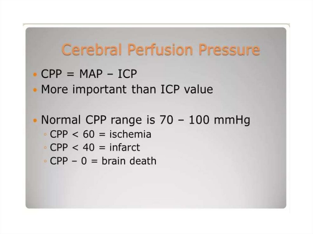
• CCP = MAP-ICP
Guide of severe TBI
• Ventriculostomy
• Hyperventilation
• Sedation + pain
control
• Hyperosmolar
therapy = hypertonic
saline Mannitol
• Paralysis +
barbiturate coma
46.
47. Injuries to the Neck
• Neck injuries are uncommon but result in the highestmortality rate of all body regions (20.0% mortality)
• Penetrating injuries from gunshot and stab wounds are
the most common mechanism of injury. Penetrating
injuries can directly lacerate vascular and aerodigestive
structures.
48.
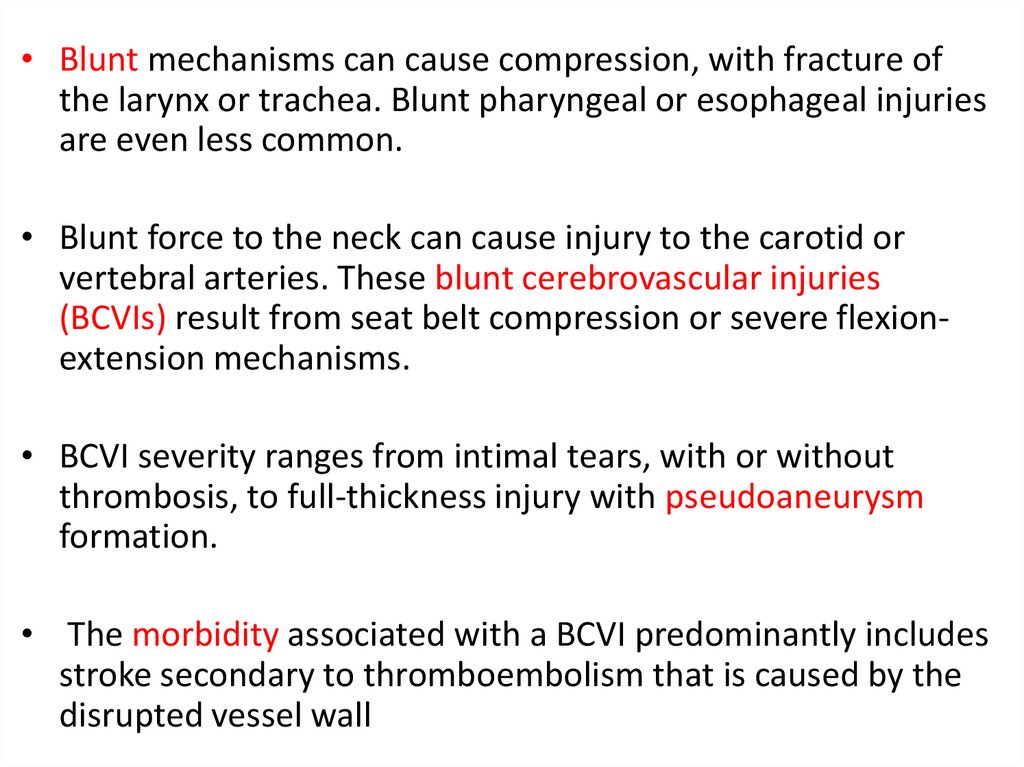
• Blunt mechanisms can cause compression, with fracture ofthe larynx or trachea. Blunt pharyngeal or esophageal injuries
are even less common.
• Blunt force to the neck can cause injury to the carotid or
vertebral arteries. These blunt cerebrovascular injuries
(BCVIs) result from seat belt compression or severe flexionextension mechanisms.
• BCVI severity ranges from intimal tears, with or without
thrombosis, to full-thickness injury with pseudoaneurysm
formation.
• The morbidity associated with a BCVI predominantly includes
stroke secondary to thromboembolism that is caused by the
disrupted vessel wall
49.
50.
51. Neck Injury
• Zone I: from the thoracicinlet to the cricoid
cartilage and contains
large vascular structures
as well as the trachea and
esophagus.
• Zone II: contains the
carotid and vertebral
arteries, jugular veins, and
structures of the
aerodigestive tract.
• Zone III: blood vessels that
are difficult, to expose
surgically.
52.
53. CHEST INJURY
• With more than 65% of blunt trauma patientssustaining one or more rib fractures, chest wall
injuries are the most common thoracic injury.
• The mortality rate associated with chest wall injuries
after blunt trauma is approximately 7%, whereas it
exceeds 19% for penetrating injuries
54.
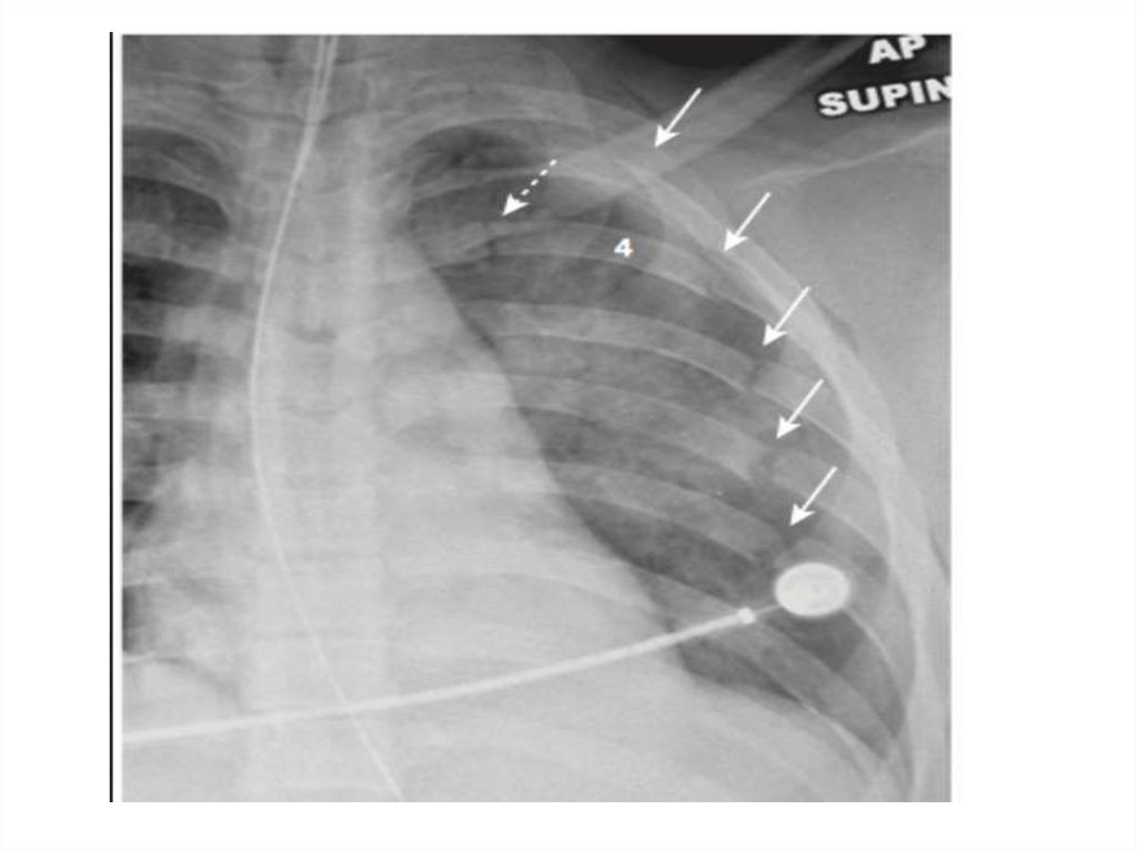
• Flail Chest : This condition usually results fromtrauma associated with multiple rib fractures—that
is, two or more adjacent ribs fractured in two or
more places.
• The presence of a flail chest segment results in
disruption of normal chest wall movement. Although
chest wall instability can lead to paradoxical motion
of the chest wall during inspiration and expiration,
this defect alone does not cause hypoxia.
55. Flail chest
Management:> Solitary independent movement of the Fx
Paradoxical breathing
Hypoxemia due to pulmonary contusion
High mortality rate - 33% - especially old patients
Pain management –
epidural ?
Consider – good ventilation
and oxygenation
Indications for an
Intubation:
Tachypnea > 40/min
PaO2 <60%
Multiple injuries, multiple
Fx
56.
57. Thoracic injury
58. Thoracic injury
• Tension pneumothorax is a clinical diagnosisreflecting air under pressure in the affected
pleural space.
• Treatment should not be delayed to wait for
radiologic confirmation.
59.
60.
61.
• Cardiac tamponade is indicated by thepresence of the classic diagnostic Beck’s triad:
venous pressure elevation, decline in arterial
pressure, and muffled heart tones.
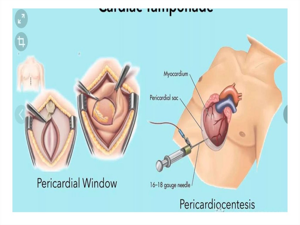
• If surgical intervention is not possible,
pericardiocentesis can be diagnostic as well
as therapeutic, but it is not definitive
treatment for cardiac tamponade
62.
63.
• Massive hemothorax results from the rapidaccumulation of more than 1500 mL of blood or onethird or more of the patient’s blood volume in the
chest cavity
• Patients who have an initial output of less than 1500
mL of fluid, but continue to bleed, may also require
thoracotomy.
• This decision is not based solely on the rate of
continuing blood loss (300 mL/hr for 3hours), but
also on the patient’s physiologic status
64.
65. Thoracic injury
• Pulmonary injuries. Lung injuries are commonafter chest trauma, with 31.9% of patients
• Mortality after pulmonary contusion is
approximately 10%, predominantly as a result
of respiratory failure from the acute
respiratory distress syndrome or pneumonia.
66.
• Cardiac injuries uncommon, but most severe injuriessustained by patients after penetrating and blunt
trauma.
• Penetrating injury to the heart occurred in 1.8% of
patients with penetrating trauma and in 8.7% of the
subset with penetrating chest trauma alone.
• These statistics likely underestimate the true incidence
of penetrating cardiac injuries because most are
immediately lethal and never present to a hospital.
• For those penetrating cardiac injuries that do present
to the emergency department the mortality rate is
72.9%.
67.
• Blunt injury to the heart occurs lesscommonly, being seen in only 2.2% of blunt
chest trauma cases.
• Most of these cases represent a contusion of
the myocardium that results in arrhythmias
and are frequently self-limited.
• In rare cases, blunt cardiac injury results in
heart failure with cardiogenic shock.
68.
Tracheobronchial injuriesTracheobronchial tree injuries are uncommon but are
associated with significant morbidity and mortality.
Penetrating mechanisms are the most common
cause, although these injuries still represent only
0.4% of all penetrating chest.
Despite this low incidence, the associated mortality
was significant at 57.9%.
Blunt injury to the tracheobronchial tree can occur
but is extraordinarily rare, representing only 0.07% of
blunt thoracic injuries
69.
Esophageal injuries:• The thoracic esophagus is uncommonly injured
• Penetrating injury is more common, but only 1.6% of
penetrating chest injuries had involvement of the esophagus.
Most of these are caused by gunshot wounds, followed by stab
wounds in less than 20% of cases
• The mortality associated with penetrating esophageal injuries
is substantial at 35.6% as a result of mediastinal sepsis and
because of the adjacent vital structures that can also be injured
along with the esophagus.
• Blunt esophageal injury is exceedingly rare, identified in only
0.02% of blunt trauma patients
70.
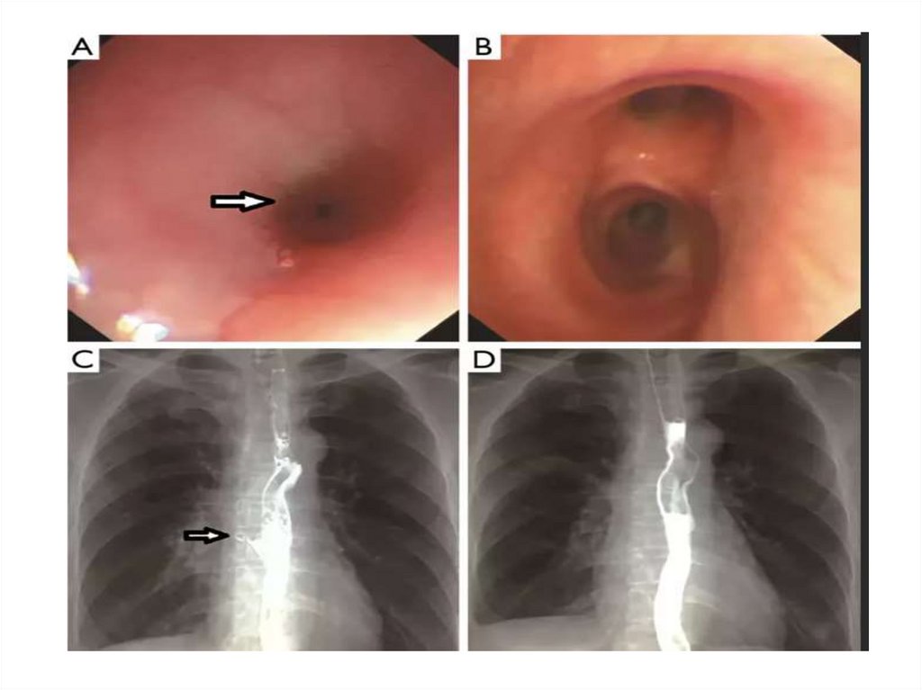
• The esophagus is best evaluated through a combinationof contrast esophagography and esophagoscopy
• Together these two modalities result in a sensitivity of
almost 100% for esophageal injury
• Esophageal injuries with associated mediastinal
contamination require immediate identification and
repair because delays are associated with worse
outcomes.
71.
72.
• The upper and midthoracic esophagus is bestapproached through a right posterolateral
thoracotomy through the fourth or fifth
interspace, whereas the lower esophagus is
exposed from the left through the sixth or seventh
interspace.
• Creation of a vascularized intercostal muscle flap
• Wide drainage of the mediastinum and chest is extremely
important to control any leak that may develop.
• A gastrostomy and feeding jejunostomy are frequently
performed to allow gastric decompression and early
nutritional support.
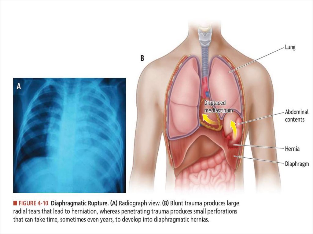
73. Diaphragmatic injury
- 1.6% of blunt trauma- 20% mortality (due to high
energy?)
- Rapid increase in IAP drug
on anterior impact blow
out of the diaphragm
- Usually LT (75%)
- Rt side covered by liver
Diagnostic: CXR / CT
74.
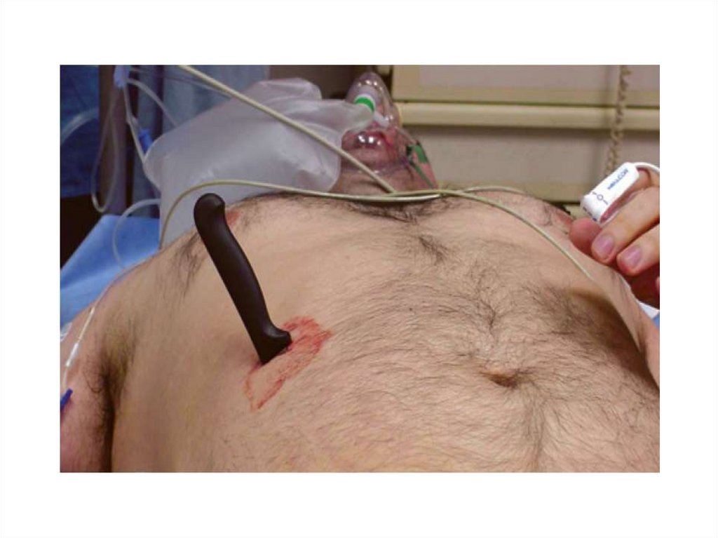
75. Abdominal trauma
76.
77.
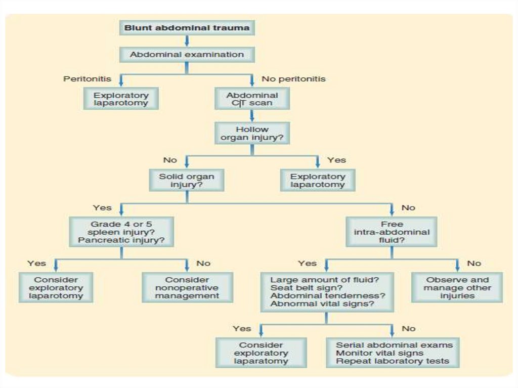
78. Indication for OR
Penetration of fascia
Unstable patient
NGT-blood
PR- blood
79.
80. SPLEEN
• The spleen is the most commonly injuredabdominal organ with 23.8% of patients with
abdominal trauma demonstrating splenic injuries.
• Many splenic injuries are self-limited,
demonstrating no evidence of ongoing bleeding;
• The mortality after blunt splenic injury 9.3%.
81.
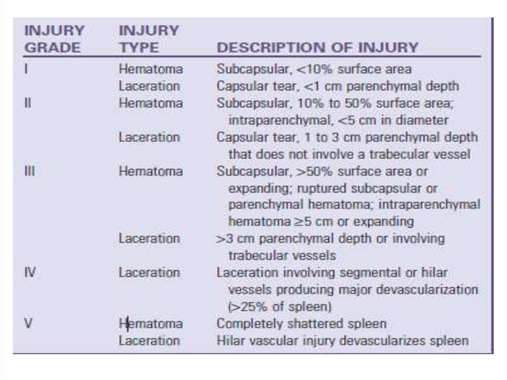
82. AAST Spleen Injury Scale
83.
84. Management of spleen Injury
• No preferable management• Depends on surgeon’s preference
• Recommended:
– I, II, III nonoperative + Angio for bleeding
– IV , V OP!
• post splenectomy vaccinations: Encaupsulated
bacteria:
– Streptococcus pneumoniae
– Hemophilus influanzea
– Neiseria meningitidis
85.
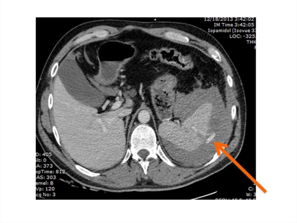
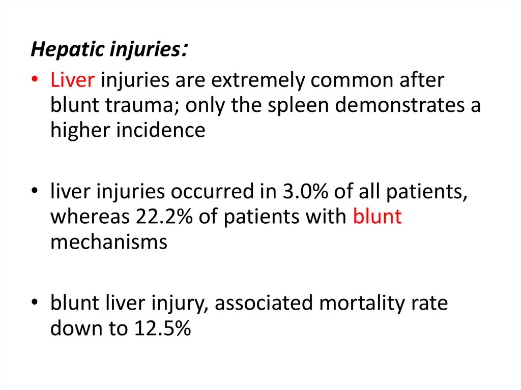
Hepatic injuries:• Liver injuries are extremely common after
blunt trauma; only the spleen demonstrates a
higher incidence
• liver injuries occurred in 3.0% of all patients,
whereas 22.2% of patients with blunt
mechanisms
• blunt liver injury, associated mortality rate
down to 12.5%
86.
87.
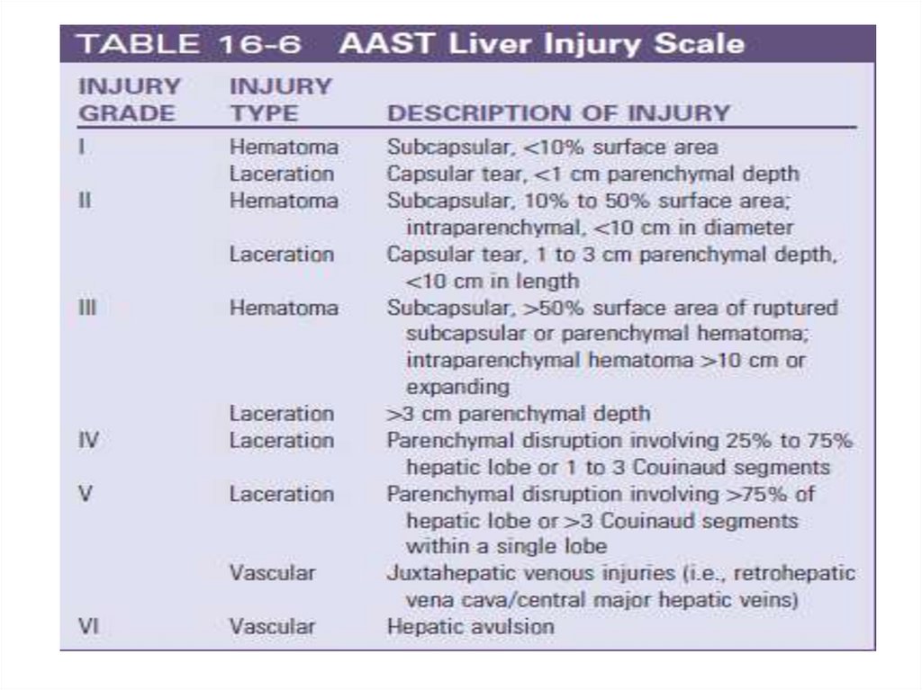
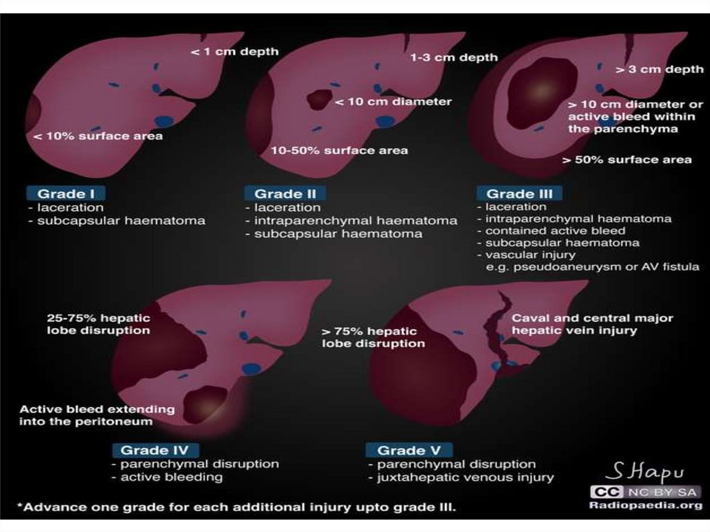
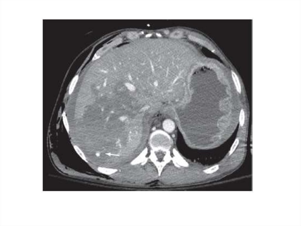
88. Hepatic Injury
• Mechanism: compression with direct parenchymal damageand shearing forces tears in hepatic tissues disruption
of vascular and ligamentous attachements.
• 2nd mechanism: penetrating trauma – higher morbidity
with vascular /biliary injury
Diagnostic:
FAST ,
instability,
CT ,
expl. Laparotomy due to + FAST
89.
90.
91. Hepatic Injury - Management
• Hemodynamically instable patient OPERATION• Conditions or non-operative management:
No tachycardia, no hypotension, no Metabolic acidosis
No evidence of shock
– Slow decreasing in Hb-levels with HD stability
transfusions
– Angioembolization for blush when stable
92. Non-surgical hepatic injury treatment Complications
• abdominal compartment syndrome• bile duct injury leading to bile peritonitis
or biloma
• delayed hemorrhage
• intra-abdominal abscess formation
• acute acalculous cholecystitis
• hemobilia
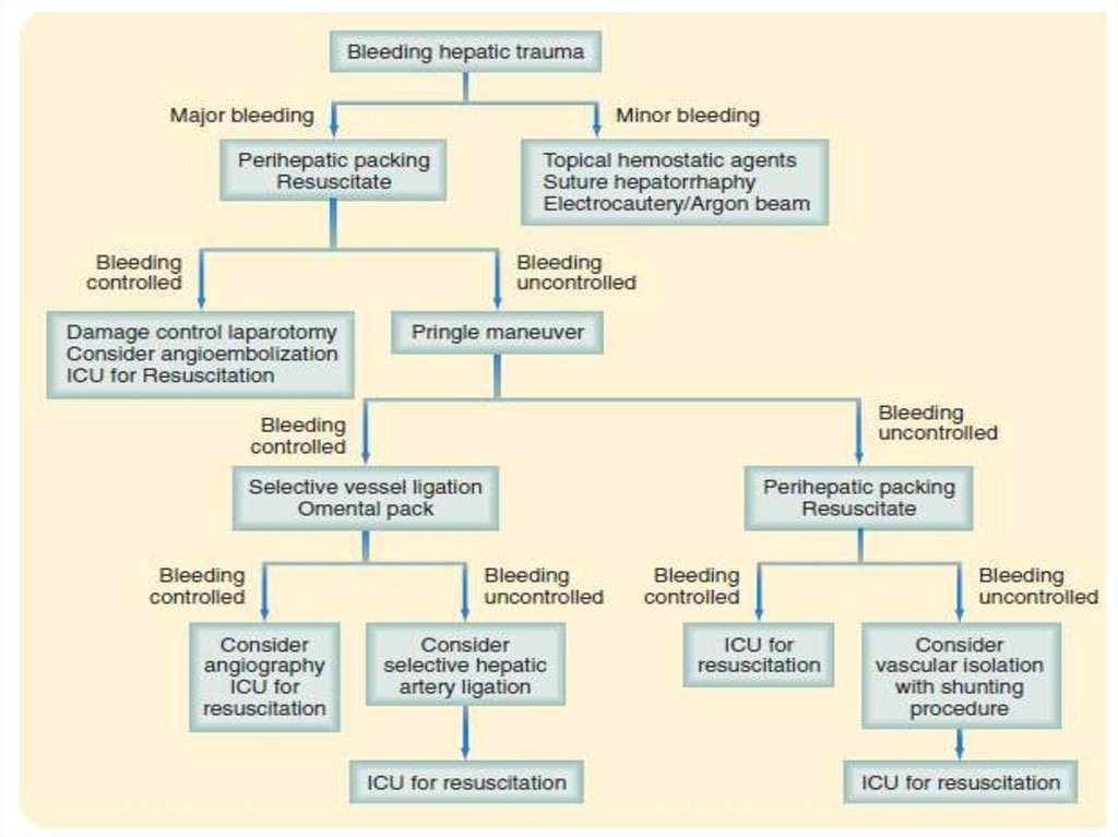
93. Surgical management of liver Injury options
Packing
Pringle
Push
Plug
94. Surgical management of liver Injury
• Perihepatic sponges• Manual pressure
• When stable – remove packing and reevaluate
• Mild injuries:
– compression
– Topical hemostasis agents
– Suture hepatorrhaphy
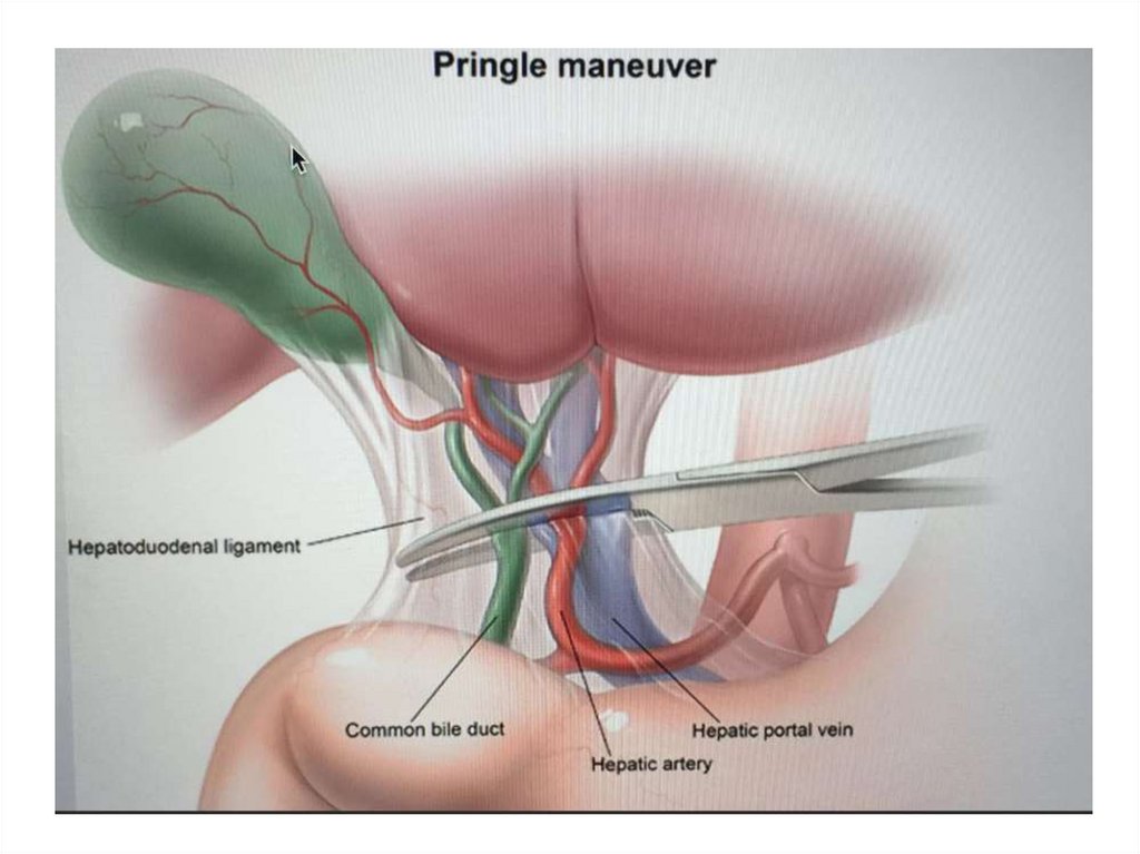
• severe bleeding:
– Pringle maneuver: encircling of the hepatoduodenal ligament (hepatic
artery + portal vein)
• Hepatic vein bleeding will continue after binding! (helps distinguishing)
• Post Packing – consider angioembolization
95.
96. Gastric injuries
• Penetrating mechanisms are the most common cause ofinjuries to the stomach, with these being present in 1217.6%. With associated mortality is 21.5%.
• Frequently, penetrating gastric injuries cause fullthickness perforations with likely spillage of gastric
contents into the abdomen.
• Conversely, blunt gastric injuries are rare, occurring in
0.05% of all blunt trauma patients and 4.3% of patients
with any blunt hollow visceral
97. Traumatic Gastric Injury
• Blunt injury – due to high energymechanism
• Clinical signs: Peritonitis
• Diagnostic – CT (but limited)
• Management:
– If perforation: absorbable suture and
another layer of non absorbable
– Hematoma – evacuation
– More challenging: GEJ, lesser
curvature, fundus, posterior wall
– Major tissue loss – partial or total
agstrectomy
98. Duodenal injuries
• Duodenal injuries are uncommon after blunt andpenetrating trauma but can pose a diagnostic and
therapeutic challenge.
• Because of the retroperitoneal location of the duodenum,
most injuries are due to penetrating mechanisms,
occurring in 4.0% of cases. Gunshot wounds are the
predominant cause, and the associated mortality is
significant at 24.5%
• Blunt duodenal injuries are much less common, occurring
in 0.1% of cases
99. Duodenal injury
• Children bicycle handlebar orsteering wheel stucking in
drivers
• Clinic:
– Do not expect peritonitis!
(extraperi)
• Diagnostic: CT (Also for low
grade injuries – hematoma)
Management:
perforation: immediate surgery
Hematoma: resolve without
intervention
if GOO – NGT, TPN
5-7 D reevaluation
100. Duodenal injury
Management:Duodenal injury
Operation:
Kocher-Maneuver – mobilization of
the duodenum
Primary repair in single or double layer
Greater injuries / tissue loss :
Ampulla not involved resection
and primary anastomosis
Ampulla involved: enteric bypass with
Roux-en Y
PE – pyloric exclusion
GJ- gastrojejunostomy
PE /o GJ- pyloric exclusion without
gastrojejunostomy
3 tubes: gastrojejunostomy,
duodenostomy for drains
+jejunostomy for feeding
/
O
101. Small bowel injuries
• The small intestine is one of the more frequently injuredorgans after penetrating abdominal trauma.
• Incidence to be as high as 60%
• Mortality rates range from 15% to 20%, with most caused by
associated vascular injuries
• Penetrating injuries can range from tiny perforations to large
destructive injuries that devitalize circumferential segments
of small bowel.
• Blunt injuries of the small bowel are less common, present in
1.7% of all blunt abdominal are associated with a significant
mortality rate of 14.0%.
102. Small bowel injury
• Management:– Primary repair – when no stricture
– Resection with anastomosis
– Resection without anastomosis (instable, shock)
• Temporarily abdominal closure
103. Colon injuries
• Colon and rectal injuries occur most commonly after penetratingabdominal trauma and rarely after blunt mechanisms.
• After penetrating abdominal trauma, injury to the colon is second
only to small bowel trauma, occurring in 36.4%
• Despite this, the associated mortality for colon and rectal injuries is
the lowest of all the abdominal viscera in the NTDB at 12.3%.
• Colon and rectal injuries occur in less than 1% of all blunt trauma
patients, demonstrating an associated mortality of 13%. When only
patients with blunt hollow visceral injury are considered, the colon
or rectum is involved in 30.2%
104. Colon Injury
• Retroperitoneal location ofasc. Desc. obscure indings
and injury
• CT has limited capability
• Usually detecting by
laparotomy in unstable
patient
• Rectum injury – may need
rigid recto-sigmoidoscopy
• Management:
– Primary
• Stable
• < 50% of circumference
– Resection + anastomosis !
• >50% circumeference
• Injuries prox to MCA : rt hemi +
ileocolostomy anastomosis
– Resection + colostomy
• Unstable
• Rectal injury: fecal
diversion+ presacral drain +
colostomy
– Pelvic sepsis
105. Pancreatic injuries
• Pancreatic injuries commonly occur in association with injuryto the duodenum because of their proximity.
• A penetrating mechanism is more commonly the cause,
4.4%.
• Mortality rates of 15.3% and 29.8% for blunt and penetrating
mechanisms, respectively.
• Delays in diagnosis and management are believed to
contribute to these significant mortality rates. Pancreatic
enzymes are caustic, making delays in management of the
injuries a source of massive systemic inflammation and
subsequent poor outcomes.
106.
• Pancreas tissue injury can result from direct laceration of theorgan or through the transmission of blunt force energy to the
retroperitoneum
• A common mechanism of blunt pancreatic injury involves the
crushing of the body of the pancreas between a rigid
structure, such as a steering wheel or seat belt, and the
vertebral column
• The impact to the pancreas causes injury that ranges from
mild contusion to complete transection with ductal disruption
107.
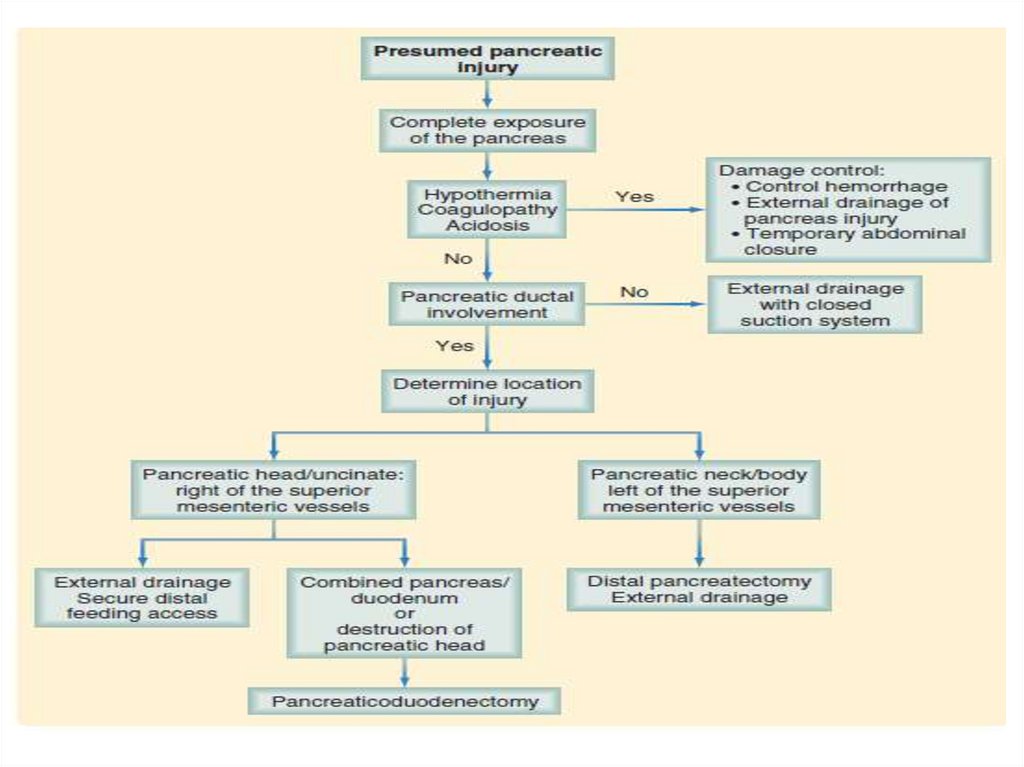
108. Management of Pancreatic Injury
• Operation in any significance• Location of injury determines the
surgical plan:
• Left to SMA distal
pancreatectomy
• Pancreas head :
• Limited tissue destruction:
controlled fistula
• Massive destruction of the
head / additional duodenal
Injury whipple pancreaticoduodenectomy
/
O
109. Abdominal great vessel injuries
110.
• The major blood vessels of the abdomen arepredominantly located within the retroperitoneum,
with some larger vessels also in the intestinal
mesenteries
• Most commonly, major abdominal vascular injuries
are secondary to penetrating mechanisms
• In the setting of blunt trauma, hematomas within the
retroperitoneum are often secondary to pelvic
fractures with bleeding from pelvic blood vessels
that dissect
111.
The retroperitoneum can be divided into threezones:
• Zone 1 hematomas require exploration because
these frequently involve the aorta, proximal
visceral vessels, or inferior vena cava, although
an exception may be the dark hematoma
behind the liver, which suggests a retrohepatic
vena cava injury.
• Injuries to the retrohepatic vena cava are best
served by not exposing the contained, lowpressure injury and by gently packing the
surrounding area
112.
• A hematoma in the region of zone 2, whichpredominantly contains the kidneys, should be
explored only if it appears that the hematoma is
expanding and continuing to lose blood
• A hematoma in zone 3 is usually secondary to pelvic
fracture bleeding and should not be explored unless
exsanguinating hemorrhage is obvious
113. Abdominal great vessel Injuries
• I – central hematoma– Aorta, prox. visceral vessels, inferior
vena cava
– Always demands exploration
– But not when dark retrohepatic –
vena cava – no exposure- packing
• II - Kidney
– Exploration only if hematoma is
expanding + blood loss
• III - pelvic fracture no
exploration unless
exsanguinating hemorrhage is
obvious (↓ Hb)
114.
Genitourinary injuries• The genitourinary organs include the kidneys,
ureters, bladder, and urethra, all of which are
contained within the retroperitoneum
• Bleeding and extravasation of urine are the major
concern with injuries to these structures
• Blunt mechanisms can result in renal laceration or
bladder rupture
115.
• Intraperitoneal bladder injuries can berepaired in two layers of absorbable suture
and the bladder drained with a Foley catheter
or suprapubic cystostomy tube.
• Extraperitoneal bladder ruptures require only
decompression with a urinary catheter,
followed by cystography to confirm healing
after a period of recovery.
116. Genitourinary Injury
• Kidney ureter bladderurethra
• Clinic: (gross hematuria),
Bleeding , extravasation
of urine
• Mechanism: energy
transmission to urinefilled bladder
• Diagnostic:
– Usually during laparotomy
– CT cystogram
• Management:
– Ongiong bleeding in
shock: nephrectomy
– Intraperitoneal Bladder
wall injury suturing
– Extraperitoneal bladder
rupture : Foley catheter
– Pseudoaneurysm –
angioembolization
– Expanding hematoma in
Zone II – expl lap.
117. Pelvis and extremities injuries
• Danger of retroperitonealhematoma
• Low mortality but longterm morbidity and
functional implications
• Pelvic Fx – commonly after
MV-Accidents and falls
• Diagnostic – physical
examination
– X-RAY
– CT
• Extremity –
– compartment syndroms!
• 6P’s rules:
–
–
–
–
–
–
Pale
Pulseless
Pain
Parasthesia
Paralysis
Poikilothermia (temp
difference)
– Peripheral vascular injury –
CTA
118. Injuries to the Pelvis and Extremities
119. In which of the following the pulse pressure is normal? A. Shock class I B. Shock class II C. Shock class III D. Shock class IV
120. A 30 - year - old male is brought to the trauma unit due to chest trauma . Blood pressure is low and heart rate of 122 / minute
. Chest X - ray shows widemediastinum . What is the most likely diagnosis ?
a. Cardiac tamponade
b. Tension pneumothorax
c. Sepsis
d. Head trauma
121. 22 - year - old male is brought to the trauma unit following a gun - shot wound to the pelvis . He rapidly deteriorates and one
of your colleagues isconcerned that the patient may suffer from the
lethal triad . What is the lethal triad ?
a. Acidosis , hyperthermia , coagulopathy
b. Acidosis , hypothermia , coagulopathy
c. Alkalosis , hypothermia , coagulopathy
d. Alkalosis , hyperthermia , coagulopathy
122.
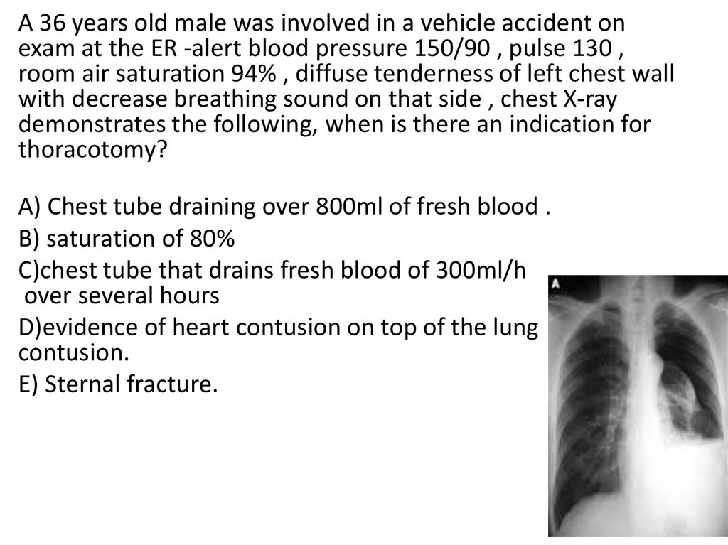
A 36 years old male was involved in a vehicle accident onexam at the ER -alert blood pressure 150/90 , pulse 130 ,
room air saturation 94% , diffuse tenderness of left chest wall
with decrease breathing sound on that side , chest X-ray
demonstrates the following, when is there an indication for
thoracotomy?
A) Chest tube draining over 800ml of fresh blood .
B) saturation of 80%
C)chest tube that drains fresh blood of 300ml/h
over several hours
D)evidence of heart contusion on top of the lung
contusion.
E) Sternal fracture.
123.
21-yearsold man arrives in the emergency department with a stab w
ound to the left chest located 1 cm to the middle line
of the left nipple. He is awake, diaphoretic, tachycardic and
hypotensive. Bilateral breath sounds are present and equal.
Chest Xray show no evidence of a hemopneumothorax, pericardial
ultrasound
shows pericardial fluid. What is the next best step?
a. CT of the chest
b. Pericardiocentesis
c. Bilateral chest tube placement
d. Sternotomy
e. Pericardial window
124. A 40 years old male is status post-splenectomy following a motor vehicle accident 10 years ago, present to the ER with high
fever, chills andproductive cough. During his treatment he rapidly
deteriorates with decrease in blood pressure. Which
of the following immunizations could have prevented
his current presentation?
A.
B.
C.
D.
Varicella
Pertussis
Pneumococcus
Tetanus
125. A 15-years-old girl fall while cycling. in the ER. 8 hours later she complains of left upper quadrant and shoulder pain. Her BP
is110/70, HR 95 /min RR 18 breaths per minute. She has tenderness
to palpation in the left upper quadrant without peritoneal sign .
FAST shows some fluid in the left upper quadrant window and trace
of fluid in the pelvic. A CT scan shows a grade 3 spleen laceration
with no blush. What is the next step in her management?
a. Splenectomy
b. Splenorrhaphy
c. Angioembolization
d. Observation
126.
A 40 year old male is admitted to the ER following stabwound to the abdomen. on exam alert without distress
hemodynamic stable stab wound 5 cm lateral and inferior
to the umilicus with omentum protrouding out of skin. no
signs of peritonitis, what is the most appropriate next step
in management?
A. Abdominal CT
B. Explorative laparotomy
C. Local wound exploration
D. Focused assessment with sonography in trauma (fast)
E. Diagnostic peritoneal lavage.
127.
45 years old female is involved as a pedestrian in motor vehicleaccident and is brought to the ER, her blood pressure 130/80 pulse
100 .focused assessment with sonography in trauma (fast) is negative
for abdominal fluid, pelvic X-ray demonstrated multiple pelvic
fractures (open book).
All of the following can assist in patient stabilization except :
A. Pelvic binder
B. Angiography
C. Extra peritoneal packing
D. External Pelvic fixation
E. Explorative laparotomy