


")


Похожие презентации:


")

")


Digestive tract
1. Digestive tract
2. DIAGNOSTIC APPROACH TO COLIC IN ADULT HORSES
• PAIN – degree, duration, and type• PULSE – rate and character
• PERFUSION – mucous membranes, skin tent, jugular fill, etc.
• PERISTALSIS – gut sounds, fecal production
• PINGS – simultaneous auscultation/percussion
• PASSING A TUBE – amount and character of reflux, if present
• PALPATION – rectal exam
• PAUNCH – a word for obvious abdominal distention that begins with “P”
• PCV/TP
• PERITONEAL FLUID
3. MEDICAL MANAGEMENT OF COLIC IN ADULT HORSES
• Non-steroidal anti-inflammatory drugs (NSAIDs )- Flunixinmeglumine, ketoprofen, and phenylbutazone are non-selective
inhibitors of COX1 and 2, whereas carprofen and etodolac are
somewhat COX-2 selective.
• α-2 adrenergic agonists - xylazine, romifidine and detomidine,
can provide excellent sedation, analgesia, and muscle relaxation
for horses with severe abdominal pain.
• Opiods: butorphanol
• Anti-spasmodics: N-butylscopolammonium bromide has both
anticholinergic and antispasmodic properties
4.
• Decompression - gastric decompression via a nasogastric tube,cecal enterocentesis in the right paralumbar fossa
• Alternate analgesia: Intravenous lidocaine has been used in
horses both as an analgesic and as a treatment/preventative for
post-operative ileus.
• Oral fluids - 6-8 L every 4-6 hours Always check for reflux prior
to administration, and never administer enteral fluids to a horse
with more than 1-2 liters of reflux.
• Intravenous fluids: half of the calculated fluid deficit within the
first 1-2 hours, with replacement of the remaining deficit (plus
maintenance and ongoing losses) over the next 12-24 hours.
5.
• LAXATIVES –mineral oil at 0.5-1 gallon via NGT in an adult horse.
magnesium sulfate (Epsom salts; 0.5-1 g/kg in 8 L water)
Psyllium mucilloid
6. stomach
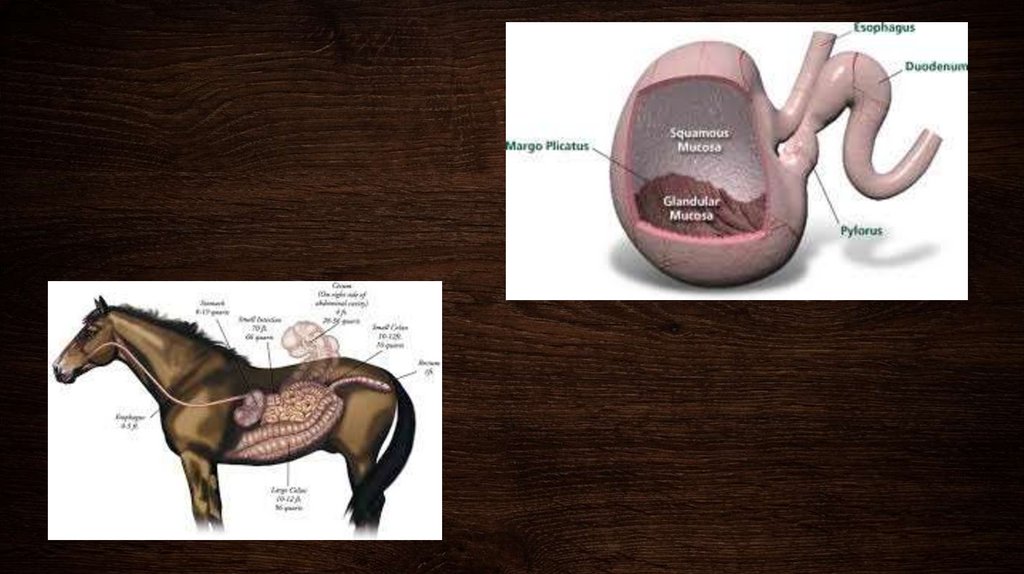
• The horse’s stomach is relatively small, with a capacity ofapproximately 10 to 15 liters, and is situated dorsocranially in
the left side of the abdomen within the confines of the ribcage.
• The oblique nature of the cardia renders it virtually impossible
for the horse to vomit and thus makes passage of a nasogastric
tube a critical component of the diagnostic work up of horses
with colic.
7.
8. Stomach
DO PREZENTACJI UŻYTO ZDJĘĆ Z ATLAS OF EQUINE ENDOSCOPY N.M.SLOVIS WYD MOSBY9. Gastritis
• Gastritis is an inflammation and irritation of the lining of thestomach.
• Unlike a stomach ulcer, gastritis involves large areas. The
mucosa--inner lining --throughout much of the stomach appears
red and swollen, and contains many small ulcerations or areas of
erosion
10. Acute Gastritis
• Acute gastritis is caused by ingesting moldy or spoiled feed,sand, chemicals and toxins, or by overeating.
• Laminitis--a metabolic and vascular disease which involves the
inner sensitive structures of the feet--can accompany or follow an
episode of acute gastritis.
11. Chronic Gastritis
• Chronic gastritis is associated with the long-standing ingestionof poor quality feeds or foreign materials such as wood shavings,
sand, or stones. These ingestible materials irritate the lining of
the stomach and often remain for long periods, during which
they combine with feed to form bezoars--impacted feed balls. The
bezors are too large to pass into the small intestines but are
small enough to intermittently block the outlet of the stomach.
12.
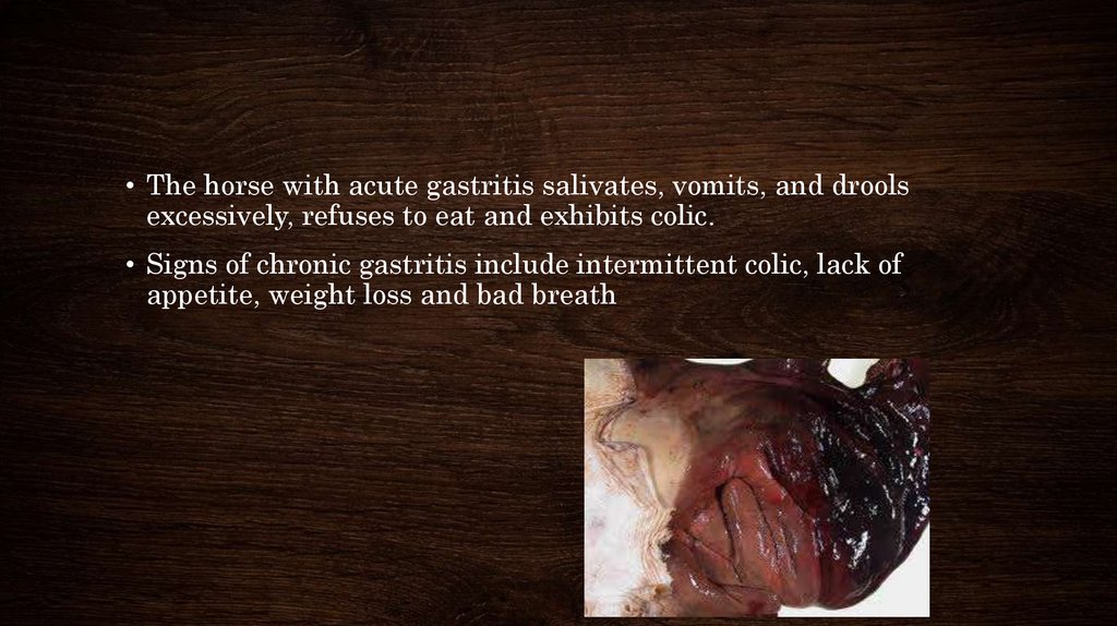
• The horse with acute gastritis salivates, vomits, and droolsexcessively, refuses to eat and exhibits colic.
• Signs of chronic gastritis include intermittent colic, lack of
appetite, weight loss and bad breath
13. treatment
• H2 receptors blockers – ranitidin 6-7 mg/kg every 8 hours,cymetidin 10-20 mg/kg
• Proton pump inhibitors – omeprazol 2-4 mg/kg
• Good hey , pasturage
14. Gastric ulcers
• Ulcers are a common medical condition in horses and foals. It isestimated that almost 50% of foals and 1/3 of adult horses
confined in stalls may have mild ulcers.
• Up to 60% of show horses and 90% of racehorses may develop
moderate to severe ulcers.
• Because they are so common, and can occur as a result of a
number of factors, the condition is often called "equine gastric
ulcer syndrome" (EGUS) or "equine gastric ulcer disease"
(EGUD).
15.
• Stomach is divided into two distinct parts. The non-glandularportion (also called the esophageal region) is lined by tissue
similar to the lining of the esophagus. The glandular portion is
lined with glandular tissue, which produces hydrochloric acid
and pepsin, an enzyme needed for the digestion of food.
• In the horse, however, hydrochloric acid is constantly being
produced. So, if a horse does not eat, the acid accumulates in the
stomach, and can start to irritate the stomach, especially the
non-glandular portion.
16. Causes of gastric ulcers
• Fasting (not eating) - Horses evolved to graze, eating many small meals frequently. Thisway, the stomach is rarely empty and the stomach acid has less of a damaging effect. If
horses and foals do not eat frequently, the acid builds up and ulcers are more likely to
develop.
• Type of feed - The type and amount of roughage play a role in ulcer development.
Roughage, because it requires more chewing, stimulates the production of more saliva. The
swallowed saliva helps to neutralize stomach acid. There is an increase in acid production
when concentrates are fed. The type of roughage is also important. Alfalfa is higher in
calcium, and it is thought that this may help decrease the risk of ulcers.
• Amount of exercise - As the amount of exercise increases, there is often a change in feeding
(e.g., more times of fasting, less roughage), which increases the risk of ulcer development.
In addition, exercise may increase the time it takes for the stomach to empty, so large
amounts of acid can remain in an empty stomach for a prolonged period of time. Stress
itself may decrease the amount of blood flow to the stomach, which makes the lining of the
stomach more vulnerable to injury from stomach acid.
• Medications - Chronic use of non-steroidal anti-inflammatory drugs (NSAIDS) such as
phenylbutazone (Bute) and flunixin meglumine (Banamine) blocks the production of a
particular chemical called PgE2. PgE2 decreases acid production, so when PgE2 levels are
low, acid levels are high, contributing to the development of ulcers.
17. Signs of gastric ulcers in horses
In foals, signs of gastric ulcersinclude:
In adult horses, signs of gastric
ulcers include:
• Intermittent colic, often after
nursing or eating
• Poor appetite
• Poor appetite and nursing for
only very short periods
• Teeth grinding
• Excessive salivation
• Weight loss and poor body
condition
• Poor hair coat
• Mild colic
• Diarrhea
• Mental dullness or attitude
changes
• Lying on the back
• Poor performance
• Lying down more than normal
18. Stomach ulceration
• I gradeIII grade
DO PREZENTACJI UŻYTO ZDJĘĆ Z ATLAS OF EQUINE ENDOSCOPY N.M.SLOVIS WYD MOSBY
19. Treatment of gastric ulcers in horses
• H2 blockers: These are medications that block the action of histamine.Histamine stimulates the production of stomach acid. Example:
Cimetidine, ranitidine.
• Proton pump inhibitors: These are medications that inhibit the
production of acid by the stomach
• Buffers: Antacids buffer the action of the stomach acid. Because acid is
constantly being produced in the horse, antacids are effective for only
a short time (less than an hour) and require large amounts be given.
This makes them relatively impractical in the horse, though their use
on the day of performance or a stressful event may be beneficial.
• Protectants: Certain drugs can block acid from coming into contact
with the stomach lining. Unfortunately, these do not appear to be as
effective in the esophageal portion of the stomach. Example:
Sucralfate.
20.
In addition to medications, changes in management are almostalways necessary including:
• Increasing the amount of roughage in the diet.
• Increasing the number of feedings by increasing the amount of
time the horse is actually eating. Putting the horse on pasture
would be the best alternative.
• Avoiding or decreasing the amount of grain. Use supplements to
add the vitamins and minerals, and vegetable oils to add the
calories the horse may need.
• Giving probiotics to aid in digestion.
21. Gastric parasites
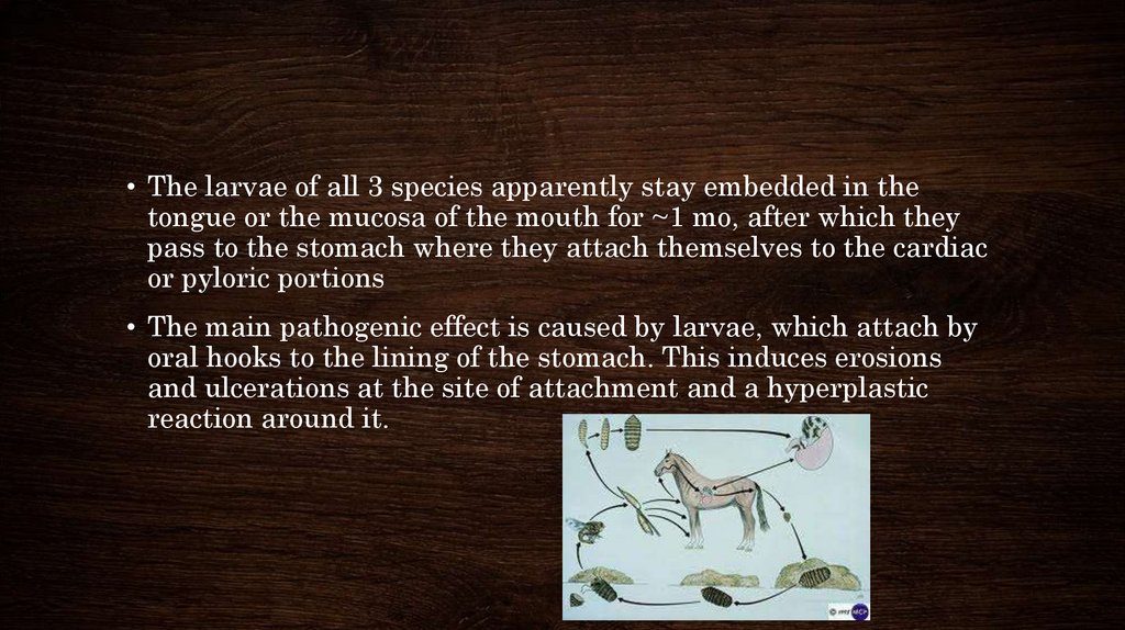
• Horse bots, which are found in the stomach, are the larvae ofbotflies, Gasterophilus spp.
• The eggs of G intestinalis (the common bot) are glued to the
hairs of almost any part of the body but especially the forelimbs
and shoulders. The eggs of G haemorrhoidalis (the nose or lip
bot) are attached to the hairs of the lips. G nasalis (the throat
bot) deposits eggs on the hairs of the submaxillary region.
22.
• The larvae of all 3 species apparently stay embedded in thetongue or the mucosa of the mouth for ~1 mo, after which they
pass to the stomach where they attach themselves to the cardiac
or pyloric portions
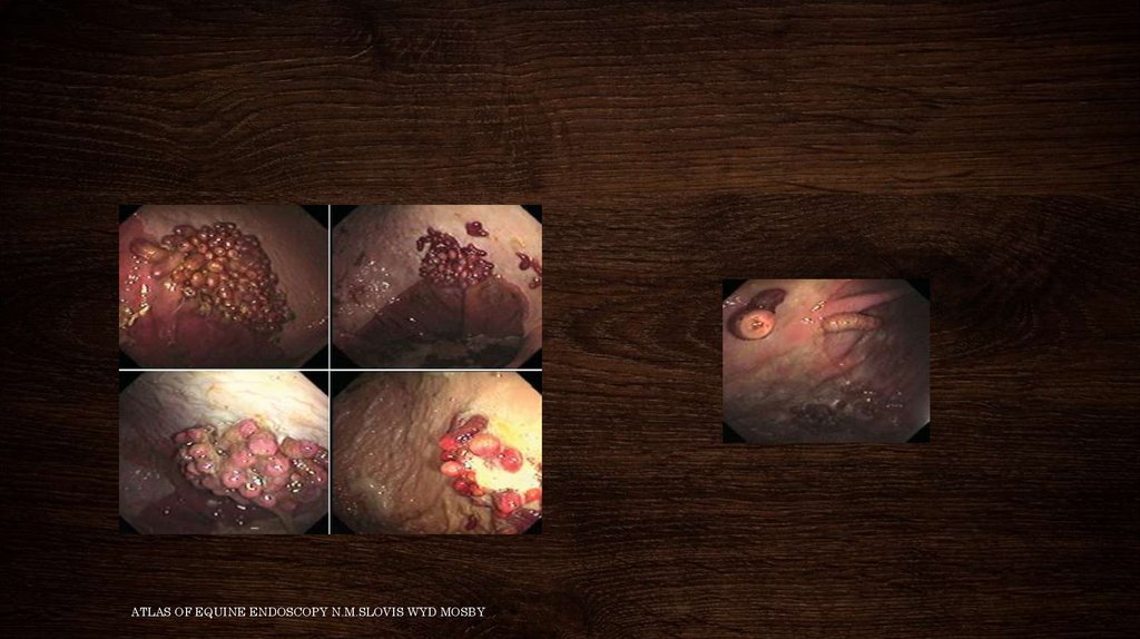
• The main pathogenic effect is caused by larvae, which attach by
oral hooks to the lining of the stomach. This induces erosions
and ulcerations at the site of attachment and a hyperplastic
reaction around it.
23.
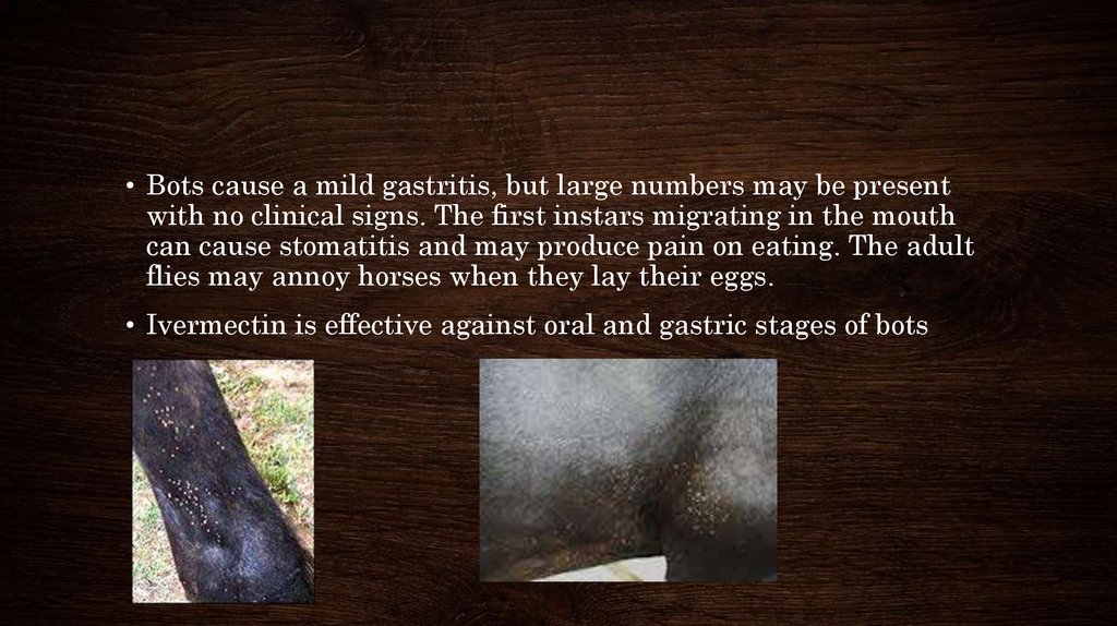
• Bots cause a mild gastritis, but large numbers may be presentwith no clinical signs. The first instars migrating in the mouth
can cause stomatitis and may produce pain on eating. The adult
flies may annoy horses when they lay their eggs.
• Ivermectin is effective against oral and gastric stages of bots
24.
ATLAS OF EQUINE ENDOSCOPY N.M.SLOVIS WYD MOSBY25. Gastric dilatation and rupture
• Gastric dilatation can be classified as primary, secondary, oridiopathic.
• Causes of primary gastric dilatation include gastric impaction,
grain engorgement, excessive water intake after exercise,
aerophagia, and parasitism.
• Excessive consumption of fermentable feeds (grains, lush grass,
and beet pulp) causes a large increase in the production of
volatile fatty acids which is thought to delay gastric emptying.
26.
• Secondary gastric dilatation occurs more commonly and canresult from primary intestinal ileus or small or large intestinal
obstruction. Fluid from the obstructed small intestine
accumulates in the stomach, causing nasogastric reflux. Gastric
dilation may also occur with certain colonic displacements,
especially right dorsal displacement of the colon around the
caecum. Gastric fluid accumulation is also characteristic of
proximal enteritis-jejunitis.
• Time to development of reflux is proportional to the distance to
the segment involved, with duodenal obstruction resulting in
reflux within 4 hours.
27.
Gastric dilation usually produces:Gastric rupture typically results in:
• Acute, severe colic
• Relief
• Tachycardia
• Depression
• Pale mucous membranes
• Retching
• Ingesta at the nares in severe cases (rare)
• Gastric reflux
The inevitable peritonitis and endotoxic
shock will lead to:
• Reluctance to move
• Tachypnoea
• Tachycardia
• Sweating
• Muscle fasciculations
• Blue or purple mucous membranes
28.
Primary gastric dilation should besuspected :
Secondary gastric dilation should
be considered:
copious amounts of gastric
reflux in the absence of small
intestinal distension on rectal
examination and the absence of
endotoxaemia.
persistent colic, repeated
retrieval of nasogastric reflux,
intestinal distension on rectal
examination and clinical signs
of endotoxaemia.
colic signs cease following
decompression, and other
clinical parameters return to
normal.
indications for exploratory
laparotomy to look for an
intestinal obstruction.
does not cause any significant
change in peritoneal fluid
parameters until rupture occurs.
29.
Gastric rupture results in septic peritonitis which will be reflected in the natureof fluid collected by abdominocentesis:
• Foetid, turbid sample containing particulate matter
• White cell count >40 x 109/l
• Protein content >30g/l.
Findings on rectal examination may include:
• A 'gritty feeling' on the serosal surfaces of intestine due to adherent food
material
• An impression of 'space' in the abdomen due to gas in the peritoneal cavity.
Laboratory findings may include:
• Haemoconcentration
• Hypokalaemia
• Hypochloraemia
30. treatment
• Gastric lavage (water or oil)• Treat underlying disease
31. Gastric Impaction (Obstruction)
• Gastric impaction can result in either acute or chronic signs ofcolic.
• Although a specific cause is not always evident, ingestion of
coarse roughage (straw bedding, poor quality forage), foreign
objects (rubber fencing material), and feed that may swell after
ingestion or improper mastication (persimmon seeds, mesquite
beans, wheat, barley, sugar beet pulp) have been implicated.
• Possible predisposing factors include poor dentition, poor
mastication and rapid consumption of feedstuffs, and inadequate
water consumption.
32. Clinical signs
The colic associated with gastric impaction varies from mild and chronic toacute and severe.Other signs reported include:
• Anorexia
• Lethargy
• Prolonged recumbency
• Dysphagia
• Dropping of feed
• Bruxism
• Salivation
• Insidious weight loss (if chronic)
• Spontaneous reflux with gastric contents visible at the nares (in severe cases)
33. treatment
• gastric lavage with water• IV fluid therapy and analgesia
• the impacted stomach can be felt extending back midway between the
xiphisternum and the umbilicus
• Infusion of balanced polyionic fluids such as saline either directly into the
impaction through the gastric wall (adjacent to the greater curvature) or via a
nasogastric tube
• Massage of the stomach to reduce the impaction and aid movement of fluid
into the ingesta
• Impactions diagnosed at surgery may benefit from bethanechol to stimulate
gastric motility.
• The stomach should be lavaged by nasogastric tube post-operatively and the
horse starved for 48-72 hours.
34. prevention
• Regular dental care• Ensure sugar beet nuts are adequately soaked prior to feeding
• Secure storage of roughage and hard feeds
• Ensure free access to water at all times
• Good pasture management to prevent ragwort poisoning
35.
36. Rectal examination
37. Obstruction
• normal movement of ingesta is restricted or prevented but nochange occurs in the blood supply to the intestine
• occur when ingesta fails to move from a portion of the bowel
having a large diameter into a portion with a smaller diameter
• impaction of the large colon at the pelvic flexure, enterolithiasis,
38. Pelvic Flexure Impaction
• occur when dry or inadequately digested feed fails to movethrough the pelvic flexure, the region connecting the large left
ventral colon with the smaller left dorsal colon
• additional ingesta fills the entire left ventral colon
39. Signs and treatment
• mild abdominal pain• heart rate slightly increased
• intestinal sounds usually can be heard
• oral and intravenous administration of fluids
• mild analgesics
• laxatives
40. Adhesions
• develop as a complication of previous small intestinal surgery orbecause of parasite migration, abdominal abscesses,penetrating
abdominal wounds, or serosal inflammation
41.
• history of a gradual onset of colic and weight loss, and in manyinstances the pain occurs after the horse eats
• diet to facilitate movement of ingesta, or, more often,
• surgery to remove the affected segments of intestine
42. Distention
• occurs when excess gas in the intestinal lumen stretches the wallof the intestine
• cecal tympany and gastric dilation
43. Cecal Tympany
• occurs commonly in horses with colonic displacements, colonvolvulus, or obstruction of the small colon
• as a primary disease due to the rapid fermentation of lush
pasture grasses or an abrupt change in diet
44.
• distention of the abdomen• tight paralumbar fossae
• pain
• tachycardia and tachypnea
• high-pitched pinging sound in the right
• removal of the gas through a trocar
45. Spasm
• abnormal, uncoordinated contractions of smooth muscle cells inthe wall of the intestine
• the blood supply to the intestine is normal, and there is no
obstruction to the movement of ingesta
46. Spasmodic Colic
• occurs due to spasm or cramping of intestinal musculature• diagnosis is based on the lack of other findings
• abdominal pain is relieved by administration of mild analgesics
or spasmolytic agents
47. Strangulation Obstruction
• occur when both the flow of ingesta and the intestinal bloodsupply are interrupted
• occur if the intestine moves through an opening, such as a tear
in the mesentery, or if the intestine twists enough to occlude the
lumen and the vessels
• large colon volvulus, inguinal hernia, and incarceration of small
intestine through a mesenteric rent
48. Small Intestinal Strangulation through Mesenteric Rent
• defect in the small intestinal mesentery called a mesenteric rent• a loop of the jejunum to pass through a mesenteric rent
• outflow of blood and lymph from the intestinal loop is impeded
• horses are painful, toxemic, hemoconcentrated, and dehydrated
49. Enteritis and Colitis
• Enteritis refers to inflammation of the small intestine. Thisinflammation results in thickening of the intestinal wall,
secretion of fluid into the intestinal lumen, and distention of the
intestine with gas and fluid
• Colitis refers to inflammation of the colon. The inflamed colonic
wall becomes edematous, and large volumes of fluid are secreted
into the colonic lumen
50. Nonstrangulating Infarction
• Loss of blood supply to part of the intestine in the absence of adisplacement or incarceration
51.
• thromboembolism or a reduction in local blood flow• secondary to parasitism
• postoperative
Signs:
• chronic intermittent episodes of mild to moderate abdominal pain
• deterioration of the systemic circulation
• depresion
• when complete infarction of the intestine
• Very strong pain and distended colon
Treatment:
• analgesics
• intravenous fluid replacement
• larvicidal
• aspirine/heparine
52. Ulceration
• Loss of mucosal epithelial cells• may result in bleeding into the intestinal lumen and even
perforation of the intestinal wall
• gastric ulcer disease, which occurs in the stomach, and right
dorsal colitis, which occurs in the right dorsal colon
53. Peritonitis
• occurs secondary to strangulated or severely inflamed intestineand results in the movement of large numbers of white blood
cells into the peritoneal cavity