


")


")
")


 Медицина
Похожие презентации:


")

")


Clinical anatomy of abdominal cavity
1. Clinical anatomy of abdominal cavity
2. Abdomilal cavity
liver in the upper right quadrant ofthe cavity. It is separated into
right and left lobes by the
falciform ligament (fl).
the tip of the gall
bladder (gb) hanging down under
the margin of the liver
stomach (st) in the upper left
quadrant
a small edge of the spleen (sp) in
the upper left quadrant
greater omentum (go) covering
most of the abdominal structures
small intestines (ileum) (il) in the
lower right quadrant
sometimes the transverse
colon (tc) can be seen through a
thin portion of the greater
omentum.
3. Upper storey
borders:superior: inferior surface of diaphragm
Inferior: mesocolon transversum
Contents: hepatic bursa, pregastric bursa,
omental bursa, liver, stomach, gall bladder,
spleen, adrenal glands, superior poles of the
kidneys, superior part of duodenum,
abdominal aorta, inferior vena cava
4. Inferior storey
Borders:Superior: mesocolon transversum
Inferior: inlet of the lesser pelvis
contents:
Right & left paracolic canals
Right & left mesenteric sinuses
Mesentry
Sigmoid mesocolon
Duodenojejunal recess
Superior and inferior ileocaecal recesses
Large and small intestines
5. peritoneum
After cutting through the abdominal wall, if youput your hand under the wall, you will be touching
parietal peritoneum. If you start by putting your
finger as high as possible (1), then run it along the
inner aspect of the abdominal wall (2) until you
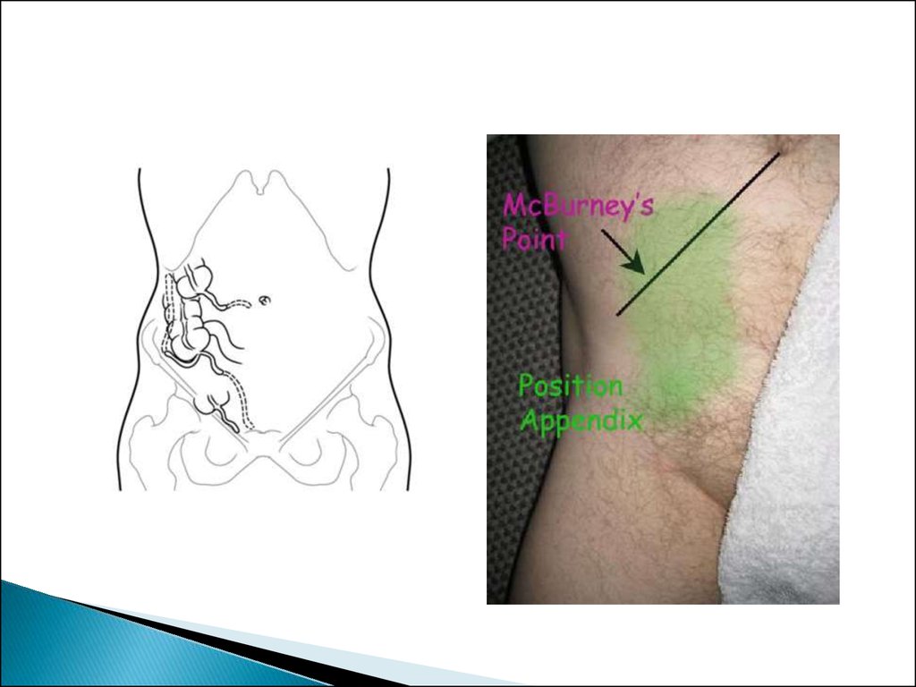
reflect onto the superior surface of the urinary
bladder (3), then over the uterus in the female (4),
then down into the pouch of Douglas (5), again in
the female, up along the anterior surface of the
rectum onto the posterior abdominal wall (6) until
you reach the root of the mesentery of the small
intestine.
From here you follow the mesentery of the small
intestine (7) going around its coils until you reach
the other side of the mesentery back down to the
posterior abdominal wall where you will cross over
the horizontal part of the duodenum (8). Your
finger will then travel along the inferior aspect of
the gastrocolic ligament (9), down the posterior
surface of the greater omentum (go) to its lower
border and back up along its anterior surface(11).
Your finger then passes over the anterior surface of
the stomach (12), along the anterior lamina of the
lesser omentum (13). At this time you probably
couldn't continue the trip because you would have
to enter the epiploic foramen (ef) to enter the
lesser peritoneal cavity (lpc) where visceral
peritoneum lines this space anteriorly and parietal
peritoneum posteriorly.
6. ligaments
lig.lig.
lig.
lig.
lig.
lig.
lig.
lig.
lig.
lig.
lig.
lig.
lig.
lig.
lig.
lig.
lig.
lig.
lig.
falciforme
coronarium hepatis
triangulare
hepatogastricum
hepatoduodenale
hepatocolicum
hepatorenale
gastrophrenicum
gastrolienale
gastrocolicum
gastropancreaticum
phrenicoesophageale
phrenicocolicum
phrenicorenale
phrenicolienale
pancreaticolienale
lienorenale
pyloropancreaticum
duodenorenale
7. Recesses - pouches formed by the peritoneal folds
duodenojejunal recesssuperior ileocaecal recess
inferior ileocaecal recess
retrocaecal recess
intersigmoid recess
8. Folds – reflection of the peritoneum arised from the abdominal wall by uderlying structures
PlicaPlica
Plica
Plica
Plica
Plica
Plica
gastropancreatica
ileocecalis
duodenalis superior
duodenalis inferior
umbilicalis mediana
umbilicalis medialis
umbilicalis lateralis
9. sinuses
RIGHT MESENTERIC SINUSborders:
medial-root of the mesentry
Lateral – ascending colon
Superior – transverse colon
LEFT MESENTERIC SINUS
Borders
Medial – descending colon
Lateral – root of the mesentry
Inferior – sigmoid colon
10. Paracolic canals
Right paracolic canal communicates with righthepatic bursa
Borders:
Medial – ascending colon
Lateral – parietalperitoneum
inferior – caecum
Left paracolic canal communicates with lesser
pelvis
Borders:
Medial – descending colon
Lateral – parietal peritoneum
Superior – phrenicocolic ligament
11. Bursae of the abdominal cavity
HEPATIC BURSABorders:
Superior – diaphragm
Inferior – transverse mesocolon
Anterior – anterior abdominal wall
Medial – falciform ligament
Pathology: abscess from the inferior storey of
the abdominal cavity may spread here and
cause subphrenic abscess through the right
paracolic canal
12. Bursae of the abdominal cavity
Pregastric bursaBorders:
Anterior – left lobe of the liver and anterior
abdominal wall
Posterior – lesser omentum
Pathology: abscess from this bursa may
spread to the omental bursa
13. Omental bursa (bursa omentalis)
BORDERS:Superior – lobus caudatus hepatis
Inferior – mesocolon transversum
Anterior – stomach & lesser omentum
Posterior – parietal peritoneum
Pathology: inflammation from this bursa may
spread to the general peritoneal cavity through
the epiploicc foramen.
FORAMEN EPIPLOICUM
BORDERS
Superior – lobus caudatus hepatis
Inferior – superior part of duodenum
Anterior – lig.hepatoduodenale
Posterior – lig.hepatorenale, parietal peritoneum
which covers v.cava inferior
14. stomach
The branches to the stomach arise from the above: celiac (C)◦ left gastric (LG) - supplies the lesser curvature of the stomach and lower
esophagus
esophageal (E)
splenic (S) which gives rise to:
common hepatic (CH)
◦ short gastric (SG) - supplies area of the fundus
◦ left gastroepiploic (LGE) - supplies the left part of greater curvature of the
stomach
◦ gastroduodenal (GD)
right gastric (RG) - supplies right side of lesser curvature of the stomach
right gastroepiploic (RGE) - supplies the right part of the greater curvature of
the stomach
15. Venous drainage from stomach
The stomach drains eitherdirectly or indirectly into the
portal vein as follows:short
gastric veins (SG) from the
fundus to the splenic vein (S)
left gastroepiploic (LGE) along
greater curvature to superior
mesenteric vein (SM)
right gastroepiploic (RGE) from
the right end of greater
curvature to superior
mesenteric vein (SM)
left gastric vein (LG) from the
lesser curvature of the stomach
to the portal vein (PV)
right gastric vein (RG) from the
lesser curvature of the stomach
to the portal vein (PV)
16. Nerve supply
17.
18. Gastritis (acute or stress)
Produces inflammationof the mucosa.
Can be associated with
erosions and bleeding.
Causes:
◦ H. pylori, NSAIDS, bile
reflux, Etoh, radiation,
local trauma, physiologic
stress.
19. Menetrier’s Disease (aka Hypertrophic Gastritis)
20. Gastric Polyps
21. Bezoars
22. The “Culprit”
H. pyloriTreatment:
◦ Triple therapy
23. Gastric ulcers
24. Gastric Ulcers
25. History of Peptic Ulcer Surgery
Harberer 1882- first gastric resection forulcer
Billroth 1885- Billroth II gastrectomy
Hofmeister 1896- Retrocolic anastamosis
Dragstedt 1943- Truncal vagotomy
Visick 1948- vagotomy and drainage
Johnson 1970- highly selective vagotomy
26. Laser Coagulation of Bleeding Ulcer
27. Coil Embolization of Bleeding Ulcer
28. Pyloroplasty for Bleeding Ulcer
29. Open Surgical Procedures
Truncal vagotomy and pyloroplastyTruncal vagotomy and gastrojejunostomy
Truncal vagotomy and antrectomy
Highly selective vagotomy
30. Operations on stomach
GASTROSTOMYTemporary gastrostomy
Minimal gastrostomy
Vitzel’s gastrostomy
Stamm-Kader’s gastrostomy
Permanent gastrostomy
Toprover’s gastrostomy
Beck Jian’s gastrostomy
PARTIAL RESECTION OF THE STOMACH
Billroth I – the stump of the stomach is
anastomosed with that of the duodenum
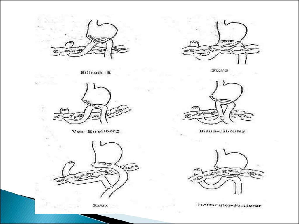
Billroth II - the stump of the stomach is
anastomosed with the initial portion of the ileum
Modifications of Billroth II
31.
32. Roux -en -Y Reconstruction
33. Antecolic and Retrocolic BII
34. Truncal Vagotomy
Resect 1-2cm of each vagal trunk on distalesophagus.
Reduces acid by 80%.
Denervates parietal cells, antral pump,
pyloric sphincter mechanism.
Delays gastric emptying, so need drainage.
With pyloroplasty recurrence 3-10%
With pyloroplasty morbidity 1-2%
35. Antrectomy and Truncal Vagotomy with BI
36. Truncal Vagotomy and Antrectomy
Entails distal gastrectomy of 50-60% ofstomach.
Removes parietal cell mass.
Requires a BI or BII reconstruction.
Recurrence rate 0.6-4%
Morbidity rate 0.9-1.6%
37. Selective Vagotomy
Total denervation of the stomach fromdiaphragmatic crus to pylorus.
Procedure still needs drainage, but advantage
is other organs are spared, liver, gallbladder,
small bowel, colon.
38. Highly Selective Vagotomy
Spares nerves of Latarjet, but divides vagalbranches to proximal 2/3 of stomach.
Antral innervation is thus preserved, gastric
emptying preserved, so drainage procedure
unnecessary.
Recurrence rate 10-15%
Lowest morbidity of all
39. Types of Vagotomies
40. Gastric Adenocarcinoma
41. Duodenum
4 partsMetabolically active
◦ Produces many
enzymes
D2: site of pacemaker
D2: posterolateral
insertion of ampulla.
Becomes jejunum at
the _____________?
42. Duodenum
Brunner’s glandsBlood supply:
◦ GDA- superior
pancreaticoduodenal
◦ SMA- inferior
pancreaticoduodenal
43. duodenum
Blood Supply of theDuodenum
superior
pancreaticoduodenal
◦ anterior and posterior
branches
inferior
pancreaticoduodenal
◦ anterior and posterior
branches
44. Duodenal Ulcers
45. Obstruction
46. Small Bowel Obstruction
HistorySigns and Symptoms
◦ Prior surgery
◦ Hernias
◦
◦
◦
◦
Colicky abdominal pain
Nausea and vomiting
Abdominal distension
Rectal exam
No peritoneal signs
47. Intestinum Crasum
48. Large Bowel Obstruction
49. colostomy
50. Anastamosis
Stapled vs. Hand-Sewn◦ Brundage et al. J trauma.
1999
◦ Multicenter retrospective
cohort design
“anastamotic leaks and
intra-abdominal
abscesses appear to be
more likely with stapled
bowel repairs compared
with sutured
anastamoses in the
injured patient. Caution
should be exercised in
deciding to staple a
bowel anastomosis in the
trauma patient.”
51. Anastamosis
Burch et al. Ann Surg. 1999Burch et al. Ann of Surg.
1999.
Prospective randomized
trial of single-layer
continuous vs. two layer
interrupted intestinal
anastamosis
NB: Important to invert,
4-6mm seromuscular
bites, 5mm advances,
larger bites at mesenteric
border
Single layer – similar leak
rate (approx 2%),
cheaper, faster
52. Appendix vermiformis
53.
54.
The caecum was at McBurney's point in 245(80.9%) patients, pelvic in 45 (14.9%) and high
lying in 13 (4.3%). The appendix was pelvic in
155 (51.2%) patients, pre-ileal in 9 (3.0%), paracaecal in 11 (3.6%), post-ileal in 67 (22.1%) and
retrocaecal in 61 (20.1%) patients.
The average length was 8.9 cm in males and 9.4
cms in females. The appendix was commonly
found to be retrocaecal (58.3%) on pelvic (21.7%)
or paracaecal (11.7%). Anomalies of the appendix
were more common in children than adults and
occurred in 47% of cases.
55. Topography of appendix vermiformis and ceacum
56.
57. Ulcerative Colitis
Disease SeverityMild colitis: 20%
Moderate colitis: 71%
Severe colitis: 9%
Acute disease
complications
Toxic colitis or megacolon
Perforation
Hemorrhage
Langholz 1991
58. Subtotal Colectomy
59. Liver
60. Liver
61. Liver Structure
Slide 61Mosby items and derived
items © 2006 by Mosby,
Inc.
62. Porto-caval anastomoses
63. Caput Medusa
64.
65. Varices on EGD
66. Varix Banding
67. Gall bladder
68. Arteries of the gall bladder
69. Innervation of gall bladder
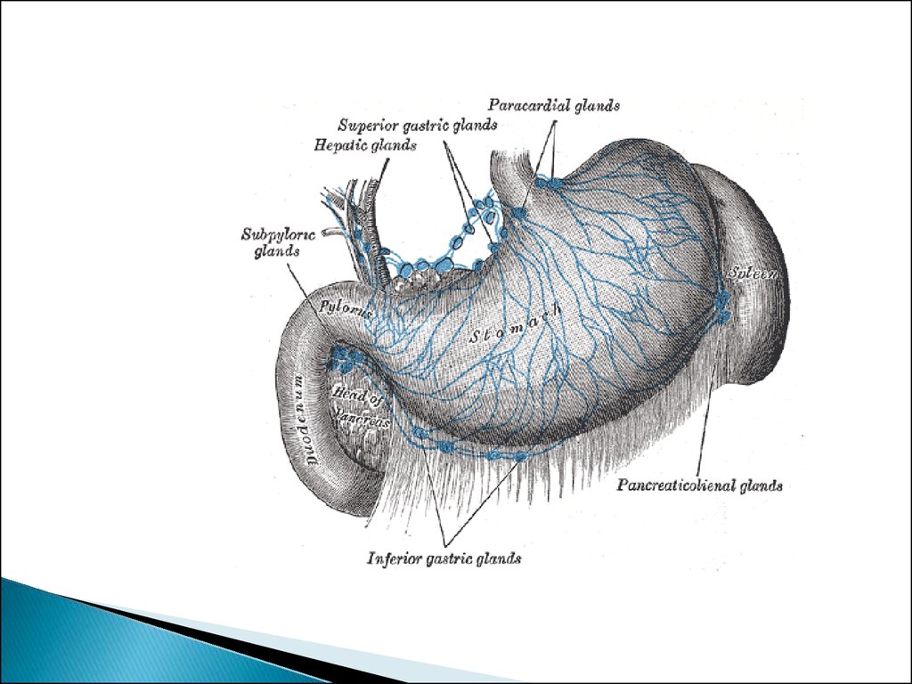
70. Lymphatic drainage of the gallbladder
71. Harvest Time
72. CT Scan
73. Plain Films
74. Ultrasound
75. Laparoscopic Cholecystectomy
76. cancer
77. Surgical Options
Simple cholecystectomyRadical cholecystectomy
Radical chole w/ anatomic liver resection
Radical chole w/ Whipple