


 Биология
Похожие презентации:


")


")
. Occurrence and classification")


Premalignant conditions of the cervix
1. Premalignant conditions of the cervix
2. cervix
Thecervix is a tubular structure. It is
composed of stromal tissue which is lined by
sequamous epithelium in the vagina
(ectocervix) and columnar epithelium within
the cervical canal (endocervix).
The meeting of the two types of the
epithelium is called squamocolumnar
junction SCJ and this is usually at the
ectocervix.
3.
The position of the SCJ changes throughout thereproductive years.
In children it lies at the ectocervix that is
just at the external os.
At puberty and during pregnancy it extends
outwards as the cervix enlarges and in adult
life it returns to the ectocervix through the
process of metaplasia
4. Transformation zone:
isan important area on the cervix which is
defined as the area where the original SCJ
was to the current SCJ and it includes areas
of metaplasia.
Occasionally, when the mucous columnar
epithelium is covered by the squamous
epithelium there is retention of the mucusthis is called a nabothian follicle.
The transformation zone TZ is the site where
pre-malignancy and
malignancy develop.
5. Definitions and terminology:
CIN:cervical intraepithelial neoplasia,
Dysplasia: a histological term describing
architectural abnormalities within the tissue.
Dyskaryosis: a cytological term describing
the nuclear abnormalities- not synonymous
with dysplasia
6.
CINI: minimal dysplasia.
CIN II: moderate dysplasia.
CIN III: sever dysplasia or CIS carcinoma in
situ ( CIN III, sever dysplasia and CIS are all
different names for the same thing that is
early cervical cancer)
7.
Metaplasia:a physiological process whereby
columnar epithelium is replaced by squamous
tissue in response to the acid environment of
the vagina.
Squamocolumnar
junction SCJ: where
squamous and columnar tissue meets, this is
not fixed, but is affected by metaplasia.
8.
Papssmear: or cervical smear- cytological
test described by Papanicolaou.
9. CIN
is a condition characterized by newcellular growth (neoplasia) in a normal tissue
Once CIN is diagnosed this alarm us that an
abnormal tissue has been diagnosed in the
cervix of that lady.
The most important causative factor is HPV
which could cause other combined genital
and anal cancer.
10.
However,CIN is much more common than the
other types of genital neoplasia.
The tissue changes associated with CIN
signify presence of premalignant or
precancerous condition i.e. CIN is essentially
a precursor to invasive cervical cancer and is
collectively composed of cells that have
undergone abnormal individual changes that
is with abnormal mitotic activity and leads to
formation of a lesion in the cervix.
11. Classification of CIN:
A revised classification has been introduced:Low – grade lesion CIN I and HPV associated
changes with unknown but a likely low
progressive potential.
High-grade lesion CIN II and CIN III that is likely
to behave as cancer precursors.
Simpler classification is according to Bethesda
divided to:
Low grade squamous intraepithelial lesion (LSIL)
= CIN I.
High grade squamous intraepithelial lesion (HSIL)
= CIN II and CIN III
12.
13. Aetiology
Humanpapillomavirus HPV infection is the
essential prerequisite for the development of
cervical malignancy.
HPV infection is extremely common with up
to 80% of sexually active women being HPV
positive at some point during their lifetime.
14.
Usingthe incidence of genital wart as a
marker, the incidence appears to be raising
five fold in the female population and eight
fold in male population with approximately
15% prevalence of the oncogenic HPV types
16 and 18.
However most infections are usually
transient with 90% of women clearing the
infection within 2 year and young competent
women are able to eliminate the infection.
15. Risk factors
Smokingreduces local cervical immunity.
Multiple sexual partners.
Having a partner with multiple sexual
partners or with sexually transmitted
disease.
Presence of other sexually transmitted
disease like HIV and genital herpes.
16. Risk factor
Longterm use of contraceptive pill.
Immunosupression or use of anticancer
drugs.
Being born to mother used diethylstilbestrol.
17. Clinical feature:
Oftenit’s a symptomatic and diagnosed
during routine annual Pap smear,
non-specific:
Genital lesion (wart)
Abnormal lower genital bleeding.
Abnormal vaginal discharge.
Vague lower abdominal pain.
18. Pathophysiology:
Metaplasiais a normal finding but this may
be disrupted by some factors like HPV,
smoking or immunosupression and etc.. And
lead to development of disorder squamous
epithelium called dysplasia which
characterized by:
Lack of normal maturation of cell as they
move from basal layer to superficial layer.
Large nuclei more variablle in size and
shape.
High mitotic activity means more rapidly
dividing cell.
19.
20. These cellular changes are divided to CIN I, II and III depending on :
Severityof atypia.
Thickness of the epithelium involved.
CIN I means 1/3 of the epithelium from the
basal layer is involved.
CIN II means 2/3 of the epithelium from the
basal layer is involved.
CIN III means no maturation throughout the
full thickness.
21. Natural history
Regression and progression of CIN may occur. Spontaneousregression of low grade disease is common and is likely to
occur through the patient’s own cell mediated immunity.
High grade lesion is less likely to regress spontaneously and
requires treatment as there is risk of progression to cancer.
22. Natural History of HPV Infection and Cin
1 yearUp to 5 years
Persistent
infection
Initial
HPV
infection
CIN* 1
CLEARED HPV INFECTION
*cervical intraepithelial neoplasia
Up to 20 years
CIN* 2/3
CANCER
23.
If left untreated 20% of patients with high gradeabnormalities may develop cancer of cervix.
Reasons for this remain unclear but may include
reduced host immunity, oncogenic HPV and
smoking.
24. Screening:
. Even the most sever CIN III take several manyyears to change to cancer, this mean we can
apply screening test to do early detection of
premalignant condition and do appropriate
treatment and follow up.
25. Screening is done by using Pap cytological test
Since 1988, the UK has offered populationbased cervical screening for women. Women
aged 25 and 64 are tested routinely as
follow:
25
first test
25-49
every 3 years
50-64
every 5 years
64+
only screen those who have not
screened since age 50 or has recent
abnormal test
26. Test performance:
Originallythe “Pap” smear was introduced by
Papanicolou, where cell removed from the
cervix using a wooden spatula and placed on
glass slide and fixed. This was then examined
by a cytologist for the immature squamous
cells sheds from the area of the CIN.
27.
NowPap smear is superseded by liquid based
cytology where a small brush is used to
sample cells from the transformation zone
and the brush head placed in the fixative.
This is then spun down and read by
cytologist.
Normal cervical cell has small nuclei that is
flattened and pyknotic but abnormal cell has
large nuclei, cytological atypia and high N/C
28.
Anabnormal smear can show cells in
different degree of maturity (dyskaryosis)
and is divided into:
Mild dyskaryosis and borderline changes (low
grade)
Moderate and sever dyskaryosis (high grade)
Abnormal smears act as a mean of referring
the patient to the colposcopic clinic for
further assessment.
29.
Thesensitivity of cervical smear in picking
up women with CIN is around 70 percent,
however, as there is slow progression for
most women with CIN to cancer, if a lesion is
missed then this should be picked up on
subsequent smear. The specifity is 90%.
If the test is negative the patient is re-placed
on routine recall.
30.
Ifthe smear shows low grade changes the
patient offered repeated test in next 3-6
months and managed accordingly and if test
shows high grade lesion the patient is
urgently referred to colposcopy.
31. Technique of smear:
Patientin lithotomy position under good
light,
start by inspection (spread labia and look for
any discharge or abnormal growth and ulcer)
then
insert warm vaginal speculum (not too hot),
do not use any lubricant , Vaseline or K-Y
jelly. The blades of speculum is kept closed
until is fully inserted.
32.
Identify the SCJ that is the junction of pinkcervical skin and red endocervical canal then
use Ayres spatula is used to sample the cervix
33.
theconcave end is used to fit the cervix and
should be rotated 360 degree
do not use too much force as it may cause
bleeding and pain
or too little force as it may lead to in
adequate sample).
The smear should be as thin as possible,
properly labeled,
allow fully drying before packaging and
spraying with fixative within 10-15 seconds.
34. Colposcopy:
Colposcopyis the outpatient examination of
the magnified cervix using a light source. It is
used for both diagnosis and treatment. After
inserting a speculum the cervix is examined
using Binocular operative microscope under
magnification (5-20 time).
35. colposcopy
5%acetic acid is applied, as it causes
nucleoproteins within the cells to coagulate.
Therefore areas of increased cell turnover,
for example CIN will appear white.
36. colposcopy
Schiller’stest: by application of iodine,
areas of CIN lack the presence of
intracellular glycogen and therefore are stain
yellow as opposed to normal which stain
brown when iodine is applied.
37. colposcopy
Abnormalvascular pattern like punctuate or
mosiasim.
Biopsy is taken from the most abnormal site.
Colposcopy is deemed unsatisfactory if TZ is
not viewed adequately.
38. HPV DNA testing:
AsHPV is the main causative factor of CIN
and cervical cancer, recently detection of
HPV DNA in serum has been introduced to
screening program but this is still used under
research.
39. Treatment of CIN:
Theaim of treatment is to make the posttreatment test negative while minimizing
harm to the patient.
Low grade lesion will regress spontaneously
in over 60% of cases and usually they require
no treatment but careful follow up by with
colposcopy and cytology in next six month
after initial diagnosis.
If CIN is not resolve on follow up tests or
progress to high grade then treatment is
needed to avoid development of active
disease.
40. treatment
Could be out patient or in patientExcisional methods like:
Loop electrosurgical excision (LEEP) and
large loop excision of TZ (LLETZ)
Laser TZ excision
Knife, laser or loop cone biopsy.
Hysterectomy.
41.
Ablative methods:Cryocautery.
Electrodiathermy
Coagulation
Laser.
42.
Thefavored method is LLETZ which is done
as outpatient under local anesthesia and
take 15 minutes and should go 10 mm deep
down cervical stroma,
43.
theadvantage is that its effective (95% test
negative post treatment), cost-effective and
provide specimen for histology. The
disadvantage may lead to poor obstetric
outcome as it may weaken the cervix
44.
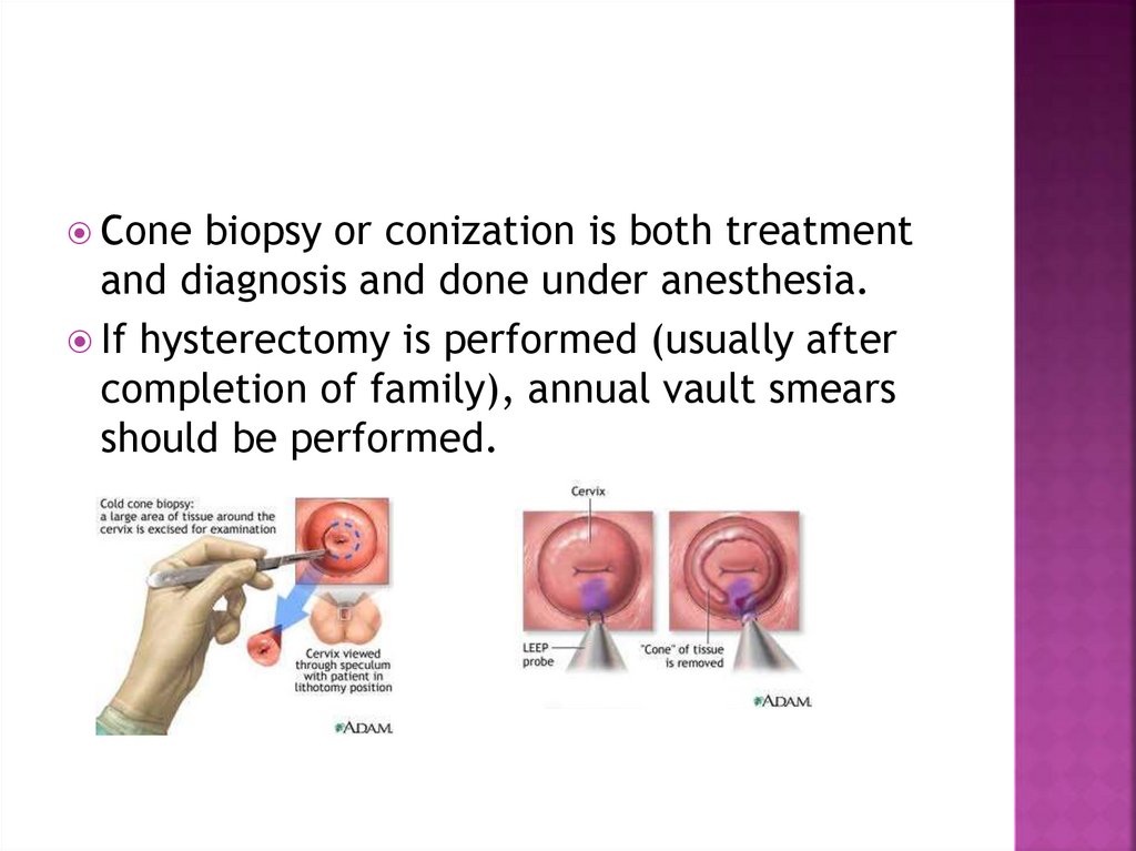
Conebiopsy or conization is both treatment
and diagnosis and done under anesthesia.
If hysterectomy is performed (usually after
completion of family), annual vault smears
should be performed.
45. Follow up:
Closefollow up after initial treatment by
regular cervical smear is needed after six
month then yearly for ten year, as the risk of
recurrence and cancer is remains.
46. HPV VACCINES
RecentlyHPV vaccines have been developed
to prevent primary infection with certain
oncogenic HPV types (16,18,31,33).
Many countries have a national program with
the sole aim to reduce death rate .
The evidence to date suggests that the
vaccination is not only effective in
preventing the development of high grade
CIN, but is safe to be given.
Its debatable.