


 Биология
Похожие презентации:


")


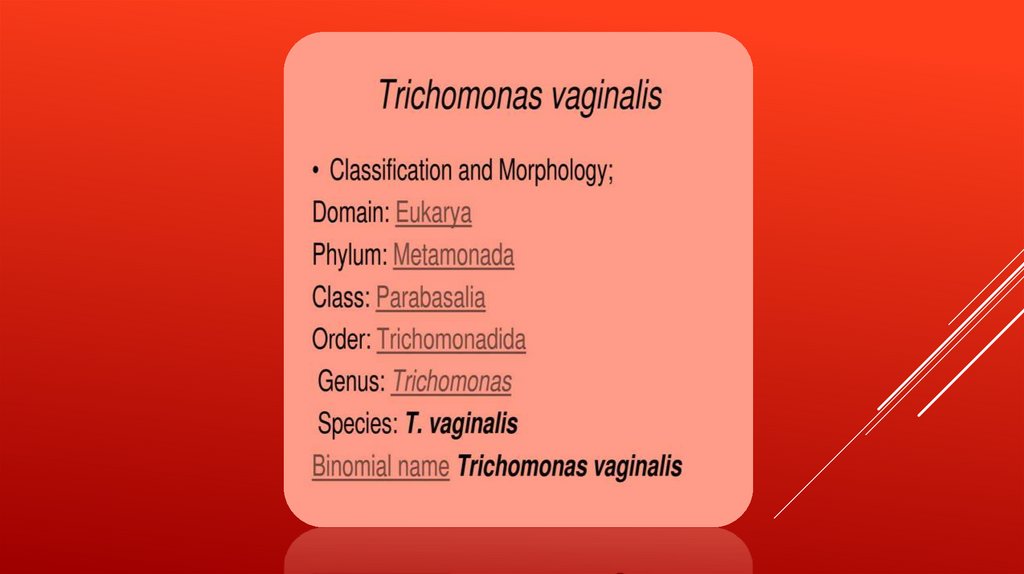
Trichomonas vaginalis
1.
2.
3.
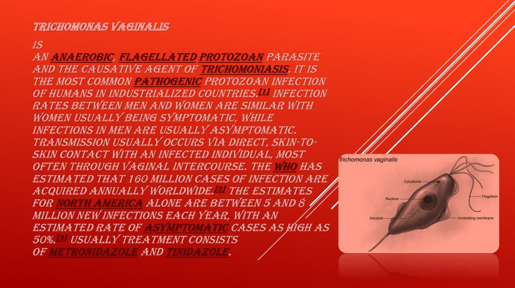
Trichomonas vaginalisis
an anaerobic, flagellated protozoan parasite
and the causative agent of trichomoniasis. It is
the most common pathogenic protozoan infection
of humans in industrialized countries.[1] Infection
rates between men and women are similar with
women usually being symptomatic, while
infections in men are usually asymptomatic.
Transmission usually occurs via direct, skin-toskin contact with an infected individual, most
often through vaginal intercourse. The WHO has
estimated that 160 million cases of infection are
acquired annually worldwide.[2] The estimates
for North America alone are between 5 and 8
million new infections each year, with an
estimated rate of asymptomatic cases as high as
50%.[3] Usually treatment consists
of metronidazole and tinidazole.
4.
5.
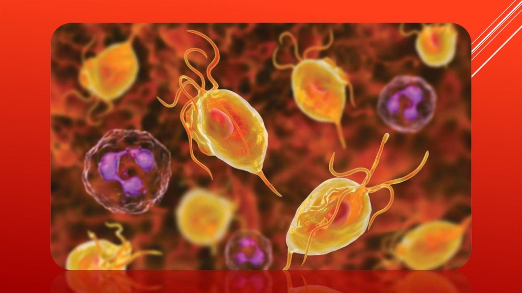
MORPHOLOGYUnlike other parasitic protozoa (Giardia lamblia, Entamoeba
histolytica etc.), Trichomonas vaginalis exists in only
one morphological stage, a trophozoite, and cannot encyst. The T.
vaginalis trophozoite is oval as well as flagellated, or "pear" shaped
as seen on a wet-mount. It is slightly larger than a white blood cell,
measuring 9 × 7 μm. Five flagella arise near the cytostome; four of
these immediately extend outside the cell together, while the fifth
flagellum wraps backwards along the surface of the organism. The
functionality of the fifth flagellum is not known. In addition, a
conspicuous barb-like axostyle projects opposite the four-flagella
bundle. The axostyle may be used for attachment to surfaces and may
also cause the tissue damage seen in trichomoniasis infections.
While T. vaginalis does not have a cyst form, organisms can survive for
up to 24 hours in urine, semen, or even water samples.
6.
Mechanismof infection
TRICHOMONAS VAGINALIS, A PARASITIC PROTOZOAN, IS
THE ETIOLOGIC AGENT OF TRICHOMONIASIS, AND IS
A SEXUALLY TRANSMITTED INFECTION.[2][6] MORE THAN
160 MILLION PEOPLE WORLDWIDE ARE ANNUALLY
INFECTED BY THIS PROTOZOAN
7.
HISTORYAlfred
Francois Donné (1801–1878) was the first to
describe a procedure to diagnose trichomoniasis through
"the microscopic observation of motile protozoa in
vaginal or cervical secretions" in 1836. He published this
in the article entitled, "Animalcules observés dans les
matières purulentes et le produit des sécrétions des
organes génitaux de l'homme et de la femme" in the
journal, Comptes rendus de l'Académie des sciences.[5] As
a result, the official binomial name of the parasite is
Trichomonas vaginalis DONNÉ.
8.
COMPLICATIONSSome of the complications of T. vaginalis in women include: preterm
delivery, low birth weight, and increased mortality as well as
predisposing to HIV infection, AIDS, and cervical cancer.[11] T.
vaginalis has also been reported in the urinary tract, fallopian tubes,
and pelvis and can cause pneumonia, bronchitis, and oral
lesions. Condoms are effective at reducing, but not wholly preventing,
transmission.[12]
Trichomonas vaginalis infection in males has been found to cause
asymptomatic urethritis and prostatitis.[13] It has been proposed that
it may increase the risk of prostate cancer; however, evidence is
insufficient to support this association as of 2014.
9.
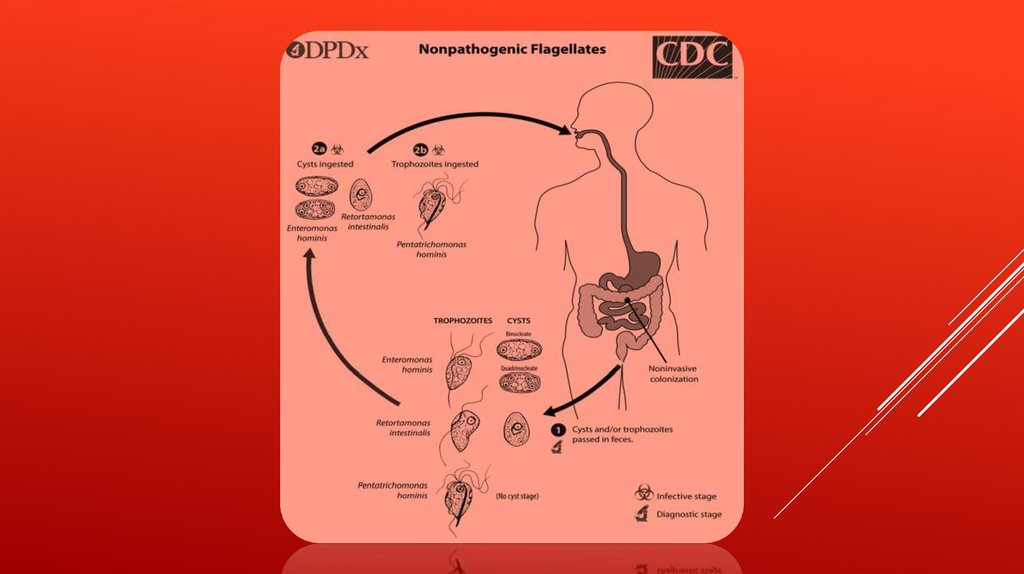
LIFE CYCLETrichomonas
vaginalis resides in the female lower genital
tract and the male urethra and prostate
where
it replicates by binary fission
The
parasite does not appear to have a cyst form, and
does not survive well in the external environment.
Trichomonas
vaginalis is transmitted among humans, its
only known host, primarily by sexual intercourse
10.
11.
GENETIC DIVERSITYRecent studies into the genetic diversity
of T.vaginalis has shown that there are two
distinct lineages of the parasite found
worldwide; both lineages are represented
evenly in field isolates. The two lineages differ
in whether or not T.vaginalis virus (TVV)
infection is present. TVV infection in T.vaginalis is
clinically relevant in that, when present, TVV
has an effect on parasite resistance to
metronidazole, a first line drug treatment for
human trichomoniasis.
12.
GENOME SEQUENCING AND STATISTICSThe T. vaginalis genome was found to be approximately 160 megabases in size[26] – ten times
larger than predicted from earlier gel-based chromosome sizing.[27] (The human genome is
~3.5 gigabases by comparison.[28]) As much as two-thirds of the T. vaginalis sequence consists of
repetitive and transposable elements, reflecting a massive, evolutionarily recent expansion of the
genome. The total number of predicted protein-coding genes is ~98,000, which includes ~38,000
'repeat' genes (virus-like, transposon-like, retrotransposon-like, and unclassified repeats, all with
high copy number and low polymorphism). Approximately 26,000 of the protein-coding genes
have been classed as 'evidence-supported' (similar either to known proteins, or to ESTs), while the
remainder have no known function. These extraordinary genome statistics are likely to change
downward as the genome sequence, currently very fragmented due to the difficulty of ordering
repetitive DNA, is assembled into chromosomes, and as more transcription data
(ESTs, microarrays) accumulate. But it appears that the gene number of the single-celled parasite T.
vaginalis is, at minimum, on par with that of its host H. sapiens.
In late 2007 TrichDB.org was launched as a free, public genomic data repository and retrieval
service devoted to genome-scale trichomonad data. The site currently contains all of the T.
vaginalis sequence project data, several EST libraries, and tools for data mining and display.
TrichDB is part of the NIH/NIAID-funded EupathDB functional genomics database project
13.
14.
VIRULENCE FACTORSOne
of the hallmark features of Trichomonas vaginalis is the
adherence factors that allow cervicovaginal
epithelium colonization in women. The adherence that this
organism illustrates is specific to vaginal epithelial
cells (VECs) being pH, time and temperature dependent. A
variety of virulence factors mediate this process some of
which are the microtubules, microfilaments, bacterial
adhesins (4), and cysteine proteinases. The adhesins are four
trichomonad enzymes called AP65, AP51, AP33, and AP23 that
mediate the interaction of the parasite to the receptor
molecules on VECs.[24] Cysteine proteinases may be another
virulence factor because not only do these 30 kDa proteins
bind to host cell surfaces but also may
degrade extracellular
matrix proteins like hemoglobin, fibronectin or collagen IV.
15.
PROTEIN FUNCTIONTrichomonas
vaginalis lacks mitochondria and therefore
necessary enzymes and cytochromes to conduct oxidative
phosphorylation. T. vaginalis obtains nutrients by transport
through the cell membrane and by phagocytosis. The organism
is able to maintain energy requirements by the use of a small
amount of enzymes to provide energy
via glycolysis of glucose to glycerol and succinate in
the cytoplasm, followed by further conversion
of pyruvate and malate to hydrogen and acetate in
an organelle called the hydrogenosome.
16.
INCREASED SUSCEPTIBILITY TO HIVThe
damage caused by Trichomonas vaginalis to the
vaginal epithelium increases a woman's susceptibility
to an HIV infection. In addition to inflammation, the
parasite also causes lysis of epithelial cells and RBCs
in the area leading to more inflammation and disruption
of the protective barrier usually provided by the
epithelium. Having Trichomonas vaginalis also may
increase the chances of the infected woman
transmitting HIV to her sexual partner(s).
17.
DIAGNOSISClassically, with a cervical smear, infected women may have a
transparent "halo" around their superficial cell nucleus but more
typically the organism itself is seen with a slight cyanophilic
tinge, faint eccentric nuclei, and fine acidophilic granules.[14] It is
unreliably detected by studying a genital discharge or with a
cervical smear because of their low sensitivity. T. vaginalis was
traditionally diagnosed via a wet mount, in which "corkscrew"
motility was observed. Currently, the most common method of
diagnosis is via overnight culture,[15][16] with a sensitivity range of
75–95%.[17] Newer methods, such as rapid antigen
testing and transcription-mediated amplification, have even
greater sensitivity, but are not in widespread use.[17] The presence
of T. vaginalis can also be diagnosed by PCR, using primers specific
for GENBANK
18.
TREATMENTInfection
is treated and cured
with metronidazole[19] or tinidazole. The CDC
recommends a one time dose of 2 grams of either
metronidazole or tinidazole as the first-line
treatment; the alternative treatment
recommended is 500 milligrams of metronidazole,
twice daily, for seven days if there is failure of the
single-dose regimen.[20] Medication should be
prescribed to any sexual partner(s) as well
because they may be asymptomatic carriers.
19.
FOR BETTERUNDERSTANDING
https://www.youtube.com/watch?v=SYd4lLed3CI
https://www.youtube.com/watch?v=yk0P7IpSiIg
https://www.youtube.com/watch?v=TlNBQx9rH20&list=TLPQMTMwNjIwMjBRQCLpCu
aiHQ&index=3