


 Медицина
Похожие презентации:
 – Digestion and Organs of digestive system")


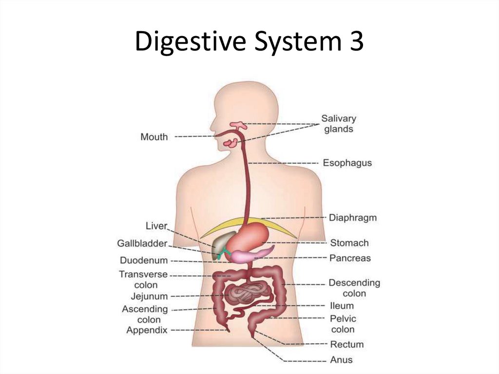
Digestive System
1.
Digestive System 32.
Small IntestinePROPERTIES OF SUCCUS ENTERICUS
• Volume : 1800 mL/day
• Reaction : Alkaline
• pH : 8.3
3.
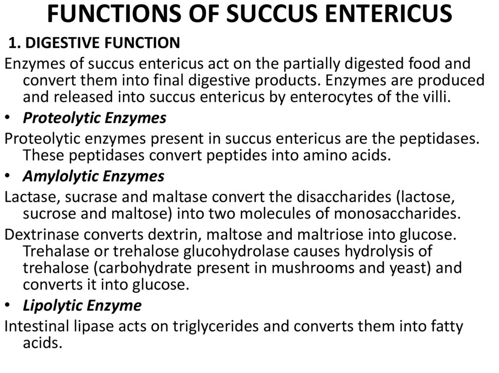
FUNCTIONS OF SUCCUS ENTERICUS1. DIGESTIVE FUNCTION
Enzymes of succus entericus act on the partially digested food and
convert them into final digestive products. Enzymes are produced
and released into succus entericus by enterocytes of the villi.
• Proteolytic Enzymes
Proteolytic enzymes present in succus entericus are the peptidases.
These peptidases convert peptides into amino acids.
• Amylolytic Enzymes
Lactase, sucrase and maltase convert the disaccharides (lactose,
sucrose and maltose) into two molecules of monosaccharides.
Dextrinase converts dextrin, maltose and maltriose into glucose.
Trehalase or trehalose glucohydrolase causes hydrolysis of
trehalose (carbohydrate present in mushrooms and yeast) and
converts it into glucose.
• Lipolytic Enzyme
Intestinal lipase acts on triglycerides and converts them into fatty
acids.
4.
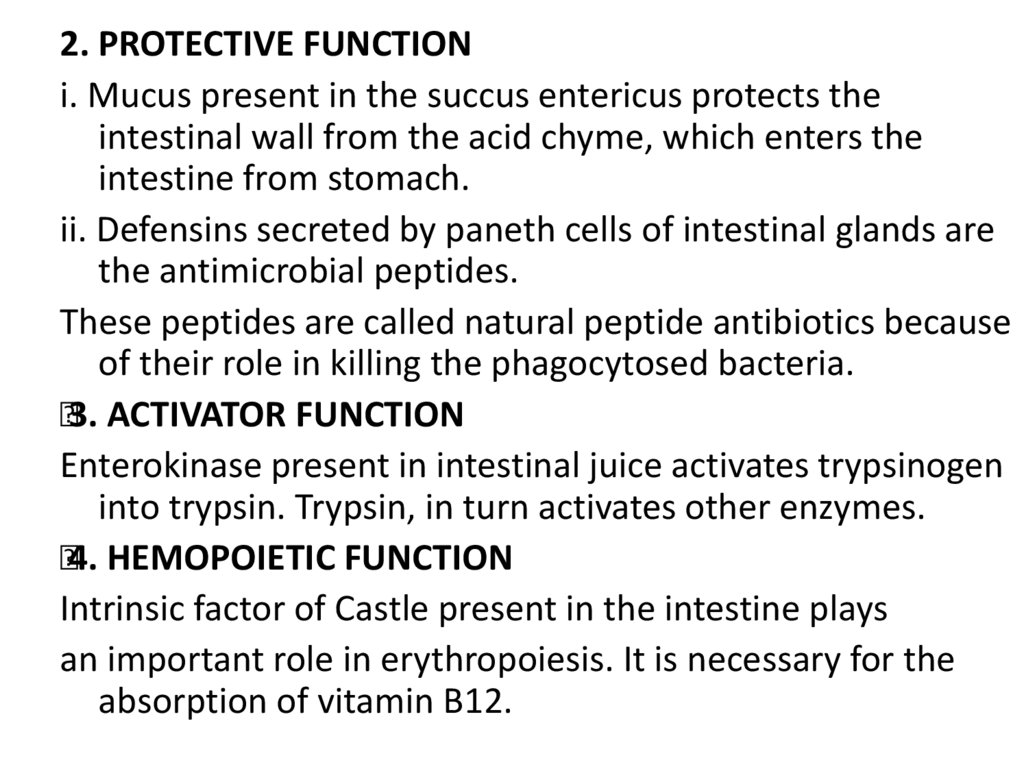
2. PROTECTIVE FUNCTIONi. Mucus present in the succus entericus protects the
intestinal wall from the acid chyme, which enters the
intestine from stomach.
ii. Defensins secreted by paneth cells of intestinal glands are
the antimicrobial peptides.
These peptides are called natural peptide antibiotics because
of their role in killing the phagocytosed bacteria.
3. ACTIVATOR FUNCTION
Enterokinase present in intestinal juice activates trypsinogen
into trypsin. Trypsin, in turn activates other enzymes.
4. HEMOPOIETIC FUNCTION
Intrinsic factor of Castle present in the intestine plays
an important role in erythropoiesis. It is necessary for the
absorption of vitamin B12.
5.
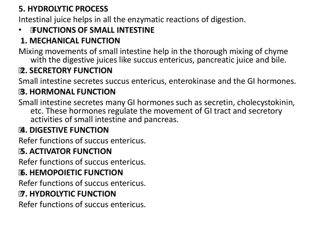
5. HYDROLYTIC PROCESSIntestinal juice helps in all the enzymatic reactions of digestion.
• FUNCTIONS OF SMALL INTESTINE
1. MECHANICAL FUNCTION
Mixing movements of small intestine help in the thorough mixing of chyme
with the digestive juices like succus entericus, pancreatic juice and bile.
2. SECRETORY FUNCTION
Small intestine secretes succus entericus, enterokinase and the GI hormones.
3. HORMONAL FUNCTION
Small intestine secretes many GI hormones such as secretin, cholecystokinin,
etc. These hormones regulate the movement of GI tract and secretory
activities of small intestine and pancreas.
4. DIGESTIVE FUNCTION
Refer functions of succus entericus.
5. ACTIVATOR FUNCTION
Refer functions of succus entericus.
6. HEMOPOIETIC FUNCTION
Refer functions of succus entericus.
7. HYDROLYTIC FUNCTION
Refer functions of succus entericus.
6.
• 8. ABSORPTIVE FUNCTIONS• Presence of villi and microvilli in small intestinal mucosa increases the
surface area of mucosa. This facilitates the absorptive function of intestine.
• Digested products of foodstuffs, proteins, carbohydrates, fats and other
nutritive substances such as vitamins, minerals and water are absorbed
mostly in small intestine. From the lumen of intestine, these substances
pass through lacteal of villi, cross the mucosa and enter the blood directly
or through lymphatics.
• Absorption of Water and Minerals
i. In small intestine, sodium is absorbed actively. It is responsible for
absorption of glucose, amino acids and other substances by means of
sodium cotransport.
ii. Water moves in or out of the intestinal lumen until the osmotic pressure of
intestinal contents becomes equal to that of plasma.
iii. In ileum, chloride ion is actively absorbed in exchange for bicarbonate. The
significance of this exchange is not known.
iv. Calcium is actively absorbed mostly in upper part of small intestine.
• Absorption of Vitamins
Most of the vitamins are absorbed in upper part of small intestine and vitamin
B12 is absorbed in ileum. Absorption of water-soluble vitamins is faster
than fatsoluble vitamins.
7.
REGULATION OF SECRETIONOF SUCCUS ENTERICUS
Secretion of succus entericus is regulated by both nervous and hormonal
mechanisms.
NERVOUS REGULATION
Stimulation of parasympathetic nerves causes vasodilatation and increases
the secretion of succus entericus. Stimulation of sympathetic nerves
causes vasoconstriction and decreases the secretion of succus entericus.
But, the role of these nerves in the regulation of intestinal secretion in
physiological conditions is uncertain. However, the local nervous reflexes
play an important role in increasing the secretion of intestinal juice.
When chyme enters the small intestine, the mucosa is stimulated by
tactile stimuli or irritation. It causes the development of local nervous
reflexes, which stimulate the glands of intestine.
HORMONAL REGULATION
When chyme enters the small intestine, intestinal mucosa secretes
enterocrinin, secretin and cholecystokinin, which promote the secretion
of succus entericus by stimulating the intestinal glands.
8.
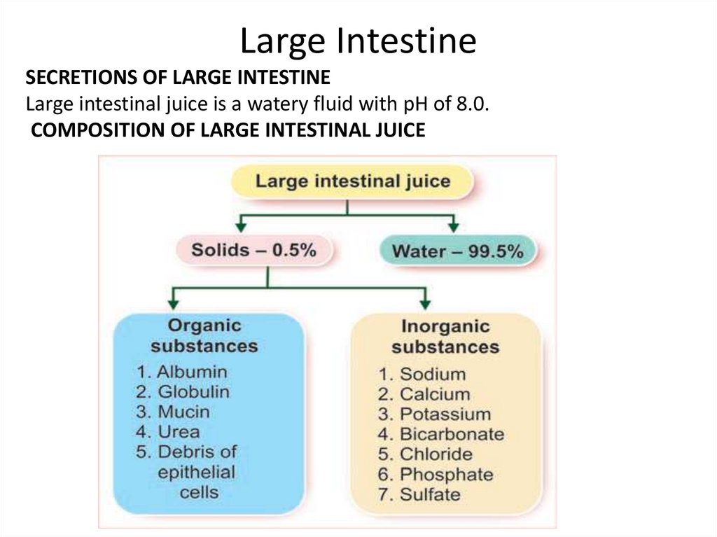
Large IntestineSECRETIONS OF LARGE INTESTINE
Large intestinal juice is a watery fluid with pH of 8.0.
COMPOSITION OF LARGE INTESTINAL JUICE
9.
FUNCTIONS OF LARGE INTESTINAL JUICENeutralization of Acids
Strong acids formed by bacterial action in large intestine
are neutralized by the alkaline nature of large intestinal
juice. The alkalinity of this juice is mainly due to the
presence of large quantity of bicarbonate.
Lubrication Activity
Mucin present in the secretion of large intestine
lubricates the mucosa of large intestine and the bowel
contents, so that, the movement of bowel is facilitated.
Mucin also protects the mucus membrane of large
intestine by preventing the damage caused by
mechanical injury or chemical substances.
10.
FUNCTIONS OF LARGE INTESTINE1. ABSORPTIVE FUNCTION
Large intestine plays an important role in the absorption of various substances such as:
a. Water
b. Electrolytes
c. Organic substances like glucose
d. Alcohol
e. Drugs like anesthetic agents, sedatives and steroids.
2. FORMATION OF FECES
After the absorption of nutrients, water and other substances, the unwanted substances in
the large intestine form feces. This is excreted out.
3. EXCRETORY FUNCTION
Large intestine excretes heavy metals like mercury, lead, bismuth and arsenic through
feces.
4. SECRETORY FUNCTION
Large intestine secretes mucin and inorganic substances like chlorides and bicarbonates.
5. SYNTHETIC FUNCTION
Bacterial flora of large intestine synthesizes folic acid, vitamin B12 and vitamin K. By this
function, large intestine contributes in erythropoietic activity and blood clotting
mechanism.
11.
Movements ofGastrointestinal Tract
MASTICATION
Mastication or chewing is the first mechanical process in the gastrointestinal (GI) tract, by which the food
substances are torn or cut into small particles and crushed or ground into a soft bolus.
Significances of mastication
• 1. Breakdown of foodstuffs into smaller particles
• 2. Mixing of saliva with food substances thoroughly
• 3. Lubrication and moistening of dry food by saliva, so that the bolus can be easily swallowed
• 4. Appreciation of taste of the food.
MUSCLES AND THE MOVEMENTS OF MASTICATION
Muscles of Mastication
• 1. Masseter muscle
• 2. Temporal muscle
• 3. Pterygoid muscles
• 4. Buccinator muscle.
Movements of Mastication
• 1. Opening and closure of mouth
• 2. Rotational movements of jaw
• 3. Protraction and retraction of jaw.
CONTROL OF MASTICATION
Action of mastication is mostly a reflex process. It is carried out voluntarily also. The center for mastication is
situated in medulla and cerebral cortex. Muscles of mastication are supplied by mandibular division of 5 th
cranial (trigeminal) nerve.
12.
DEGLUTITIONDeglutition or swallowing is the process by which food moves from mouth into stomach.
Stages of Deglutition
I. Oral stage, when food moves from mouth to pharynx
II. Pharyngeal stage, when food moves from pharynx to esophagus
III. Esophageal stage, when food moves from esophagus to stomach.
ORAL STAGE OR FIRST STAGE
Oral stage of deglutition is a voluntary stage. In this stage, the bolus from mouth passes into pharynx by means of series of actions.
Sequence of Events during Oral Stage
1. Bolus is placed over postero-dorsal surface of the tongue. It is called the preparatory position
2. Anterior part of tongue is retracted and depressed.
3. Posterior part of tongue is elevated and retracted against the hard palate. This pushes the bolus backwards into the pharynx
4. Forceful contraction of tongue against the palate produces a positive pressure in the posterior part of oral cavity. This also
pushes the food into pharynx.
PHARYNGEAL STAGE OR SECOND STAGE
Pharyngeal stage is an involuntary stage. In this stage, the bolus is pushed from pharynx into the esophagus.
Pharynx is a common passage for food and air. It divides into larynx and esophagus. Larynx lies anteriorly and continues as
respiratory passage. Esophagus lies behind the larynx and continues as GI tract. Since pharynx communicates with mouth, nose,
larynx and esophagus, during this stage of deglutition, bolus from the pharynx can enter into four paths:
1. Back into mouth
2. Upward into nasopharynx
3. Forward into larynx
4. Downward into esophagus.
However, due to various coordinated movements, bolus is made to enter only the esophagus. Entrance of bolus through other
paths is prevented as follows:
1. Back into Mouth
Return of bolus back into the mouth is prevented by:
i. Position of tongue against the soft palate (roof of the mouth)
ii. High intraoral pressure, developed by the movement of tongue.
13.
2. Upward into NasopharynxMovement of bolus into the nasopharynx from pharynx is prevented by elevation of soft palate
along with its extension called uvula.
3. Forward into Larynx
Movement of bolus into the larynx is prevented by the following actions:
• i. Approximation of the vocal cords
• ii. Forward and upward movement of larynx
• iii. Backward movement of epiglottis to seal the opening of the larynx (glottis)
• iv. All these movements arrest respiration for a few seconds. It is called deglutition apnea.
Deglutition apnea
Apnea refers to temporary arrest of breathing. Deglutition apnea or swallowing apnea is the
arrest of breathing during pharyngeal stage of deglutition.
4. Entrance of Bolus into Esophagus
As the other three paths are closed, the bolus has to pass only through the esophagus. This
occurs by the combined effects of various factors:
• i. Upward movement of larynx stretches the opening of esophagus
• ii. Simultaneously, upper 3 to 4 cm of esophagus relaxes. This part of esophagus is formed
by the cricopharyngeal muscle and it is called upper esophageal sphincter or
pharyngoesophageal sphincter
• iii. At the same time, peristaltic contractions start in the pharynx due to the contraction of
pharyngeal muscles
• iv. Elevation of larynx also lifts the glottis away from the food passage.
All the factors mentioned above act together so that, bolus moves easily into the esophagus.
The whole process takes place within 1 to 2 seconds and this process is purely involuntary.
14.
ESOPHAGEAL STAGE OR THIRD STAGE• Esophageal stage is also an involuntary stage. In this stage, food from esophagus enters the stomach.
• Esophagus forms the passage for movement of bolus from pharynx to the stomach. Movements of
esophagus are specifically organized for this function and the movements are called peristaltic waves.
Peristalsis means a wave of contraction, followed by the wave of relaxation of muscle fibers of GI tract,
which travel in aboral direction (away from mouth). By this type of movement, the contents are propelled
down along the GI tract. When bolus reaches the esophagus, the peristaltic waves are initiated. Usually, two
types of peristaltic contractions are produced in esophagus.
• 1. Primary peristaltic contractions
• 2. Secondary peristaltic contractions.
• 1. Primary Peristaltic Contractions
When bolus reaches the upper part of esophagus, the peristalsis starts. This is known as primary peristalsis.
After origin, the peristaltic contractions pass down through the rest of the esophagus, propelling the bolus
towards stomach. Pressure developed during the primary peristaltic contractions is important to propel the
bolus. Initially, the pressure becomes negative in the upper part of esophagus. This is due to the stretching
of the closed esophagus by the elevation of larynx. But immediately, the pressure becomes positive and
increases up to 10 to 15 cm of H2O.
• 2. Secondary Peristaltic Contractions
If the primary peristaltic contractions are unable to propel the bolus into the stomach, the secondary peristaltic
contractions appear and push the bolus into stomach. Secondary peristaltic contractions are induced by the
distention of upper esophagus by the bolus. After origin, these contractions pass down like the primary
contractions, producing a positive pressure.
• Role of Lower Esophageal Sphincter
Distal 2 to 5 cm of esophagus acts like a sphincter and it is called lower esophageal sphincter. It is constricted
always. When bolus enters this part of the esophagus, this sphincter relaxes so that the contents enter the
stomach. After the entry of bolus into the stomach, the sphincter constricts and closes the lower end of
esophagus. The relaxation and constriction of sphincter occur in sequence with the arrival of peristaltic
contractions of esophagus.
15.
Stages of deglutition.A. Preparatory stage; B. Oral stage; C.
Pharyngeal stage; D. Esophageal
stage.
16.
DEGLUTITION REFLEXThough the beginning of swallowing is a voluntary act, later it becomes
involuntary and is carried out by a reflex action called deglutition reflex. It occurs
during the pharyngeal and esophageal stages.
Stimulus
When the bolus enters the oropharyngeal region, the receptors present in this
region are stimulated.
Afferent Fibers
Afferent impulses from the oropharyngeal receptors pass via the
glossopharyngeal nerve fibers to the deglutition center.
Center
Deglutition center is at the floor of the fourth ventricle in medulla oblongata of
brain.
Efferent Fibers
Impulses from deglutition center travel through glossopharyngeal and vagus
nerves (parasympathetic motor fibers) and reach soft palate, pharynx and
esophagus. The glossopharyngeal nerve is concerned with pharyngeal stage of
swallowing. The vagus nerve is concerned with esophageal stage.
Response
• The reflex causes upward movement of soft palate, to close nasopharynx and
upward movement of larynx, to close respiratory passage so that bolus enters the
esophagus. Now the peristalsis occurs in esophagus, pushing the bolus into
stomach.
17.
MOVEMENTS OF STOMACHActivities of smooth muscles of stomach increase during gastric digestion (when stomach is filled with food) and
when the stomach is empty.
Types of movements in stomach
• 1. Hunger contractions
• 2. Receptive relaxation
• 3. Peristalsis.
1. HUNGER CONTRACTIONS
Hunger contractions are the movements of empty stomach. These contractions are related to the sensations of
hunger. Hunger contractions are the peristaltic waves superimposed over the contractions of gastric smooth
muscle as a whole. This type of peristaltic waves is different from the digestive peristaltic contractions.
• The digestive peristaltic contractions usually occur in body and pyloric parts of the stomach. But, peristaltic
contractions of empty stomach involve the entire stomach. Hunger contractions are of three types:
• Type I Hunger Contractions
Type I hunger contractions are the first contractions to appear in the empty stomach, when the tone of the gastric
muscles is low. Each contraction lasts for about 20 seconds. The interval between contractions is about 3 to 4
seconds. Tone of the muscles does not increase between contractions. Pressure produced by these
contractions is about 5 cm of H2O.
• Type II Hunger Contractions
• Type II hunger contractions appear when the tone of stomach is stronger. Tone increases in stomach if food
intake is postponed, even after the appearance of the type I contractions. Each of the type II contractions lasts
for 20 seconds like type I contractions. But the pause between the contractions is decreased. Pressure
produced by these contractions is 10 to 15 cm of H2O.
• Type III Hunger Contractions
• Type III hunger contractions are like incomplete tetanus. These contractions appear when the hunger becomes
severe and the tone increases to a great extent. Type III hunger contractions are rare in man as the food is
taken usually before the appearance of these contractions. These contractions last for 1 to 5 minutes. The
pressure produced by these contractions increases to 10 to 20 cm of H2O. When the stomach is empty, the
type I contractions occur first, followed by type II contractions. If food intake is still postponed, then type III
contractions appear and as soon as food is consumed, hunger contractionsdisappear.
18.
2. RECEPTIVE RELAXATION• Receptive relaxation is the relaxation of the upper portion of the stomach
when bolus enters the stomach from esophagus. It involves the fundus
and upper part of the body of stomach. Its significance is to
accommodate the food easily, without much increase in pressure inside
the stomach. This process is called accommodation of stomach.
3. PERISTALSIS
• When food enters the stomach, the peristaltic contraction or peristaltic
wave appears with a frequency of 3 per minute. It starts from the lower
part of the body of stomach, passes through the pylorus till the pyloric
sphincter. Initially, the contraction appears as a slight indentation on the
greater and lesser curvatures and travels towards pylorus. The
contraction becomes deeper while traveling. Finally, it ends with the
constriction of pyloric sphincter. Some of the waves disappear before
reaching the sphincter. Each peristaltic wave takes about one minute to
travel from the point of origin to the point of ending.
• This type of peristaltic contraction is called digestive peristalsis because
it is responsible for the grinding of food particles and mixing them with
gastric juice for digestive activities.
19.
VOMITINGVomiting or emesis is the abnormal emptying of stomach and upper part of intestine through esophagus and
mouth.
CAUSES OF VOMITING
• 1. Presence of irritating contents in GI tract
• 2. Mechanical stimulation of pharynx
• 3. Pregnancy
• 4. Excess intake of alcohol
• 5. Nauseating sight, odor or taste
• 6. Unusual stimulation of labyrinthine apparatus, as in the case of sea sickness, air sickness, car sickness or
swinging
• 7. Abnormal stimulation of sensory receptors in other organs like kidney, heart, semicircular canals or uterus
• 8. Drugs like antibiotics, opiates, etc.
• 9. Any GI disorder
• 10. Acute infection like urinary tract infection, influenza, etc.
• 11. Metabolic disturbances like carbohydrate starvation and ketosis (pregnancy), uremia, ketoacidosis
(diabetes) and hypercalcemia.
• MECHANISM OF VOMITING
• Nausea
• Vomiting is always preceded by nausea. Nausea is unpleasant sensation which induces the desire for vomiting.
It is characterized by secretion of large amount of saliva containing more amount of mucus.
• Retching
• Strong involuntary movements in the GI tract which start even before actual vomiting. These movements
intensify the feeling of vomiting. This condition is called retching (try to vomit) and vomiting occurs few
minutes after this.
20.
Act of Vomiting
Act of vomiting involves series of movements that takesplace in GI tract.
Sequence of events:
1. Beginning of antiperistalsis, which runs from ileum towards the mouth through the intestine, pushing the
intestinal contents into the stomach within few minutes. Velocity of the antiperistalsis is about 2 to 3
cm/second
2. Deep inspiration followed by temporary cessation of breathing
3. Closure of glottis
4. Upward and forward movement of larynx and hyoid bone
5. Elevation of soft palate
6. Contraction of diaphragm and abdominal muscles with a characteristic jerk, resulting in elevation of intraabdominal pressure
7. Compression of the stomach between diaphragm and abdominal wall leading to rise in intragastric
pressure
8. Simultaneous relaxation of lower esophageal sphincter, esophagus and upper esophageal sphincter
9. Forceful expulsion of gastric contents (vomitus) through esophagus, pharynx and mouth.
Movements during act of vomiting throw the vomitus (materials ejected during vomiting) to the exterior
through mouth. Some of the movements play important roles by preventing the entry of vomitus through
other routes and thereby prevent the adverse effect of the vomitus on many structures.
Such movements are:
1. Closure of glottis and cessation of breathing prevents entry of vomitus into the lungs
2. Elevation of soft palate prevents entry of vomitus into the nasopharynx
3. Larynx and hyoid bone move upward and forward and are placed in this position rigidly. This causes the
dilatation of throat, which allows free exit of vomitus.
21.
VOMITING REFLEX• Vomiting is a reflex act. Sensory impulses for vomiting arise from the
irritated or distended part of GI tract or other organs and are
transmitted to the vomiting center through vagus and sympathetic
afferent fibers.
• Vomiting center is situated bilaterally in medulla oblongata near the
nucleus tractus solitarius.
• Motor impulses from the vomiting center are transmitted through V, VII,
IX, X and XII cranial nerves to the upper part of GI tract; and through
spinal nerves to diaphragm and abdominal muscles.
• Center for Vomiting during Motion Sickness and Vomiting Induced by
Drugs
• Center for vomiting during motion sickness and vomiting induced by
drugs such as morphine, apomorphine, etc. is on the floor of fourth
ventricle. This area is called chemoreceptor trigger zone. During motion
sickness, the afferent impulses from vestibular apparatus reach vomiting
center through this zone.
• Center for Psychic-stimuli-induced Vomiting
• Center for vomiting due to psychic stimuli such as nauseating odor, sight
or noise is in cerebral cortex
22.
MOVEMENTS OF SMALL INTESTINEMovements of small intestine are essential for mixing the chyme with digestive juices, propulsion of food and absorption.
Types of Movements of Small Intestine
Movements of small intestine are of four types:
1. Mixing movements:
i. Segmentation movements
ii. Pendular movements.
2. Propulsive movements:
i. Peristaltic movements
ii. Peristaltic rush.
3. Peristalsis in fasting – migrating motor complex
4. Movements of villi.
1. MIXING MOVEMENTS
Mixing movements of small intestine are responsible for proper mixing of chyme with digestive juices such as pancreatic juice,
bile and intestinal juice. The mixing movements of small intestine are segmentation contractions and pendular movements.
i. Segmentation Contractions
Segmentation contractions are the common type of movements of small intestine, which occur regularly or irregularly, but in
a rhythmic fashion. So, these move ments are also called rhythmic segmentation contractions.
The contractions occur at regularly spaced intervals along a section of intestine. The segment of the intestine involved in each
contraction is about 1 to 5 cm long.
The segments of intestine in between the contracted segments are relaxed. The length of the relaxed segments is same as
that of the contracted segments. These alternate segments of contraction and relaxation give appearance of rings, resembling
the chain of sausages.
After sometime, the contracted segments are relaxed and the relaxed segments are contracted . Therefore, the segmentation
contractions chop the chyme many times. This helps in mixing of chyme with digestive juices.
ii. Pendular Movement
Pendular movement is the sweeping movement of small intestine, resembling the movements of pendulum of clock. Small
portions of intestine (loops) sweep forward
and backward or upward and downward. It is a type of mixing movement, noticed only by close observation. It helps in mixing
of chyme with digestive juices.
23.
2. PROPULSIVE MOVEMENTS
Propulsive movements are the movements of small intestine which push the chyme in the aboral direction
through intestine. The propulsive movements are peristaltic movements and peristaltic rush.
i. Peristaltic Movements
Peristalsis is defined as the wave of contraction followed by wave of relaxation of muscle fibers. In GI tract,
it always travels in aboral direction. Stimulation of smooth muscles of intestine initiates the peristalsis. It
travels from point of stimulation in both directions. But under normal conditions, the progress of
contraction in an oral direction is inhibited quickly and the contractions disappear. Only the contraction that
travels in an aboral direction persists.
Starling’s law of intestine
Depending upon the direction of the peristalsis, ‘Law of intestine’ was put forth by Starling. According to
the law of intestine, the response of the intestine for a local stimulus consists of a contractionof smooth
muscle above and relaxation below the stimulated area. Peristaltic contractions start at any part of the
intestine and travel towards anal end, at a velocity of 1 to 2 cm/sec. The contractions are always weak and
usually disappear after traveling for few centimeter. Because of this, the average movement of chyme
through small intestine is very slow and the average velocity of movement of the chyme is less than 1
cm/sec. So, the chyme requires several hours to travel from duodenum to the end of small intestine.
Peristaltic waves in small intestine increase to a great extent immediately after a meal. This is because of
gastroenteric reflex, which is initiated by the distention of stomach. Impulses for this reflex are transmitted
from stomach along the wall of the intestine via myenteric plexus.
ii. Peristaltic Rush
Sometimes, the small intestine shows a powerful peristaltic contraction. It is caused by excessive irritation
of intestinal mucosa or extreme distention of the intestine. This type of powerful contraction begins in
duodenum and passes through entire length of small intestine and reaches the ileocecal valve within few
minutes. This is called peristaltic rush or rush waves. Peristaltic rush sweeps the contents of intestine into
the colon. Thus, it relieves the small intestine off either irritants or excessive distention.
24.
• 3. PERISTALSIS IN FASTING –• MIGRATING MOTOR COMPLEX
• Migrating motor complex is a type of peristaltic contraction,
which occurs in stomach and small intestine during the periods of
fasting for several hours. It is also called migrating myoelectric
complex. It is different from the regular peristalsis because, a
large portion of stomach or intestine is involved in the
contraction. The contraction extends to about 20 to 30 cm of
stomach or intestine. This type of movement occurs once in every
1½ to 2 hours. It starts as a moderately active peristalsis in the
body of stomach and runs through the entire length of small
intestine. It travels at a velocity of 6 to 12 cm/min. Thus, it takes
about 10 minutes to reach the colon after taking origin from the
stomach.
• Significance of Peristalsis in Fasting
• Migrating motor complex sweeps the excess digestive secretions
into the colon and prevents the accumulation of the secretions in
stomach and intestine. It also sweeps the residual indigested
materials into colon.
25.
• 4. MOVEMENTS OF VILLIIntestinal villi also show movements simultaneously along
with intestinal movements. It is because of the extension
of smooth muscle fibers of the intestinal wall into the villi.
Movements of villi are shortening and elongation, which
occur alternatively and help in emptying lymph from the
central lacteal into the lymphatic system. The surface area
of villi is increased during elongation. This helps
absorption of digested food particles from the lumen of
intestine. Movements of villi are caused by local nervous
reflexes, which are initiated by the presence of chyme in
small intestine. Hormone secreted from the small
intestinal mucosa called villikinin is also believed to play
an important role in increasing the movements of villi.
26.
MOVEMENTS OF LARGE INTESTINEUsually, the large intestine shows sluggish movements. Still, these movements are important for mixing, propulsive and
absorptive functions.
Types of Movements of Large Intestine
Movements of large intestine are of two types:
1. Mixing movements: Segmentation contractions
2. Propulsive movements: Mass peristalsis.
1. MIXING MOVEMENTS – SEGMENTATION CONTRACTIONS
Large circular constrictions, which appear in the colon, are called mixing segmentation contractions. These contractions
occur at regular distance in colon. Length of the portion of colon involved in each contraction is nearly about 2.5 cm.
2. PROPULSIVE MOVEMENTS – MASS PERISTALSIS
Mass peristalsis or mass movement propels the feces from colon towards anus. Usually, this movement occurs only a few
times every day. Duration of mass movement is about 10 minutes in the morning before or after breakfast. This is because
of the neurogenic factors like gastrocolic reflex (see below) and parasympathetic stimulation.
DEFECATION
Voiding of feces is known as defecation. Feces is formed in the large intestine and stored in sigmoid colon. By the influence
of an appropriate stimulus, it is expelled out through the anus. This is prevented by tonic constriction of anal sphincters, in
the absence of the stimulus.
DEFECATION REFLEX
Mass movement drives the feces into sigmoid or pelvic colon. In the sigmoid colon, the feces is stored. The desire for
defecation occurs when some feces enters rectum due to the mass movement. Usually, the desire for defecation is elicited
by an increase in the intrarectal pressure to about 20 to 25 cm H2O.
Usual stimulus for defecation is intake of liquid like coffee or tea or water. But it differs from person to person.
Act of Defecation
Act of defecation is preceded by voluntary efforts like assuming an appropriate posture, voluntary relaxation of external
sphincter and the compression of abdominal contents by voluntary contraction of abdominal muscles.
Usually, the rectum is empty. During the development of mass movement, the feces is pushed into rectum and the
defecation reflex is initiated. The process of defecation involves the contraction of rectum and relaxation of internal and
external anal sphincters.
Internal anal sphincter is made up of smooth muscle and it is innervated by parasympathetic nerve fibers via pelvic nerve.
External anal sphincter is composed of skeletal muscle and it is controlled by somatic nerve fibers, which pass through
pudendal nerve. Pudendal nerve always keeps the external sphincter constricted and the sphincter can relax only when
the pudendal nerve is inhibited.
27.
Gastrocolic ReflexGastrocolic reflex is the contraction of rectum,
followed by the desire for defecation caused
by distention of stomach by food. It is
mediated by intrinsic nerve fibers of GI tract.
This reflex causes only a weak contraction of
rectum. But, it initiates defecation reflex.
• PATHWAY FOR DEFECATION REFLEX
When rectum is distended due to the entry of
feces by mass movement, sensory nerve
endings are stimulated. Impulses from the
nerve endings are transmitted via afferent
fibers of pelvic nerve to the defecation
center, situated in sacral segments (center)
of spinal cord. The center in turn, sends
motor impulses to the descending colon,
sigmoid colon and rectum via efferent nerve
fibers of pelvic nerve. Motor impulses cause
strong contraction of descending colon,
sigmoid colon and rectum and relaxation of
internal sphincter. Simultaneously, voluntary
relaxation of external sphincter occurs. It is
due to the inhibition of pudendal nerve, by
impulses arising from cerebral cortex