


 Медицина
Похожие презентации:


")
")


Radiation diagnostics of diseases of the thyroid and parathyroid glands
1. Radiation diagnostics of diseases of the thyroid and parathyroid glands
* Radiation diagnostics ofdiseases of the thyroid and
parathyroid glands
2. Thyroid gland
The thyroid gland is located in the neck under the larynx in frontof the trachea. In humans, it has the shape of a butterfly and is
located on the surface of the thyroid cartilage.The gland consists
of two lateral lobes (each of which is about 5 cm long and about
2.5 cm wide), connected by an isthmus (up to 1 cm wide),
covering the front 2 and 3 ring trachea. The isthmus is often
absent or represented by a strip of fibrous tissue. The volume of
the thyroid gland varies depending on age and gender.
3.
The lower pole reaches the 5-6 th cartilage of the trachea. Theisthmus is located at the level of the 2-3 th cartilage. Sometimes
there are additional lobules of the gland. Behind the thyroid gland,
in the parathyroid tissue, are the upper and lower parathyroid
glands. Their size is no more than 6 mm.
x-Ray examination of the neck area for visualization of the
thyroid gland is not informative.
4.
ThyroidAssessment of morphology:
echography
The evaluation of the function:
laboratory diagnostics
scintigraphy
5.
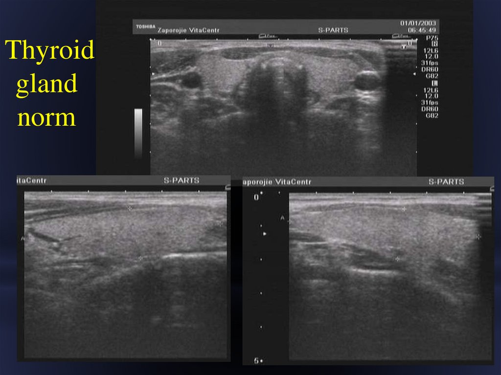
Thyroidgland
norm
6.
• To study the function of the thyroid gland, radionuclideresearch is used. To obtain the correct result of the study, it is
necessary to exclude the use of iodine preparations for a
month, so as not to block the gland. Radiopharmaceutical
containing technetium is administered intravenously. After
that, scintigraphy of the gland is performed. On the
Scintigram, the thyroid gland has an irregular shape,
resembling a "butterfly", clear convex contours. The lobes
and isthmus are usually clearly visible. The right lobe is
usually slightly larger than the left, although the position of
the gland and its size are highly variable. The density of
scintillation in the Central parts of the lobes is higher than in
the periphery, because there is a large part of the glandular
tissue.
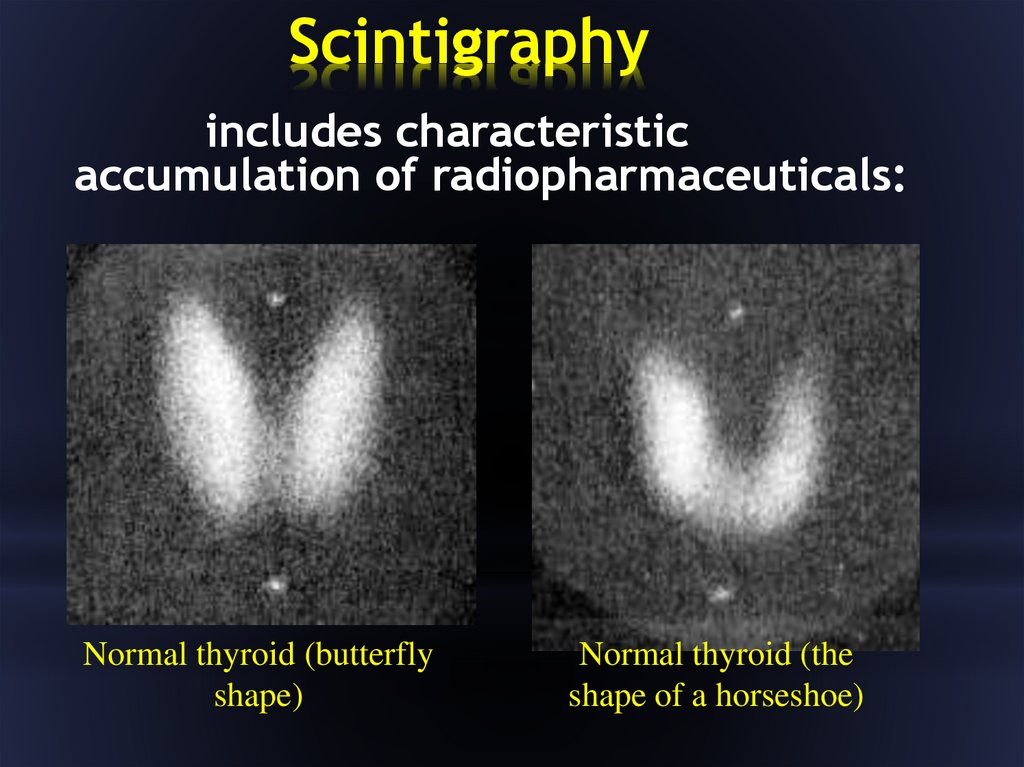
7.
Scintigraphyincludes characteristic
accumulation of radiopharmaceuticals:
Normal thyroid (butterfly
shape)
Normal thyroid (the
shape of a horseshoe)
8. Scintigraphy
High accumulation of the drug (hot spot)– increased blood flow, increased
metabolism of the studied organ, local
leukocyte infiltration, violation of the
passage of the environment that
absorbed the RFP.
Low accumulation (cold hearth) - lack of
blood flow, cyst, destructive cavity.
9.
Classification of thyroid diseases:Аномалии развития: Атиреоз;Hypoplasia (cretinism); Fragmentation; Dystopia; Ectopia;Cystic (persistent
thyroid duct).
Acquired diseases: Hypothyroidism (myxedema); Goiter (enlargement of
the thyroid gland).
Classification of goiter.
On a configuration:
diffuse;
nodal;
mixed.
By function:
hyperthyroid;
euthyroid;
hypothyroid.;
10. Diffuse goiter
- increasing the size of the gland- preserved or diffusely inhomogeneous,
without nodes, the structure.
but
The function of the gland can be normal, enhanced, or
weakened.
Scintigrams - with increased function of thyroid tissueincreased image intensity.
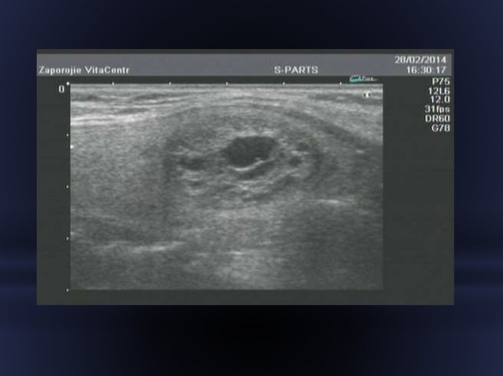
11. Nodular goiter
Sonography - diagnostics of a node,establishment of its
macromorphological structure.
Benign:
- solid - from a dense fabric,
- cystic-cavities with liquid contents
- mixed include both the dense tissue andcysts.
Follicular adenoma-the formation of a regular round
shape of reduced echogenicity with some
heterogeneity of the structure with smooth contours.
12.
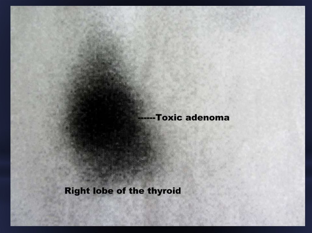
13. Toxic thyroid adenoma
- focal hyperplasia of thyroid tissue.Sonography:
single well-defined hypoechogenic node.
Scintigraphy:
fire, the other departments of the thyroid poorly
accumulate the radiopharmaceutical.
14.
15. Nodular goiter Malignant
Cancer node - single.On the Sonograms:
- low echo density node
- inhomogeneous structure
- with uneven and indistinct contours
- small calcifications on the periphery of
the node.
The scintigrams show a cold hearth.
16.
17. Parathyroid gland
Parathyroid glands number 3-4, the size of a rice grain,weighing 10-12 mg, have a denser consistency than the
thyroid gland. The parathyroid glands are attached to the
posterior surface of the lateral lobes of the thyroid gland .
From the thyroid gland are separated by a fibrous capsule.
They can also be located around the trachea or penetrate deep
into the substance of the thyroid gland.
18.
Classification of diseases of the parathyroidglands.
In the functional state:
Hyperparathyroidism: hypercalcemia after initiation of
diuretic therapy, increased levels of calcium and
parathyroidism in the serum (normal hypercalcemia leads to
a decrease in parathyroidism.), parathyroid osteodystrophy,
urolithiasis.
Hypoparathyroidism: lowering the level of calcium and
parathyroidism in the serum, calcification of soft tissues,
slowing synostosis.
19.
The following algorithm is used to establish the diagnosis ofparathyroid disease:
Blood test for calcium and parathyroid hormone (sample
with calcitonin),
• Ultrasound examination of the thyroid gland,
• Scintigraphy of the thyroid,
• Radiography to detect signs of parathyroid
osteodystrophy.
20. Hyperparathyroidism
- a disease caused by a tumor or hyperplasticchange in one or more of the 4 parathyroid
glands, which leads to hypersecretion of the
parathyroid hormone and violation of calcium
homeostasis.
Diagnostics – laboratory!:
increased parathyroid hormone levels,
accompanied by hypercalcemia.
21. Hyperparathyroidism
Increased function of the parathyroid glands leads toa violation of mineral metabolism, primarily calcium develops hyperparathyroid osteodystrophy Recklinghausen's disease.
Imaging methods are not a means of initial diagnosis
of hyperparathyroidism!
They allow you to detect parathyroid glands only in the
development of severe hyperplasia in the later stages
of development of the process.
22.
Adenoma of parathyroid glandUltrasound, CT, MRI: well-defined
educationreduced density between the
posterolateralthe edge of the thyroid gland and
the common carotid artery.
Scintigraphy: accumulation of RFP in the
adenoma + the ability to determine atypically
located parathyroid glands!
Arteriography.
Radiography: systemic osteoporosis,
delamination and thinning of the cortical layer
of bones, the appearance of single and
multiple cysts in different parts of the
skeleton.