


 Биология
Похожие презентации:


")
Gram positive and negative bacteria
1.
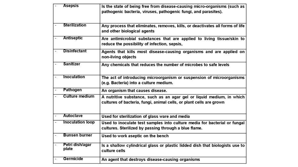
-Asepsis
Is the state of being free from disease-causing micro-organisms (such as
pathogenic bacteria, viruses, pathogenic fungi, and parasites).
-
Sterilization
Any process that eliminates, removes, kills, or deactivates all forms of life
and other biological agents
-
Antiseptic
Are antimicrobial substances that are applied to living tissue/skin to
reduce the possibility of infection, sepsis,
-
Disinfectant
Agents that kills most disease-causing organisms and are applied on
non-living objects
-
Sanitizer
Any chemicals that reduces the number of microbes to safe levels
-
Inoculation
The act of introducing microorganism or suspension of microorganisms
(e.g. Bacteria) into a culture medium.
-
Pathogen
An organism that causes disease.
-
Culture medium
A nutritive substance, such as an agar gel or liquid medium, in which
cultures of bacteria, fungi, animal cells, or plant cells are grown
-
Autoclave
Used for sterilization of glass ware and media
-
Inoculation loop
Used to inoculate test samples into culture media for bacterial or fungal
cultures. Sterilized by passing through a blue flame.
-
Bunsen burner
Used to work aseptic on the bench
-
Petri dish/agar
plate
Is a shallow cylindrical glass or plastic lidded dish that biologists use to
culture cells
-
Germicide
An agent that destroys disease-causing organisms
2.
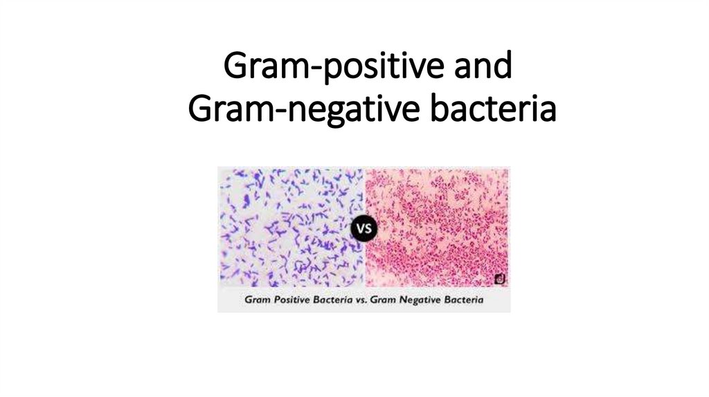
Gram-positive andGram-negative bacteria
3.
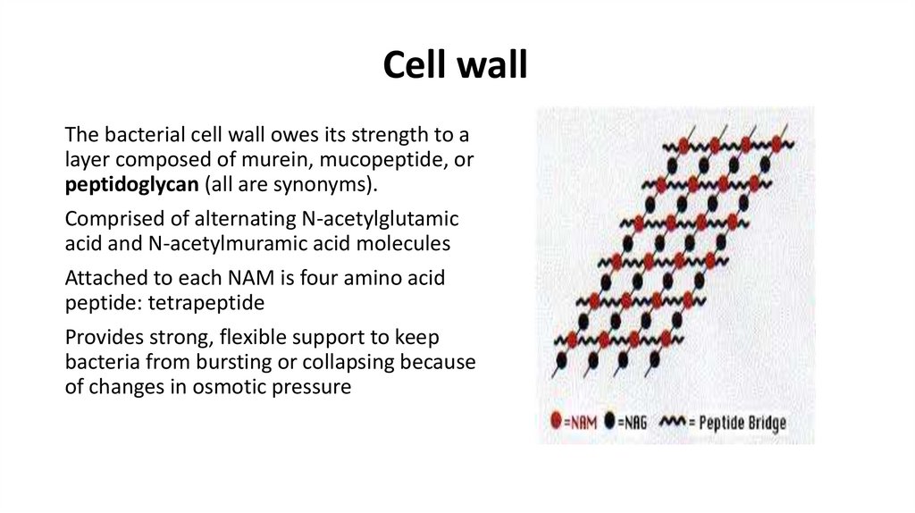
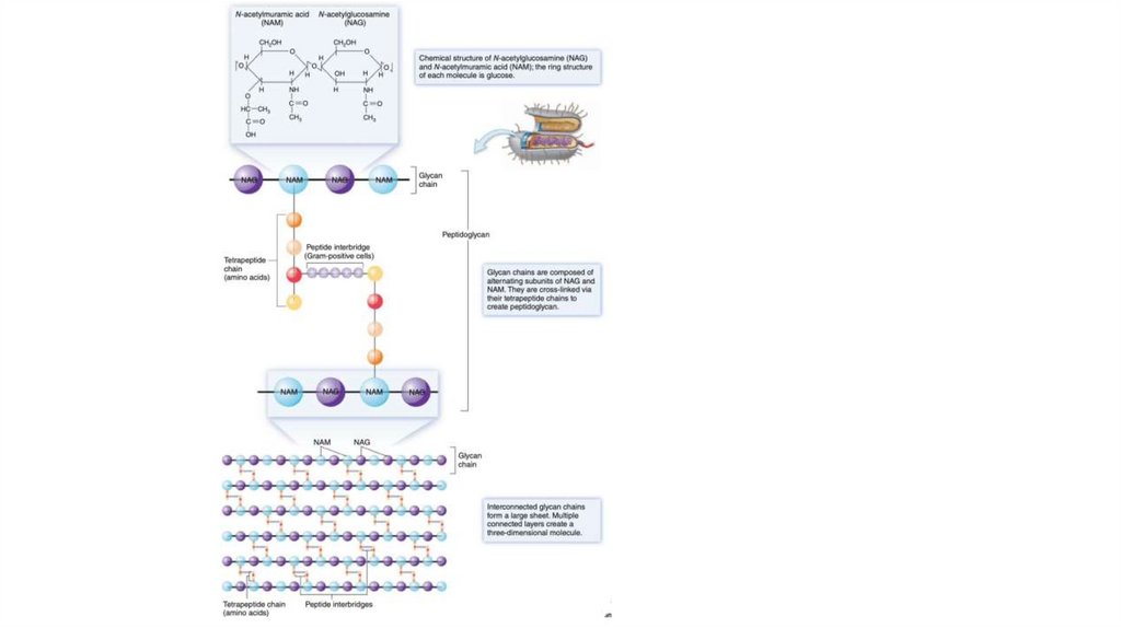
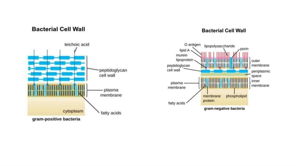
Cell wallThe bacterial cell wall owes its strength to a
layer composed of murein, mucopeptide, or
peptidoglycan (all are synonyms).
Comprised of alternating N-acetylglutamic
acid and N-acetylmuramic acid molecules
Attached to each NAM is four amino acid
peptide: tetrapeptide
Provides strong, flexible support to keep
bacteria from bursting or collapsing because
of changes in osmotic pressure
4.
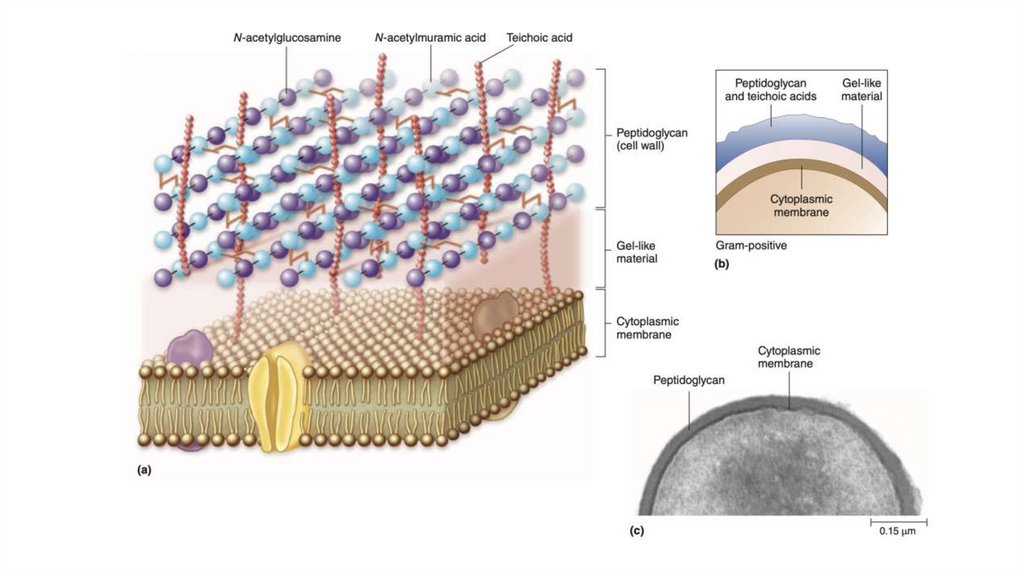
Special Components of Gram-Positive Cell Walls1. 20-30 layers of peptidoglycan
2. Teichoic and teichuronic acids,
provides functions relating to the elasticity, porosity,
tensile strength, and electrostatic properties of the
envelope
The teichoic acids constitute major surface antigens:
chains of recital-phosphate or glycerol-phosphate to
which sugars or alanine attached
• Teichoic Acid sticks out above the peptidoglycan layer
3. some gram-positive walls may contain polysaccharide
molecules
5.
6.
7.
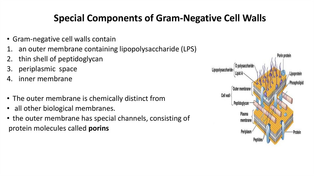
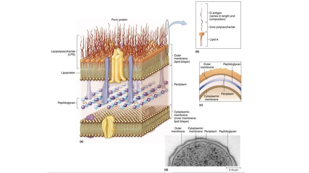
Special Components of Gram-Negative Cell Walls• Gram-negative cell walls contain
1. an outer membrane containing lipopolysaccharide (LPS)
2. thin shell of peptidoglycan
3. periplasmic space
4. inner membrane
• The outer membrane is chemically distinct from
• all other biological membranes.
• the outer membrane has special channels, consisting of
protein molecules called porins
8.
9.
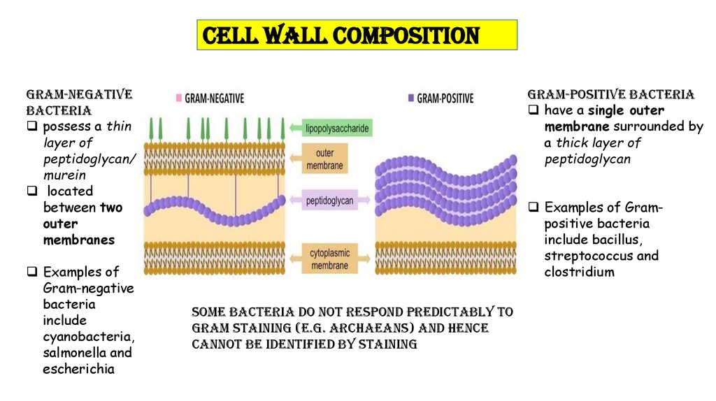
Cell Wall CompositionGram-negative
bacteria
possess a thin
layer of
peptidoglycan/
murein
located
between two
outer
membranes
Examples of
Gram-negative
bacteria
include
cyanobacteria,
salmonella and
escherichia
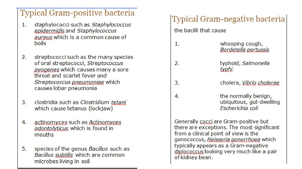
Gram-positive bacteria
have a single outer
membrane surrounded by
a thick layer of
peptidoglycan
Examples of Grampositive bacteria
include bacillus,
streptococcus and
clostridium
Some bacteria do not respond predictably to
Gram staining (e.g. archaeans) and hence
cannot be identified by staining
10.
11.
Gram staining• Basis of bacterial classification and identification, named after the
histologist Hans Christian Gram
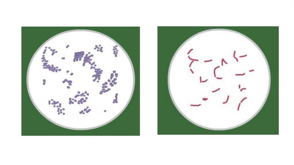
• Most bacteria are classified as
1. gram-positive or
2. gram-negative
according to their response to the Gram staining procedure.
12.
13.
14.
15.
16.
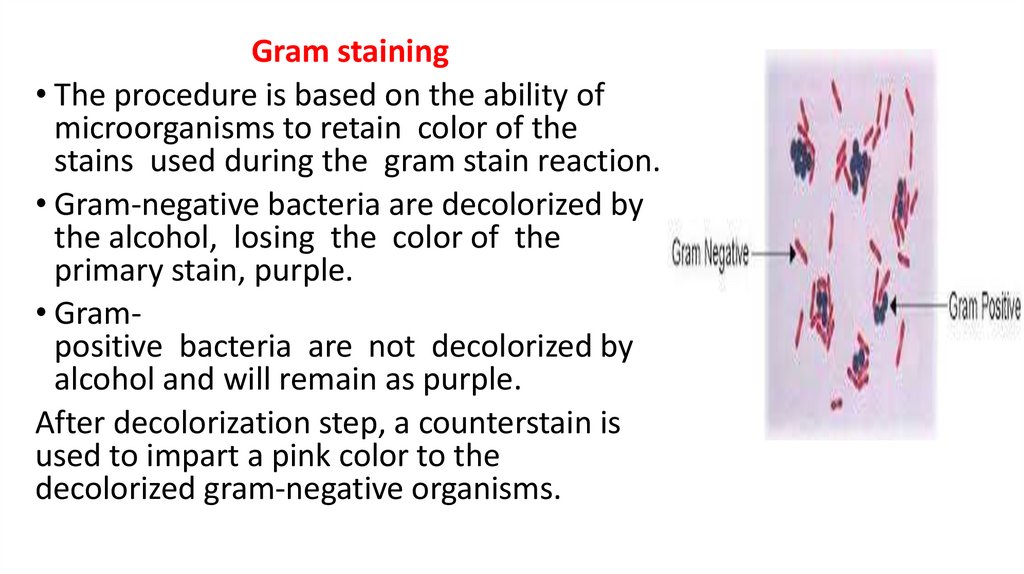
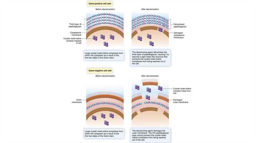
Gram staining• The procedure is based on the ability of
microorganisms to retain color of the
stains used during the gram stain reaction.
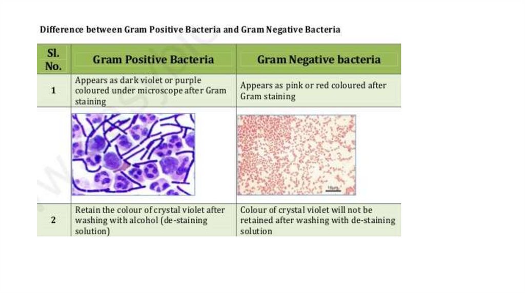
• Gram-negative bacteria are decolorized by
the alcohol, losing the color of the
primary stain, purple.
• Grampositive bacteria are not decolorized by
alcohol and will remain as purple.
After decolorization step, a counterstain is
used to impart a pink color to the
decolorized gram-negative organisms.
17.
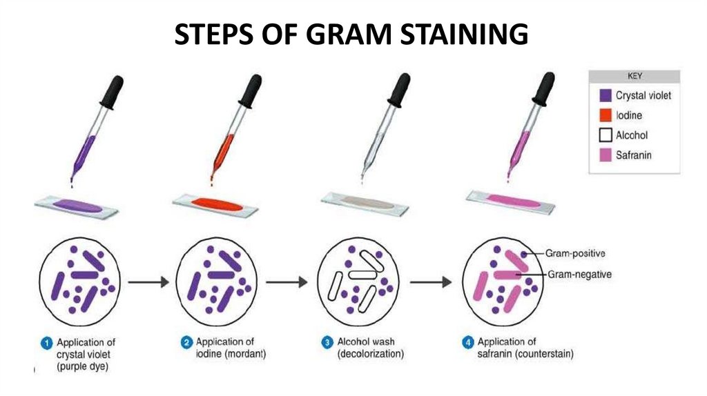
STEPS OF GRAM STAINING18.
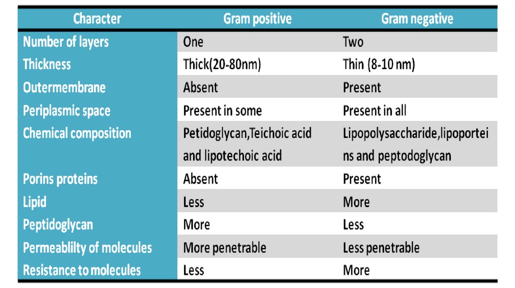
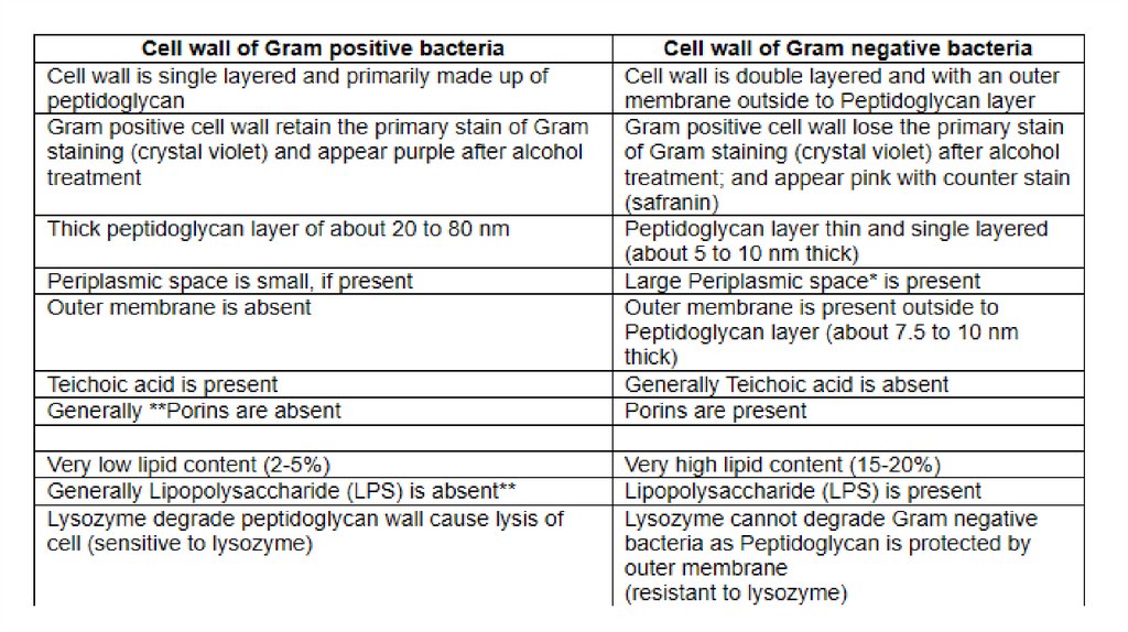
DIFFERENCE BETWEEN GRAMNEGATIVE AND GRAM-POSITIVECELL WALL
19.
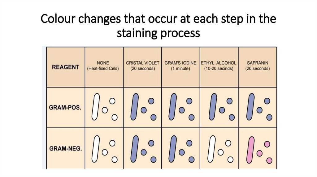
Colour changes that occur at each step in thestaining process