


 Биология
Похожие презентации:


Anatomy and phisiology of the acoustic and vestibular analyzers
1. ANATOMY AND PHISIOLOGY OF THE ACOUSTIC AND VESTIBULAR ANALYZERS
2.
3. History of ENT
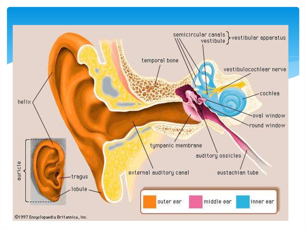
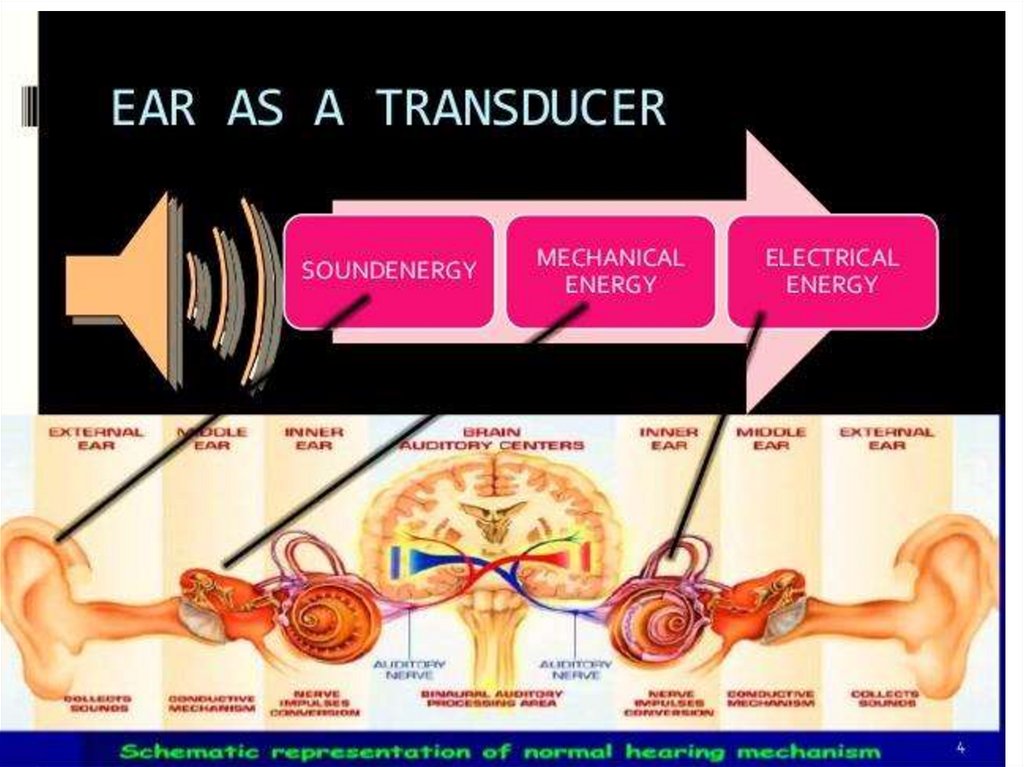
4. Parts of ear
EXTERNAL EARMIDDLE EAR
INTERNAL EAR
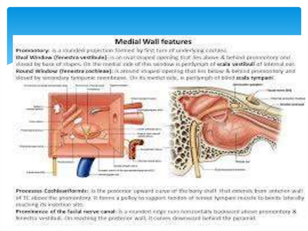
5.
6.
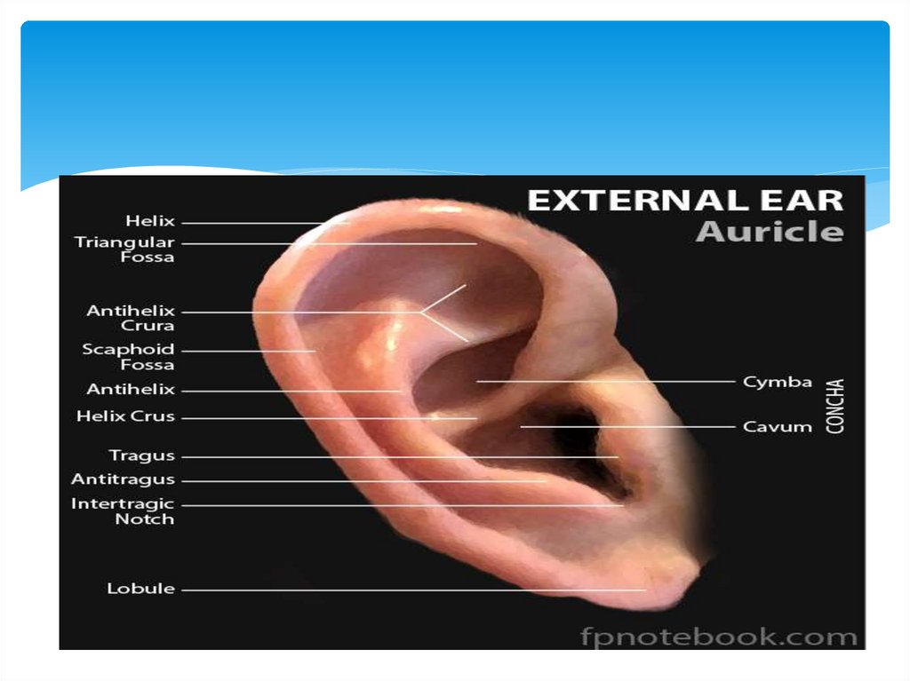
7. External ear
8.
9.
10. Ear is clinically divided from functional positions into two parts:
1-SOUND CONDUCTINGthe sound conducting
apparatus include the
concha of the auricle, the
external acoustic meatus
, the tympanic
membrane, the auditory
ossicles (the hammer,
the anvil and the stapes),
the perilymph of the
scala vestibule, and the
perilymph of the scala
tympani, these scalae
ending with the round
vestibular window.
11. 2-Sound-perceiving
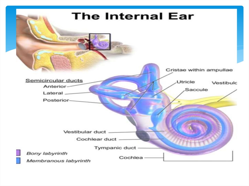
sound-perceivingapparatus include the
basal membrane and
the Corti’s organ on
it with the hair, pillar
and trophic cells,
lying in a certain
sequence and
forming tunnels (the
peripheral part of the
analyzer).
12. Functions of external ear
Sound collectionIncreasing pressure on the
tympanic membrane in a frequency
sensitive way
Sound localization
13. Functions of middle ear
Impedence matchingAttenuation
Phase differencial effect
14. Phase differential effect
Sound waves striking the tympanicmembrane do not reach the oval and round
window simultaneously
There is preferential pathway to oval window
due to ossicular chain
This acoustic separation of windows is
achieved by intact tympanic membrane and a
cushion of air around round window
This contributes 4 dB when tympanic
membrane intact
15. Sound
Sound is a form of energyIt is transmitted through a medium as a longitudinal
pressure wave
The wave consists of a series of compressions and
rarefactions of the molecules in the medium
The ear is capable of capturing this energy and perceiving
it as sound information
16.
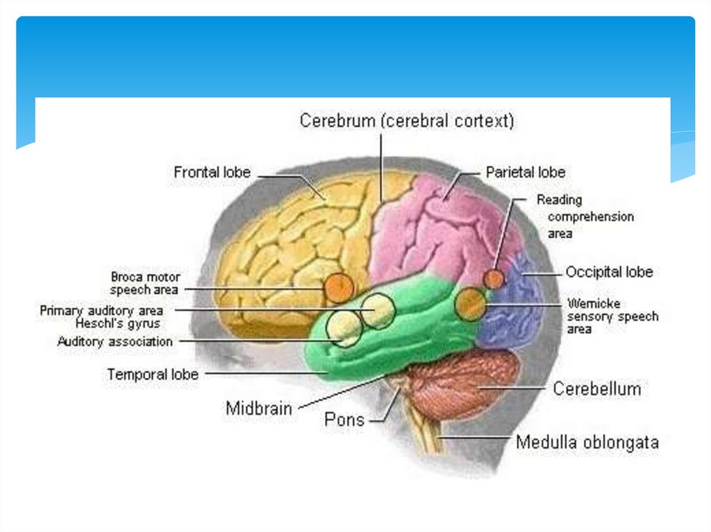
Owing to the ear, sound vibrations are reflected in the cerebralcortex in the form of acoustic sensations, they being analyzed in
compliance with three parameters:
perception of frequency
loudness
timbre.
The human ear perceives not all sound frequencies existing in the
nature, but only their certain part, the so-called acoustic scale; at the
bottom it is limited by the longest sound wave of 16 vibrations (or
cycles) per second, and at the top there is the shortest wave of
22.000 vibrations per second. The unit for measuring sound
frequency is Hertz. Thus, the human ear perceives frequencies from
16 to 22,000 Hz. Sounds lower 16 vibrations lower called infra-audible
(or infrasonic), while higher 22kHz are ultra-audible (or ultrasonic)
sounds. They are not perceived by the human ear because the waves
lower 16 vibrations per second do not reach the cochlea, whereas
higher 22,000 vibrations pass the cochlea and do not cause any
response of the receptor. Animals can have perception of high
frequencies: cats- up to 60,000 Hz, bats and dolphins – 200 and
more kHz, the working frequency in the ear of elephants is 300 Hz.
17.
Perception of the loudness (sound intensity) ismore complex, it depends upon a relationship
between the amplitude of vibration and pressure
during the passing of acoustic waves, and is
measured in physical units of loudness, i.e. Bels. In
order to determine perception of loudness by the
human ear, tenth fraction of a bell are used, i.e.
decibels (dB).
Along with frequency and intensity of sounding,
the ear also perceives a timbre of sounding in a
complex sound, i.e. its colouring. It is determined
owing to the capacity of the ear to isolate in a
complex sound both its main tone and surrounding
overtones. It makes possible to recognize familiar
18. There are two ways of conduction sounds
air (aerotympanic)osseous (divided into craniotympanic
and craniocochlear). Depending upon
the wave length, sounds go by different
waves.
19. Air and bone conduction
There are two methods by which hair cells can bestimulated
Air conduction
Sound stimulus travelling through the external and
middle ear and activating the hair cells
Bone conduction
Sound stimulus travelling though the bones of the
skull activating the hair cells
Whatever method it takes, the sound stimulus finally
activate hair cells in the cochlea
20. Physiology of hearing
21. Cross section through cochlea
22.
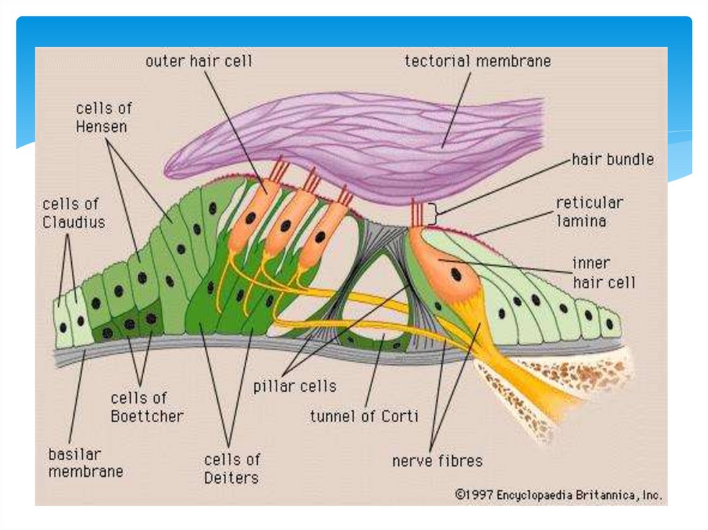
23. Corti organ
The sensory cells responsible for hearing are located on thebasilar membrane within a structure known as the organ of Corti
This is partitioned by two rows of peculiar shaped cells known as
pillar cells
The pillar cells enclose the tunnel of Corti
Situated on the basilar membrane is a single row of inner hair cells
medially and three rows of outer hair cells laterally
The hair cells and other supporting cells are connected to one
another at their apices by tight junctions forming a surface known
as reticular lamina
The cells have specialized stereocilia on their apical surfaces
24.
25.
26.
• Hair cells• Dorsal and ventral
cohlear nuclei
• Superior olivary
complex
• Nucleus of lateral
lemniscus
• Inferior colliculus
• Medial geniculate
body
• Auditory cortex
27.
28.
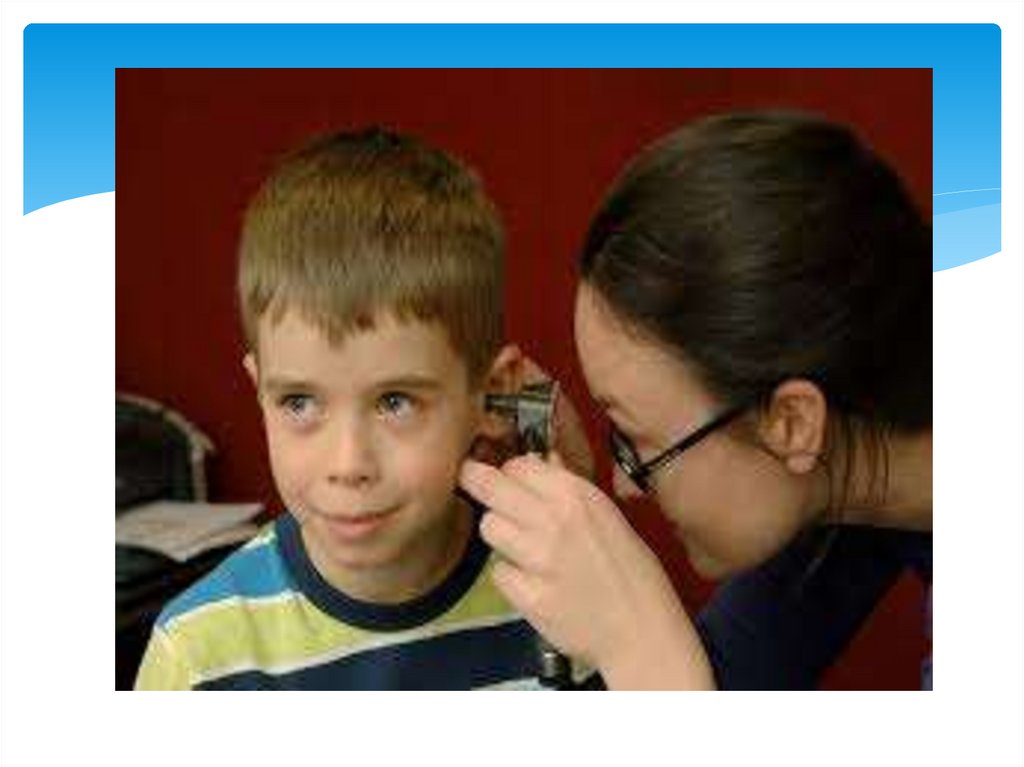
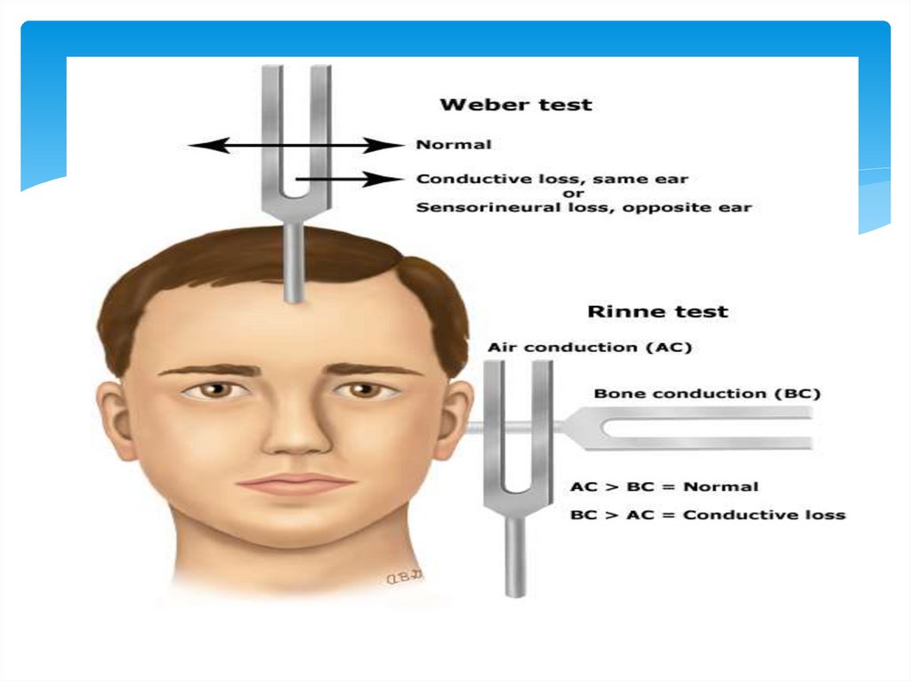
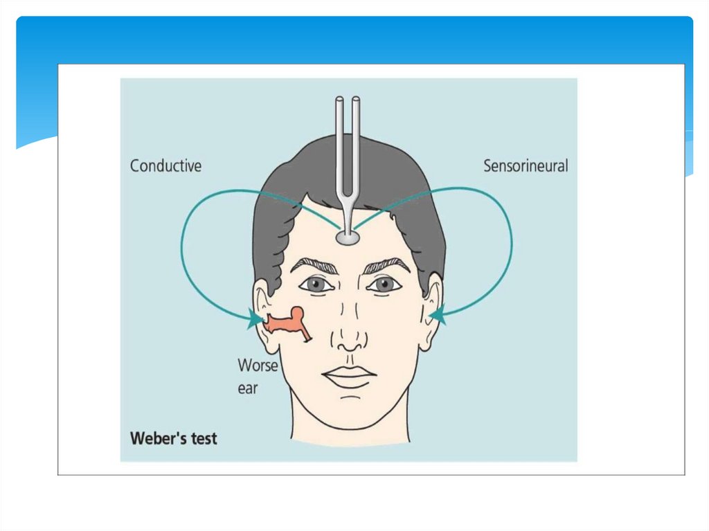
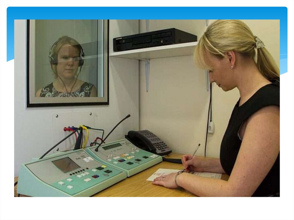
In order to clinically study the acoustic functionof the ear, there are certain methods making it
possible to determine the norm and its change in
a pathology. They include examination of the
auditory acuity with help of whisper and ordinary
speech, tests with help of tuning forks
(experiments performing by Rinne, Veber.
Schwabach. Bing and Gele), as well as by puretone, speech and game (in childhood)
audiometry.
29.
30. Internal ear
• Vestibule• Cohlea
• Semicircular
canals
31. Inner ear
The inner ear can be thought of as a series of tunnels or canalswithin the temporal bone
Within these canals are a series of membranous sacs (termed
labyrinths) which house the sensory epithelium
The membranous labyrinth is filled with a fluid termed endolymph
It is surrounded within the bony labyrinth by a second fluid termed
perilymph
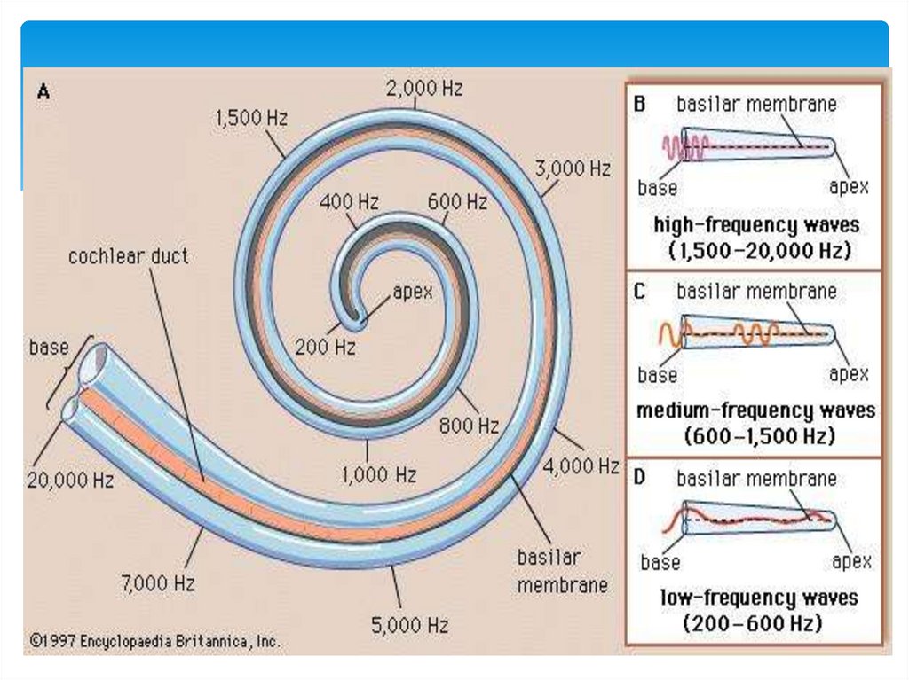
The cochlea can be thought of as a canal that spirals around itself
similar to a snail. It makes roughly 2 1/2 to 2 3/4 turns
32. Endolymph and perilymph
Endolymph is similar in ionic content tointracellular fluid (high K, low Na)
Perilymph resembles extracellular fluid (low K,
high Na)
The cochlear duct contains several types of
specialized cells responsible for auditory
perception
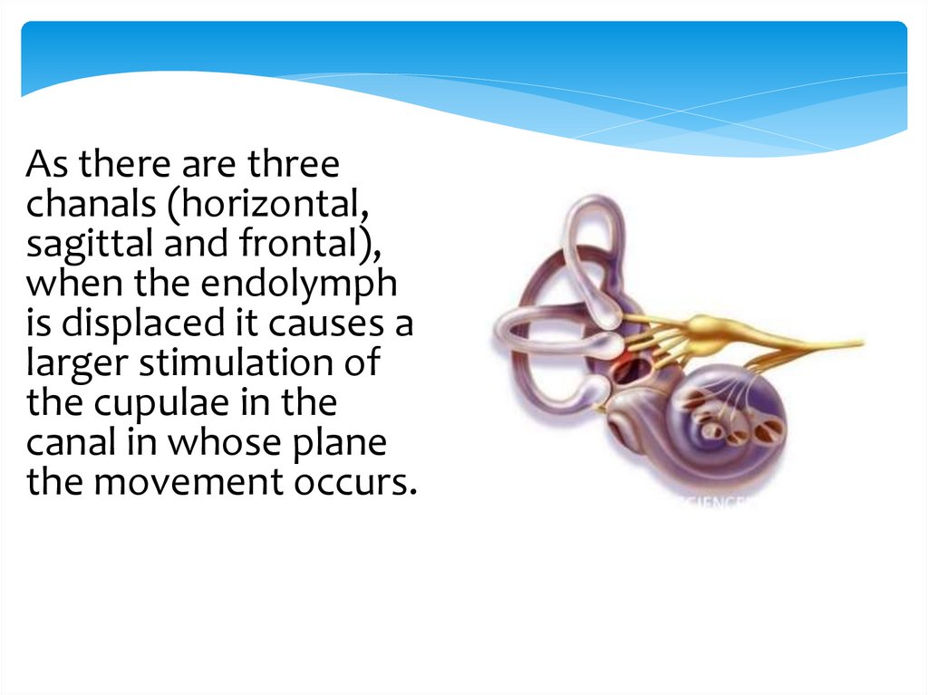
33.
As there are threechanals (horizontal,
sagittal and frontal),
when the endolymph
is displaced it causes a
larger stimulation of
the cupulae in the
canal in whose plane
the movement occurs.
34.
35. Functions of the semicircular canals
Form a mechanical link from thetympanic membrane to the oval
window
Perceive a sense of balance and
perception in space
Equalize hydraulic pressure
36. OTOLITHIC APPARATUS
37.
The statokinetic analyzer is very complex in itscomposition as it is connected by its conduction
tracts with the rachidian bulb (nuclei of the
vagus), the spinal cord, the cerebellum, nuclei of
the oculomotor nerves; later its fibres pass
through the internal capsule and run to cerebral
cortex where the nerve cells of the analyzer are
scattered along the surface of all cortical lobes.
38.
laws of nystagmus were formulated:a) the eyes slowly move in the direction where the fluid
flows;
b) the movement of the eyes will be in the plane of fluid
flow;
c) the movement of the eyes is more intensive if the
endolymph flows to the ampulla than from it. Studies
have shown that the rapid component of nystagmus is
accomplished by an order from the cortex or the
subcortex.
39.
It results in labyrinthine nystagmus with its rapidand slow components. It may be physiological
(e.g. optokinetic, or developing in case of artificial
adequate stimulation of the labyrinth); its
synonyms are “experimental” and “exogenous”.
Nystagmus may also be pathological, if it is caused
by an inadequate stimulant (e.g. inflammation of
the labyrinth, its injury, intoxication, etc.); its
synonym is “endogenous nystagmus”.
Duration of nystagmus in rotative test is 10 seconds.
40. CLASSIFICATION OF NYSTAGMUS
Direction of nystagmus by its rapid component (right,left, up, down)
Amplitude its degree (it may be of the 1st, 2nd and 3rd
degree) , the 1st degree is if nystagmus is preserved only
during look in its direction and disappears during a look
forward, the 2nd one is if in the latter case it is preserved,
and the 3rd one is if it is preserved during a look in the
direction of its slow component
plane of nystagmus (i.e. horizontal, rotatory, mixed), its
amplitude (small-medium or large-swinging)
activity (active or flaccid nystagmus)
41.
The vestibuloocular syndrome results from manifestationof evolutionally connected motions of the eyeball and
movement of the body along the surface of the medium
of inhabitancy. It is manifested by development of
nystagmus, i.e. rhythmical motions of the eyeballs in that
plane where the semicircular canal is stimulated.
Unlike the other kinds of nystagmus (in the blind,
cerebellar), the labyrinthine one always has two
components, they are: rapid and slow. The slow
component of nystagmus is connected with movement of
the fluid (endolymph) In the semicircular canal. It was
revealed by Evald as early as in 1896.
42.
• The vestibuloautonomic syndrome develops owing totransmission of an increased stimulation from the
labyrinth to the vagus, and via the latter to the
autonomic responses of the organism; its
manifesttations are paleness and not so frequently,
redness of the skin, appearance of an unpleasant
sensation of dizziness (vertigo), nausea, vomiting,
excessive sweating, hypersalivation, reduced blood
pressure, bradycardia and even diarrhea.
In hypersensitive persons, the syndrome may develop even during an
insignificant (for other people) extent of stimulation of the analyzer
(going by bus, air, etc.). Unlike dizziness in patients in hypersensitive
crisis, anaemia and intoxication, labyrinthine vertigo (synonyms:
“auditory vertigo”, “ Meniere’s desease”) is always distinct and the
patient expresses by words the direction and plane of the movement,
rather the only sensation of the movement and rotation.
43.
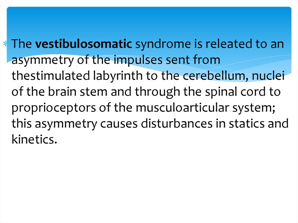
The vestibulosomatic syndrome is releated to anasymmetry of the impulses sent from
thestimulated labyrinth to the cerebellum, nuclei
of the brain stem and through the spinal cord to
proprioceptors of the musculoarticular system;
this asymmetry causes disturbances in statics and
kinetics.
44. Thank you!!!
45.
46.
47.
48.
49.
50.
DegreeSpeech voice
Whisper voice
1
26-40 db
6-3 м
2м – at the ear
2
41-55 db
3 м – у уха
0 – at the ear
3
56-70 db
Loud speech at the ear
0
4
71-90 db
Scream at the ear
0
deafness
>91 db
0
0