


")


")


 Биология
Похожие презентации:


")


Anatomy Of The Skin. Lecture 1
1. Lection 1 Anatomy Of The Skin
I.Epidermis
II.
Dermis
Lection 1
Anatomy Of The Skin
III. Hypodermis
IV. Appendixes of the
skin.
1.
Stratum basale
2.
Stratum spinosum
3.
Stratum granulosum
4.
Stratum lucidum
5.
Stratum corneum
1
2. Skin Anatomy
The skin is an organ that forms a protective barrier againstgerms (and other organisms) and keeps the inside of your
body inside your body, and keeps what's outside of your body
outside. Skin also helps maintain a constant body
temperature. Human skin is only about 0.07 inches (2 mm)
thick.
Skin is made up of two layers that cover a third fatty layer. The
outer layer is called the epidermis; it is a tough protective
layer that contains melanin (which protects against the rays of
the sun and gives the skin its color). The second layer (located
under the epidermis) is called the dermis; it contains nerve
endings, sweat glands, oil glands, and hair follicles. Under
these two skin layers is a fatty layer of subcutaneous tissue
(the word subcutaneous means "under the skin").
On average, an adult has from 18-20 square feet (about 2
square meters) of skin, which weighs about 6 pounds (2.7 kg).
2
3. Skin Anatomy
StratumStratum
papillare
sskin
Horny layer
Stratum corneum
Dermis
the true skin
lucid layer
Stratum lucidum
Granular layer
Stratum granulosum
Prikle – cell layer
Stratum spinosum
Germinatinne layer
Stratum basale
reticulare
Hypodermis
(subcutaneous
fatty tissue)
Cell
Elements
Amorphous
Intestinal
Substance
Fibrous
Substance
Skin Anatomy
Skin
Epidermis
3
4. Epidermis and it’s layers
The epidermis is the most superficial layer of theskin and provides the first barrier of protection
from the invasion of foreign substances into the
body. The principal cell of the epidermis is called
a keratinocyte. The epidermis is subdivided into
five layers or strata, the stratum germinativum
(SG), the stratum spinosum(SS), the stratum
granulosum(SGR), the stratum lucidum and the
stratum corneum(SC) in which a keratinocyte
gradually migates to the surface and is sloughed
off in a process called desquamation.
4
5. Epidermis and it’s layers
Theepidermis, the
outermost skin layer,
consists of five different
layers:
•Stratum
•Stratum
•Stratum
•Stratum
•Stratum
corneum
lucidum
granulosum
spinosum
basale
5
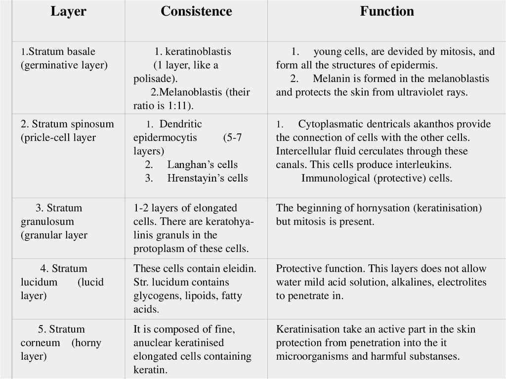
6.
Layer1.Stratum basale
(germinative layer)
2. Stratum spinosum
(pricle-cell layer
Consistence
1. keratinoblastis
(1 layer, like a
polisade).
2.Melanoblastis (their
ratio is 1:11).
1. Dendritic
epidermocytis
(5-7
layers)
2. Langhan’s cells
3. Hrenstayin’s cells
Function
1. young cells, are devided by mitosis, and
form all the structures of epidermis.
2. Melanin is formed in the melanoblastis
and protects the skin from ultraviolet rays.
Cytoplasmatic dentricals akanthos provide
the connection of cells with the other cells.
Intercellular fluid cerculates through these
canals. This cells produce interleukins.
Immunological (protective) cells.
1.
3. Stratum
granulosum
(granular layer
1-2 layers of elongated
cells. There are keratohyalinis granuls in the
protoplasm of these cells.
The beginning of hornysation (keratinisation)
but mitosis is present.
4. Stratum
lucidum
(lucid
layer)
These cells contain eleidin.
Str. lucidum contains
glycogens, lipoids, fatty
acids.
Protective function. This layers does not allow
water mild acid solution, alkalines, electrolites
to penetrate in.
5. Stratum
corneum (horny
layer)
It is composed of fine,
anuclear keratinised
elongated cells containing
keratin.
Keratinisation take an active part in the skin
protection from penetration into the it
microorganisms and harmful substanses.
7. Several cell types constitute epidermis.
Keratinocytes These epithelial cells comprise the majority of theepidermal cells. They are called keratinocytes because of the
fibrous proteins, keratins, which are the differentiated endproduct of these cells. Only the keratinocytes of the basal layer
divide, and the daughter cells migrate upwards and change from
cuboidal in appearance to squames. Eventually, the keratinocytes
lose their nuclei and become anuclear, compact, protein-dense
cells that occupy the outermost horny cell layer, the stratum
corneum. Cells of the stratum corneum ultimately desquamate.
Normally, the rate of desquamation equals the rate of the
formation of the cells in the basal layer. In certain dermatologic
diseases, like psoriasis, there is increased turnover of epidermal
keratinocytes. Keratinocytes attach to each other by structures
called desmosomes. Damage to these structures result in
separation of the keratinocytes.
7
8. Several cell types constitute epidermis.
The stratum corneum of the epidermis is responsible for the mostimportant function of the skin, namely its protective barrier
function. The mechanism by which such protection is conferred
is complex and involves the function of intracellular fibrous
proteins, dense cellular membranes, and intercellular lipid
constituents. The intercellular lipid portion is now believed to be
the crucial component responsible for barrier function.
Keratinocytes also play a role in immune responses through the
production of a variety of cytokines.
The skin is not completely impermeable. Compounds can pass
through intact skin. This permeability of the skin is used for the
delivery of topical therapeutic agents used in the treatment of
certain skin diseases, and also for delivery of medication used in
the treatment of systemic diseases. Examples of percutaneous
delivery include scopolamine, used for treatment of motion
sickness, nitroglycerine for angina pectoris and estrogen for
postmenopausal symptoms.
8
9. Several cell types constitute epidermis.
Melanocytes are cells derived from the neural crest. Theymigrate early during embryonic development and pass through
the dermal mesenchyme to reside in the basal layer of the
epidermis. There is approximately one melanocyte for every 10
basal keratinocytes. Unlike keratinocytes, melanocytes rarely
divide and normally do not migrate upwards. Melanocytes have
long, dendritic processes that reach the upper and lower layers of
the epidermis. Through these dendrites melanocytes transfer
melanosom to the keratinocytes.
Melanosomes are melanin-containing organelles that are
synthesized by the melanocytes. Dark skin individuals produce
more melanosomes and transfer more melanosomes to the
keratinocytes. However interestingly, in different races the
number of melanocytes is constant for a given cutaneous site.
Melanocytes are the cells which give rise to the development of
malignant melanoma.
9
10. Several cell types constitute the stratum bazale.
The function of melanin is to provide protection against thedamaging effects of ultraviolet light. Individuals with light
complexion and less melanin are prone to develop premature
skin aging, precancerous skin lesions, and skin cancers of
various types as a result of cumulative sun exposure.
Langerhans Cells These cells comprise 3-4% of the epidermal
cells. They are derived from the bone marrow and serve as
antigen-presenting cells to helper T Lymphocytes (CD4 positive
cells). They participate in the development of contact
hypersensitivity.
Merkel Cells are found in, or near, the basal cell layer of the
epidermis. They surround hair follicles and are speculated to
assist the touch receptors. Merkel cells resemble
neurosecretory cells that produce polypeptide hormones,
because similar to these cells, they have membrane bound
secretory-like granules.
10
11. Stratum germinativum (bazale)
The stratum germinatum(SG) provides the
germinal cells necessary
for the regeneration of
the layers of the
epidermis. These
germinal cells are
separated from the
dermis by a thin layer of
basement membrane.
After a mitotic division a
newly formed cell will
undergo a progressive
maturation called
keratinization as its
migrates to the surface.
11
12. Stratum spinosum
The cells that divide in thestatum germinativum soon
begin to accumulate many
desmosomes on their outer
surface which provide the
characteristic “prickles”
(seen on the close-up view)
of the stratum spinosum
(SS), which is often called
the prickle-cell layer.
12
13. Stratum granulosum
The progressive maturation ofa keratinocyte is charcterized
by the accumulation of
keratin, called keratinization.
The cells of the stratum
granulosum (SGR) accumlate
dense basophilic keratohyalin
granules (seen on the closeup view). These granules
contain lipids, which along
with the desmosomal
connections, help to form a
waterproof barrier that
functions to prevent fluid loss
from the body.
13
14. Stratum Lucidum
Epidermis varies inthickness throughout
the body depending
mainly on frictional
forces and is thickest
on the palms of the
hands and soles of the
feet. The stratum
lucidum is normally
only well seen in thick
epidermis and
represents a transition
from the stratum
granulosum to the
stratum corneum
The cells of the stratum lucidum
contain eleidin.
14
15. Stratum corneum
As a cell accumulateskeratinohyalin granules, it is
thought that rupture of
lysosomal membranes release
lysosomal enzymes that
eventually cause cell death. The
dead and dying cells filled with
mature keratin form the stratum
corneum (SC). The deeper cells
of the stratum corneum retain
their desmosomal junctions, but
as they are pushed to the
surface by newly forming cells of
the stratum germinativum (SG),
the dead cells gradually break
apart and are lost, a process
called desquamation
15
16. The dermal-epidermal basement membrane
Between the epidermisand the dermis there is a
basement membrane,
composed of 3
ultramicroscopically
distinct layers. The
outermost layer is called
lamina lucida.
It contains glycoproteins. The middle layer is called lamina dense.
It contains type IV collagen. The innermost layer is called the
sublamina dense zone. It contains the anchoring fibrils. The main
function of the basement membrane is to anchor the epidermis
into the dermis. Inherited or acquired defects in the basement
membrane lead to the development of blistering diseases.
16
17. Dermis
The dermis (D) assumes theimportant functions of
thermoregulation and supports
the vasular network to supply
the avascular epidermis with
nutrients. The dermis is typically
subdivided into two zones, a
papillary dermis and a reticular
layer. The dermis contains
mostly fibroblasts which are
responsible for secreting
collagen, elastin and ground
substance that give the support
and elasticity of the skin.
Also present are immune cells that are involved in defense
against foreign invaders passing through the epidermis.
17
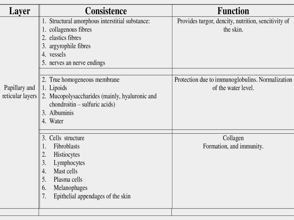
18.
LayerConsistence
1.
1.
2.
3.
4.
5.
Structural amorphous interstitial substance:
collagenous fibres
elastics fibres
argyrophile fibres
vessels
nerves an nerve endings
2. True homogeneous membrane
Papillary and 1. Lipoids
reticular layers 2. Mucopolysaccharides (mainly, hyaluronic and
chondroitin – sulfuric acids)
3. Albuminis
4. Water
3. Cells structure
1. Fibroblasts
2. Histiocytes
3. Lymphocytes
4. Mast cells
5. Plasma cells
6. Melanophages
7. Epithelial appendages of the skin
Function
Provides turgor, dencity, nutrition, sencitivity of
the skin.
Protection due to immunoglobulins. Normalization
of the water level.
Collagen
Formation, and immunity.
19. Papillary dermis
The papillary dermis(PD) contains vascular
networks that have two
important functions. The
first being to support the
avascular epidermis with
vital nutrients and
secondly to provide a
network for
thermoregulation. The
vasculature is organized
so that by increasing or
decreasing blood flow,
heat can either be conserved or dissipated. The vasculature
interdigitates in areas called dermal papillae (DP). The papillary
dermis also contains the free sensory nerve endings and structures
called Meissner’s corpuscles in highly sensitive areas.
19
20. Reticular dermis
The reticular layer of thedermis (RD) consists of
dense irregular connective
tissue, which differs from
the papillary layer (PD),
which is made up of mainly
loose connective tissue
(note the difference in the
number of cells). The
reticular layer of the
dermis is important in
giving the skin it overall
strength and elasticity, as
well as housing other
important epithelial
derived structures such as
glands and hair follicles.
20
21. Dermis
The dermis is the supporting layer of the epidermis.It consists of the fibrous components collagen and
elastin, together with the ground substance. Lying
within the dermis are the epidermal appendages,
nerves and cutaneous vasculature. The cellular
components of the dermis are fibroblasts, and
occasionally inflammatory cells are present as well.
The dermis is divided into two layers: the distal
papillary dermis and proximal reticular dermis.
21
22. Dermis
1. Fibrous Components and GroundSubstance Collagens comprise 98% of the
dermal fibrous component. They provide the
cutaneous structural stability. Elastic fibers
comprise 2% of the dermal fibrous component.
They provide cutaneous elasticity. The ground
substance in which the dermal fibrous
components are embedded, is a gel-like material
that accounts for a large proportion of the
dermal volume.
22
23. Dermis
2. Blood Vessels - The skin is richlyvascularized. The cutaneous vasculature is required
for dermal and epidermal nutrition as well as for
thermoregulation. Cutaneous blood vessels do not
enter the epidermis. This layer receives its modest
nutritional requirement from the contiguous
dermis. The skin receives an extensive blood
supply from vessels within the subcutaneous fat.
These give rise to two vascular plexuses: the deep
vascular plexus and the superficial vascular plexus
which are connected by intercommunicating
vessels. Capillary loops extend into the papillary
dermis and supply this part of the dermis as well
as the epidermis.
23
24. Dermis
3. Nerves - Unmyelinated and myelinated sensory nerves arepresent in the dermis. Free nerve endings penetrate into the
epidermis. Pain sensation is transduced by "fast" conducting
fibers, while itchiness is transduced by "slow" conducting fibers.
In addition, there are specialized sensory structures containing
myelinated fibers which mediate the sensation of touch. They are
called Meissner's corpuscles. The special structures that
mediate pressure are called Pacinian corpuscles. Motor nerves
innervate the blood vessels, sweat glands and the arrector pill
muscle. The latter is a smooth muscle which arises in the
connective tissue and inserts into the hair follicle. Its contraction
produces the phenomenon called "goose flesh".
24
25. Dermis
4. Epidermal appendages during fetaldevelopment, specialized epithelial derived
structures develop from the epidermis, towards
the dermis. These structures are called
epidermal appendages. Each performs a special
function in the skin.
Sebaceous Gland
Sweat Glands
Hair Follicle
Nail
25
26. Sebaceous Gland
Sebaceous glands are found everywhere on the human skinexcept on the palms, soles and dorsa of feet. They are part of
the pilosebaceous unit and their secretion, sebum, flows through
the sebaceous duct into the follicular canal. Sebaceous glands
are larger and more dense on the face and scalp as compared to
other areas of the body. Sebaceous gland secretion is holocrine
in nature and it is a continuous process. Their growth is under
endocrinologic
control and the major stimulus affecting
sebaceous gland growth and sebum section
are androgens. In males testosterone and its
metabolic products, like dihydrotestosterone,
provide the major stimulus. In females
sebaceous gland growth and sebum secretion
is under the control of ovarian and adrenal
androgens. Sebaceous gland growth is one of
the earliest signs of puberty.
26
27. Sweat Glands
There are two types of sweat glands: eccrine andapocrine. Eccrine glands are found in large numbers only
in man and some primates. They are distributed over the
entire skin surface. Their secretory portion is a coiled
tubular structure located deep in the dermis. From this
portion the ductal portion arises. The duct ascends straight
through the dermis, assumes a spiral configuration in the
epidermis (the acrosyringium), and opens onto the skin
surface. Eccrine glands are innervated by sympathetic
cholinergic fiber.
The function of the eccrine glands is to respond to thermal
stress by delivering hypotonic sweat to the skin surface
where it evaporates, cooling the skin, and thereby
reducing the core body temperature. Prolonged sweating
may result in dehydration and electrolyte loss.
27
28. Sweat Glands
Thermal sweating occurs over most of the body integument.Emotional stress can induce eccrine sweating, in selected
areas like the palms, soles, axillae and forehead by a
mechanism that is not well understood.
Apocrine glands are found in many mammals. In man they
are localized mainly to the axillary, areolar and genital
regions; (modified apocrine glands are found in the breast and
the external auditory canal). Like their eccrine counterparts,
apocrine glands have a coiled secretory portion deep in the
dermis. However, the duct leading from the apocrine gland,
rather than opening directly onto the skin surface, it opens
into the hair follicle above the entrance of the sebaceous duct.
Myoepithelial cells, surrounding the gland, help to force the
secretions outward.
28
29. Sweat Glands
Apocrine glands produce an odorless, oily fluid which acquires ascent only after it interacts with skin surface bacteria
(diphtheroids). Apocrine glands continuously produce their
secretions.
The gland is largely under
hormonal influences but its duct
is innervated by sympathetic
adrenergic neurons. Apocrine
glands are androgen responsive
structures that become active at
puberty. They contain large
quantities of 5 alpha-reductase.
Their function is presumed to be
the production of sexual olfactory
messages.
29
30. Hair Follicle
The hair follicle consists of the hair surrounded by anepithelial sheath that is continuous with the epidermis.
Except for its dermal papilla which contains connective tissue
and blood vessels, the rest of the hair follicle is composed of
epidermal cells.
The bulb, at the lower end of
the hair follicle, is the thickest
part. It contains the proliferating
pool of undifferentiated epidermal
cells. Differentiation begins at the
constriction above the bulb and
continues halfway up the follicle
where the cells begin to cornify.
The hair is fully hardened as it
emerges from the skin surface.
30
31. Hair Follicle
Human hair grows in cycles. The longer the hair growth phase ofan individual, the longer the hair length is. The growing stage is
called anagen, followed by a transitional stage termed catagen,
during which hair growth slows-down.
This stage is followed by the resting phase, telogen. Once a hair
has gone into telogen it is eventually shed and a new hair forms
in the same follicle. Normally ~90% of scalp hairs are in anagen
and ~10% in telogen. Hairs do not cycle together, therefore there
is a daily loss of hairs throughout the entire scalp.
During anagen the hair is firmly attached within its follicle; to
dislodge it a force must be applied which is usually sufficient to
fracture the hair in its non-keratinized zone, leaving a portion of
the root behind in the follicle. Such a hair will have a ragged end
where the fracture occurred. Anagen hairs if pulled intact will be
encapsulated by a sheath around the end of the hair.
31
32. Hair Follicle
Conversely, telogen hairs are easily dislodged and accountfor the normal loss, defluvium, that is encountered during
combing, washing, etc. On examination telogen hairs have
no encapsulating sheath but have a tiny "club" at the
terminal end and therefore they are referred to as club
hairs.
Terminal hair growth in certain areas, such as the beard,
chest, axillae and pubic triangle, is androgen-dependent.
There are racial differences in hair density and distribution
as well as structural variations in the hair shaft and
follicles, accounting for hair texture.
32
33. Nail
Nails consist of the nail plate and the supporting tissues which surroundit. The nail plate is made of horny material which is unlike the stratum
corneum in that it does not undergo desquamation. It extends in length
indefinitely until it is cut or worn away by use.
The nail plate is set in grooves that are located on the dorsal surface of
the distal part of the digit. These grooves are referred to as the lateral and
proximal nail grooves.
The grooves are covered by the lateral
and proximal nail folds. The nail plate
is a translucent and colorless
structure. Most digits display a white
semicircular lunula at the proximal end
of the nail plate. The lunula ends
distally in the nail matrix. The matrix
is the region which synthesizes the
nail plate. It extends approximately 5
mm underneath the proximal nail fold.
33
34. Skin Physiology
Protective(barrier)
function of
the skin
Secretory and
excretory
functions of the
skin
Respiratory
function of
the skin
Skin as
an organ
of sense
Skin
physiology
Resorption
function of
the skin
Skin is the
organ of
immunity
Thermoregulatory
function of the skin
Metabolic
function of
the skin
34
35. Skin Function
A. Sensation (largest sensory organ in the body)B. Protection
1. Prevents dehydration
2. Prevents infection
3. Physical barrier to injury
4. Protects against ultraviolet light injury (Melanin)
C. Thermoregulation
1. Insulation (hair and adipose tissue)
2. Heat dissipation
a. Sweat evaporation
b. Increased blood flow
D. Metabolic
1. Energy storage of Triglycerides in adipose tissue
2. Vitamin D synthesis
35
36. Protective function
Protects the organismfrom the damaging
effect of sun rays
Homogeneous
tightness of substanal
protects from
mechanical effects
(blows, friction,
compression)
An acid (pH5.0-6,0)
water-lipid mantle
which attenuatus or
neutralizis the
damaging effect of
chemical substances.
Physiology
desquamation
Protective (barrier)
functions of the skin
Bactericidal
properties of
sweat (lisocini)
and sebum
(squaleni)
Protect the
underlying
tissue
from drying
Resistance to
electric current
Immynological
function. Skin
associated limphoid
tissue. Salt
36
37. Epidermis Cell Layers (cells mature from inner to outer)
A. Stratum Corneum (Cornified Layer)1. Outermost layer of epidermis
2. Composed mostly of keratin (fibrous protein)
3. Cells desquamated (27 days after production)
B. Stratum Lucidum (present only in very thick skin)
C. Stratum Granulosum (Granular Layer)
1. Darker layer with intracellular granules
2. Produces keratin
D. Stratum Spinosum (Prickle Cell Layer)
1. Composed of keratinocytes
2. Cells produced by basal layer and growing
3. Keratin production starts
E. Stratum Germinativum (Stratum Basale, Basal Cell Layer)
1. Innermost layer of epidermis
2. Cells are produced here in the germinal layer
3. Forms the prickle cells in the layer above
37
38. Sensory Apparatus of the Skin
The skin is innervated with around one million afferentnerve fibers. Most terminate in the face and extremities;
relatively few supply the back. The cutaneous nerves
contain axons with cell bodies in the dorsal root ganglia.
Their diameters range from 0.2-20 µm. The main nerve
trunks entering the subdermal fatty tissue each divide into
smaller bundles. Groups of myelinated fibers fan out in a
horizontal plane to form a branching network from which
fibers ascend, usually accompanying blood vessels, to
form a mesh of interlacing nerves in the superficial dermis.
Throughout their course, the axons are enveloped in
Schwann cells and as they run peripherally, an increasing
number lack myelin sheaths. Most end in the dermis;
some penetrate the basement membrane, but do not
travel far into the epidermis.
38
39. Sensory Apparatus of the Skin
Sensory endings are of two main kinds: corpuscular, whichembrace non-nervous elements, and 'free', which do not.
Corpuscular endings can, in turn, be subdivided into
encapsulated receptors, of which a range occurs in the
dermis, and non-encapsulated, exemplified by Merkel's
'touch spot' which is epidermal.
Each Merkel's touch spot is composed of a battery of
Merkel cells borne on branches of a myelinated axon. A
Merkel cell has a lobulated nucleus and characteristic
granules; it is embedded in the basal layer of epidermal
cells, with which it has desmosomal connections; it
contains intermediate filaments composed of low
molecular weight keratin rather than neurofilament
protein.
39
40. Sensory Apparatus of the Skin
The Pacinian corpuscle is one of the encapsulatedreceptors. It is an ovoid structure about 1mm in length,
which is lamellated in cross-section like an onion, and is
innervated by a myelinated sensory axon which loses its
sheath as it traverses the core. The Golgi-Mazzoni
corpuscle found in the subcutaneous tissue of the human
finger is similarly laminate but of much simpler
organization. These last two lamellated end organs are
movement and vibration detectors.
The Krause end bulb is an encapsulated swelling on
myelinated fibers situated in the superficial layers of the
dermis. Meissner corpuscles are characteristics of the
papillary ridges of glabrous (hairless skin) skin; they are
touch receptors; they have a thick lamellated capsule, 2040 µm in diameter and up to 150 µm long.
40
41. Sensory Apparatus of the Skin
Ruffini endings in the human digits have severalexpanded endings branching from a single myelinated
afferent fibre; the endings are directly related to collagen
fibrils; they are stretch receptors.
'Free nerve endings', which appear to be derived from
non-myelinated fibers occur in the superficial dermis and
in the overlying epidermis; they are receptors for pain,
touch, pressure and temperature. Hair follicles have fine
nerve filaments running parallel to and encircling the
follicles; each group of axons is surrounded by Schwann
cells; they mediate touch sensation.
41
42. Composition of the lipid membrane
The most importantbarrier lipids are
ceramides,
cholesterol and free
fatty acids.
Quantitative and
qualitative changes in
the composition of these
lipids can lead to a
disturbed barrier
function.
42
43. Other functions of the skin
The skin is structured to prevent loss of essential body fluids,and to protect the body against the entry of toxic
environmental chemicals. In the absence of a stratum corneum
we would all lose significant amounts of water to the
environment, and rapidly become dehydrated. The stratum
corneum with its overlapping cells and intercellular lipid, makes
diffusion of water into the environment very difficult.
The skin is also part of the innate immunity (natural resistance)
of the body against invasion by micro-organisms. The dryness
and constant desquamation of the skin, the normal flora of the
skin, the fatty acids of sebum and lactic acid of sweat, all
represent natural defense mechanisms against invasion by
micro-organisms. Langerhans cells present in the epidermis
have an antigen-presenting capacity and might play an
important role in delayed hypersensitivity reactions.
43
44. Protective functions of the skin
4445. Natural Moisturising Factors
4546. Skin surface lipids
4647. The skin‘s immune system
They also play a role inimmunosurveillance
against viral infections.
Langerhans cells interact
with neighboring
keratinocytes, which
secrete a number of
immunoregulating
cytokines, and
epidermotropic T-cells
forming the skin immune
system: SALT (skin
associated lymphoid
tissue).
47
48. SALT
Langhan’scells
Hrenstayin’s
cells
Keratinocytes
Neutrophyle
granulocytes
Salt
Monocytes
tissue histiocytes
T-lymphocytys
Mast cells
tissue
basophyly
Vessels
endothelial cells
48
49. Other functions of the skin
Melanin pigment of the skin protects the nuclear structuresagainst damage from ultraviolet radiation.
The skin is also a huge sensory receptor for heat, cold, pain,
touch, and tickle. Parts of the skin are considered as
erogenous zones. The skin has great psychological
importance at all ages. It is an organ of emotional expression
and a site for the discharge of anxiety. Caressing favors
emotional development, learning and growth of newborn
infants.
49
50. Other functions of the skin
The skin is a vital part of the body's temperature regulationsystem, protecting us against hypothermia and hyperthermia,
both of them may be fatal (specialized vascular structures of
the dermis/insulation by fat in subcutaneous
tissue/evaporation of sweat).
The skin plays an important role in calcium homeostasis by
contributing to the body's supply of vitamin D. Vitamin D3
(cholecalciferol) is produced in the skin by the action of
ultraviolet light on dehydrocholesterol. It is then hydroxylated
in the liver and kidneys (needs parathyroid hormone to
activate alpha-hydroxylase) to 1,25 dihydroxycholecalciferol,
the active form of vitamin D. This anti-rachitic vitamin acts on
the intestine increasing calcium absorption (through
stimulation of synthesis of calcium-binding proteins in the
mucosal cells of the intestine), as well as on the kidneys
promoting calcium reabsorption.
50
51. Other functions of the skin
Fingerprints, the characteristic elevated ridge patterns onthe finger tips of humans, are unique to each individual.
The fingers and toes, the palms of the hands and soles of
the feet, are covered with a system of ridges which form
certain patterns. The term dermatoglyphics is applied to
both the configurations of the ridges, and also to the study
of fingerprints. The medicolegal importance of the ridge
patterns of fingerprints, characteristic dermatoglyphic
abnormalities frequently accompany many chromosomal
aberrations.
51