


 Syndrome")
")


 Медицина
Похожие презентации:


")


Electrical Processes of the Heart
1. Electrical Processes of the Heart
CIE Biology Jonespp 173-179
Extra For Students
A level notes
https://alevelnotes.co
m/The-MammalianHeart/171
G11 Biology 2017-2018
Learning Objective:
1. Explain the mechanism of cardiac automaticity.
2. Use and electrocardiogram to describe the cardiac cycle.
Mrs Cooper – 4 videos
https://www.youtube.
com/watch?v=X9B6df
zlvBQ
Success Criteria
1. Investigate the electrical process of in the heart .
2. Describe the structure of the heart and indicate the link between the structure of the heart
muscles and its ability to automaticity.
3. Explain the mechanism of heart automaticity.
4. Explain the essence of ElectroCardioGraphy (ECG)
2. Terminology 1 – Cardiac Mechanism
EnglishGoogle Russian
Functional syncytium
Automaticity
Myogenic action
Contraction
Cardiac fibers
Atria, atrium
Ventricles
Aorta
Pulmonary vein
Pulmonary artery
Vena cava
Voluntary – involuntary
mononucleated – multinucleated
Striated – non-striated (lines-stripes)
Функциональный синцитий
автоматизм
Миогенное действие
стягивание
Сердечные волокна
Атрия, атриум
Желудочки
аорта
Легочная артерия
Легочная артерия
Вена-кава
Добровольное - непроизвольное
мононуклеарный - многоядерный
Полосатые - нестриссированные (линии-полоски)
Разветвленная-неветвянная-коническая (форма)
Интеркалированные диски
Branched-non-branched-tapered (shape)
Intercalculated disks
https://quizlet.com/173937887/chapter-7-cardiac-cycle-conduction-system-of-the-heart-flash-cards/
Flash Cards
3.
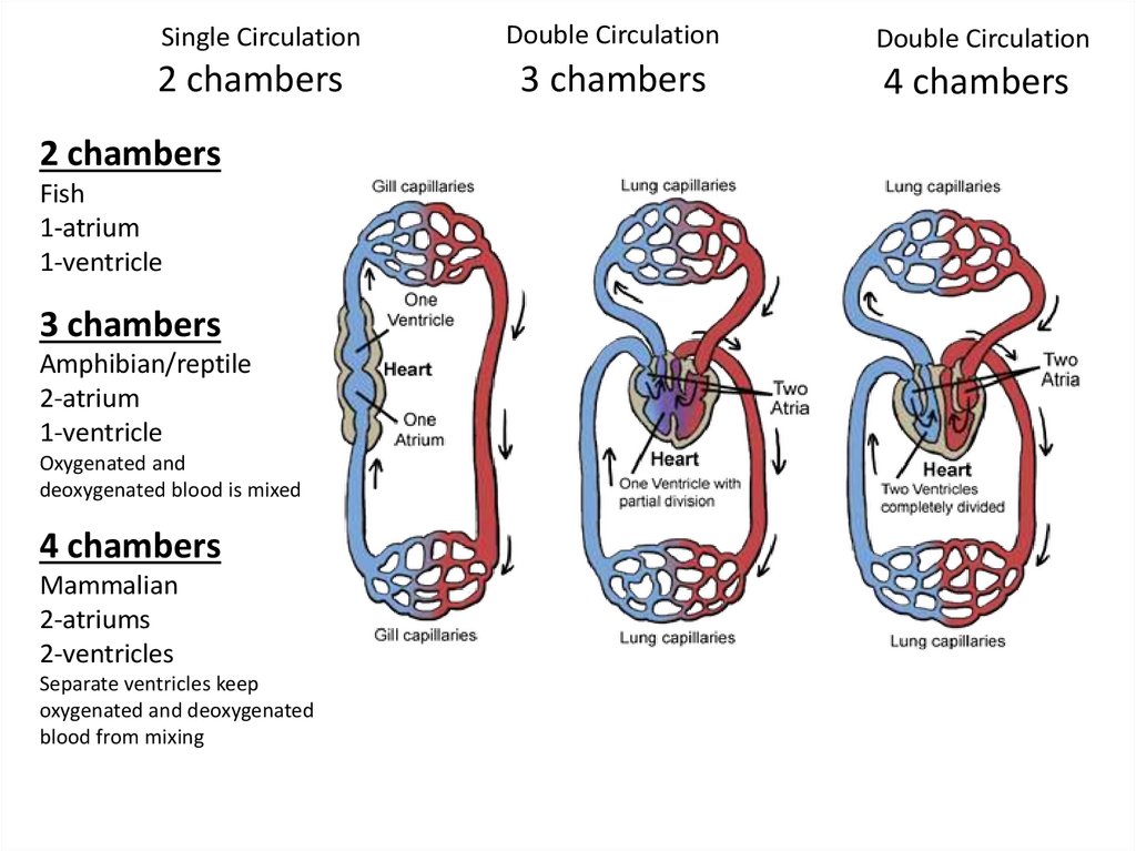
Single Circulation2 chambers
2 chambers
Fish
1-atrium
1-ventricle
3 chambers
Amphibian/reptile
2-atrium
1-ventricle
Oxygenated and
deoxygenated blood is mixed
4 chambers
Mammalian
2-atriums
2-ventricles
Separate ventricles keep
oxygenated and deoxygenated
blood from mixing
Double Circulation
Double Circulation
3 chambers
4 chambers
4. Blood travels through the heart twice before returning to the body
DoubleCirculatory
System
5.
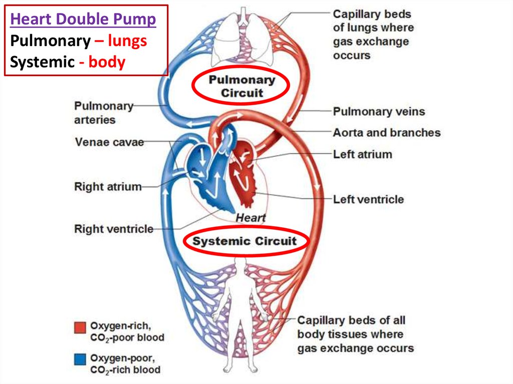
Heart Double PumpPulmonary – lungs
Systemic - body
6.
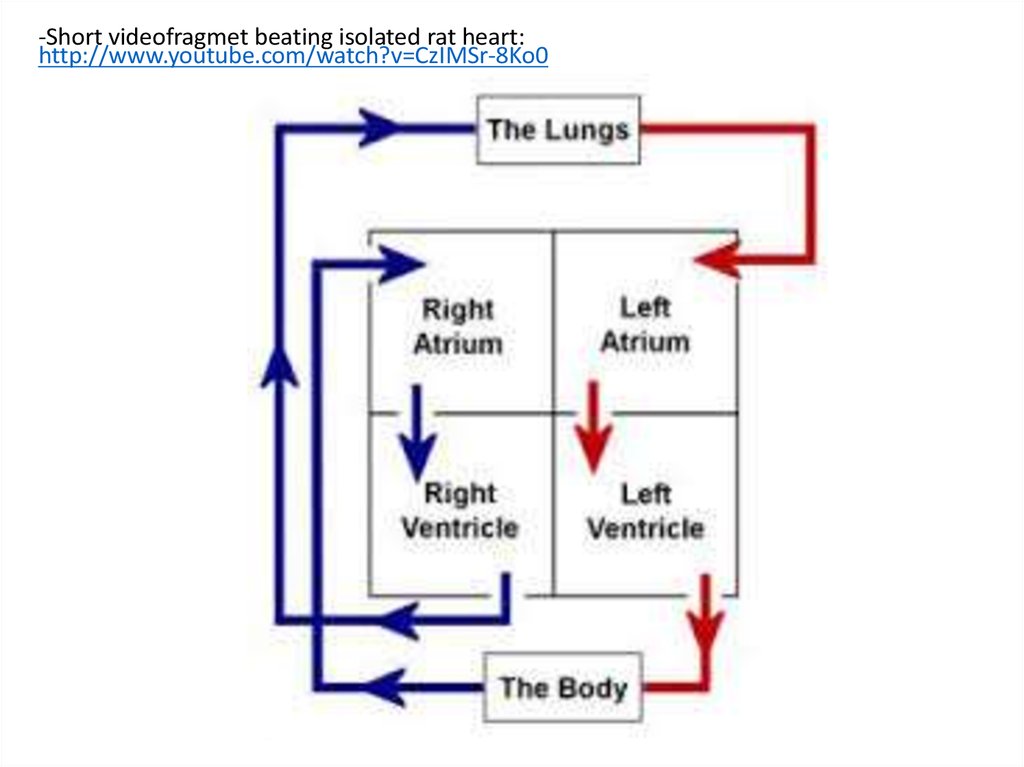
-Short videofragmet beating isolated rat heart:http://www.youtube.com/watch?v=CzIMSr-8Ko0
7.
Three Types of Muscle TissueDraw and Label
Voluntary
Striated
Multinucleated
Non-branched
Involuntary
Striated
Intercalculated disks
Mononucleated
Involuntary
Branched
Non-striated
Mononucleated Tapered
8.
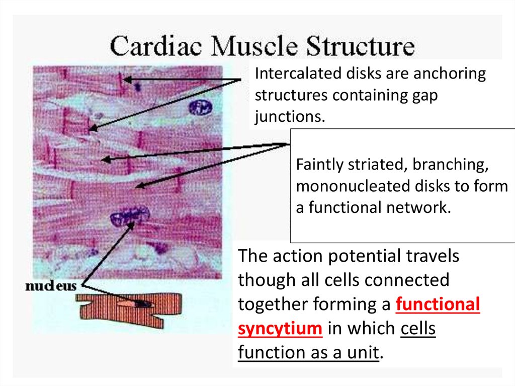
Intercalated disks are anchoringstructures containing gap
junctions.
Faintly striated, branching,
mononucleated disks to form
a functional network.
The action potential travels
though all cells connected
together forming a functional
syncytium in which cells
function as a unit.
9.
10. Define the following terms
• Functional syncytium –the heart consists ofindividual cells, the entire mass normally
responds as a unit and all of the cells contract
together.
• Myogenic – cardiac muscle can contract without
nervous input. BUT the strength and the rate of
contraction is modified by nervous input.
• Automaticity – the cardiac cell’s ability to
spontaneously generate an electrical impulse
(depolarize).
11.
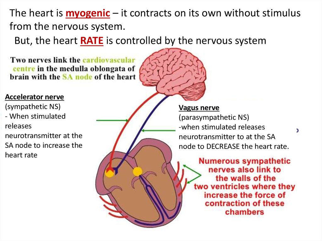
The heart is myogenic – it contracts on its own without stimulusfrom the nervous system.
But, the heart RATE is controlled by the nervous system
Accelerator nerve
(sympathetic NS)
- When stimulated
releases
neurotransmitter at the
SA node to increase the
heart rate
Vagus nerve
(parasympathetic NS)
-when stimulated releases
neurotransmitter to at the SA
node to DECREASE the heart rate.
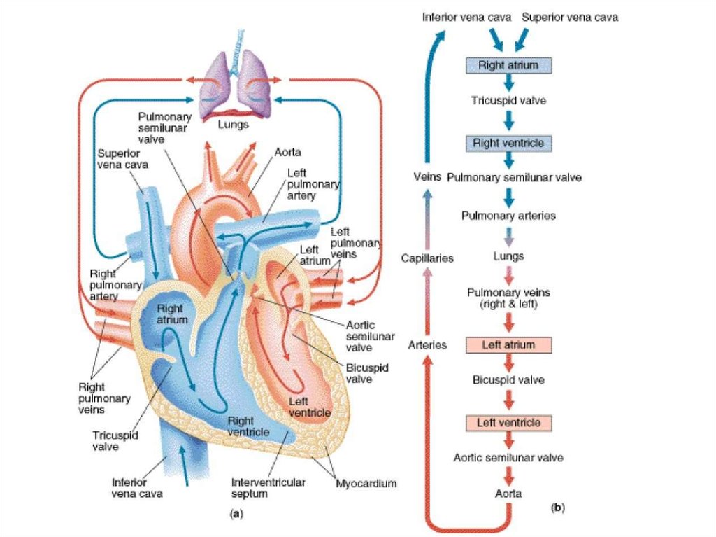
12. Heart Function- More definitions
Aorta-is connected to the left ventricle and carriesoxygenated blood to all the parts of the body except
the lungs.
Vena cava – is connected to the right atrium and brings
deoxygenated blood back from the tissues.
Pulmonary artery – is connected to the right ventricle
and carries deoxygenated blood to the lungs, where
oxygen is replenished and carbon dioxide is removed.
Pulmonary vein – is connected to the left atrium and
brings oxygenated blood back from the lungs.
13. Label Heart - 1 min
14. External Features-Label
LeftRight
Superior
Inferior
15.
RightVentricle
Septum
Left
Ventricle
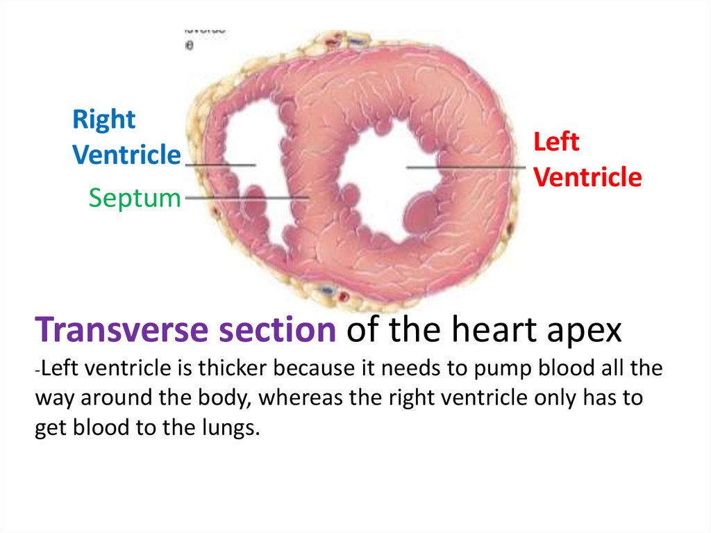
Transverse section of the heart apex
-Left
ventricle is thicker because it needs to pump blood all the
way around the body, whereas the right ventricle only has to
get blood to the lungs.
16.
17.
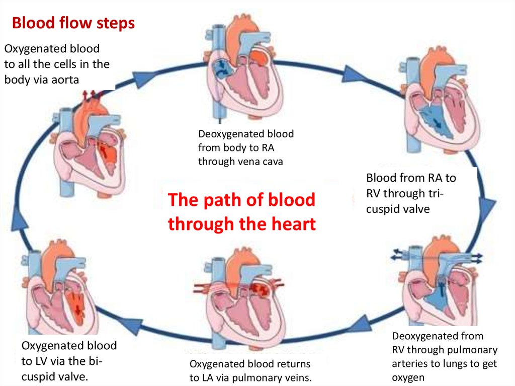
Blood flow stepsOxygenated blood
to all the cells in the
body via aorta
Deoxygenated blood
from body to RA
through vena cava
The path of blood
through the heart
Oxygenated blood
to LV via the bicuspid valve.
Oxygenated blood returns
to LA via pulmonary veins.
Blood from RA to
RV through tricuspid valve
Deoxygenated from
RV through pulmonary
arteries to lungs to get
oxygen
18.
4 Valves of the heart, open only one wayPulmonary
-high pressure behind – open
valve
-high pressure in front – closed.
Mitral
Valve
(bicuspid)
Chordae tendinae- prevent the
valves from turning inside out
under pressure.
Mitral
Valve
(bicuspid)
Aortic
Valve
Tricuspid
valve
Tricuspid
valve
19.
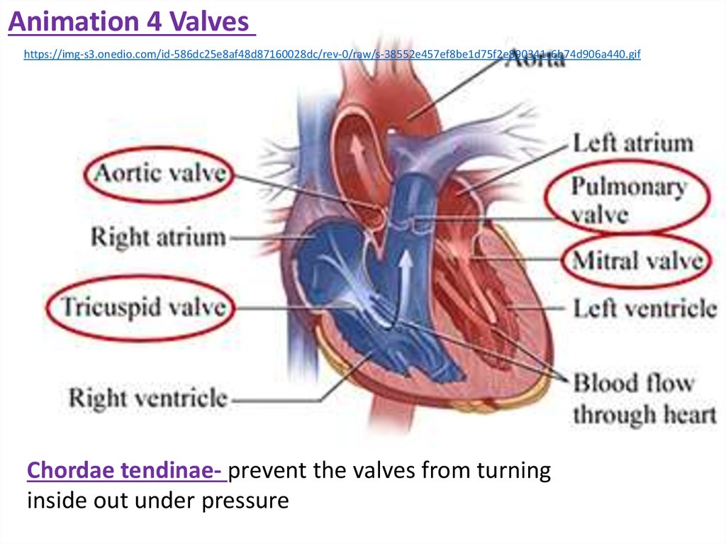
Animation 4 Valveshttps://img-s3.onedio.com/id-586dc25e8af48d87160028dc/rev-0/raw/s-38552e457ef8be1d75f2e890341c6b74d906a440.gif
Chordae tendinae- prevent the valves from turning
inside out under pressure
20. II. Cardiac Cycle
--Animation showing a cardiac cycle and the corresponding electrocardiogram wavehttp://en.wikipedia.org/wiki/File:ECG_principle_slow.gif
Information on the ECG and examples of some common anomalies
http://www.ivline.info/2010/05/quick-guide-to-ecg.html
http://www.happydoctor.ru/info/536
http://en.wikipedia.org/wiki/File:ECG_principle_slow.gif
21. Terminology 2
-Cardiac Cycle ECGEnglish
Google Russian
Myogenic / Myogenicity
SAN node
AV node
Bundles of HIS
Purkinje fibers
Septum
Depolarization
Repolarization
Миогенная / миогенность
Узел SAN
AV-узел
Связки ЕГО
Пуркинье
перегородка
деполяризация
реполяризация
Wolff-Parkinson-White Syndrome (WPW) Wolff-Parkinson-White Syndrome (WPW)
Tachycardia >100 bpm
Bradycardia < 60 bpm
Electrocardiography ECG
Тахикардия> 100 уд / мин
Брадикардия <60 уд / мин
Электрокардиография ЭКГ
22. Label heart diagram! 1 min
Label heart diagram! 1 min23.
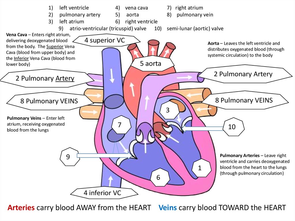
1)2)
3)
left ventricle
4) vena cava
7) right atrium
pulmonary artery
5) aorta
8) pulmonary vein
left atrium
6) right ventricle
9) atrio-ventricular (tricuspid) valve 10) semi-lunar (aortic) valve
Vena Cava – Enters right atrium,
delivering deoxygenated blood
from the body. The Superior Vena
Cava (blood from upper body) and
the Inferior Vena Cava (blood from
lower body)
4 superior VC9
Aorta – Leaves the left ventricle and
distributes oxygenated blood (through
systemic circulation) to the body
5 aorta9
2 Pulmonary Artery9
2 Pulmonary Artery9
8 Pulmonary VEINS9
8 Pulmonary VEINS9
39
Pulmonary Veins – Enter left
atrium, receiving oxygenated
blood from the lungs
79
109
99
19
69
Pulmonary Arteries – Leave right
ventricle and carries deoxygenated
blood from the heart to the lungs
(through pulmonary circulation)
4 inferior VC9
Arteries carry blood AWAY from the HEART Veins carry blood TOWARD the HEART
24. What is myogenic?
muscles or tissues that cancontract on their own, without
any external electrical stimulus
from the brain or nervous
system.
25. Electrical Activity of the Heart
Names of the numbers!1- SAN node
2 – AV node
3 - Bundle of HIS
4 – Right / Left
Branch Bundles
5 - Purkinje fibers
Your text here
Electrical Activity of the Heart 1.0 min
https://www.youtube.com/watch?v=te_SY3MeWys
Electrical Activity 3 min https://www.youtube.com/watch?v=RYZ4daFwMa8
26.
Bundle of His-heart muscle cells
specialized for
electrical conduction
-transmit electrical
impulses from AV
node to apex via
bundle branches.
Purkinje fibers
-cardiomyocytes that
are able to conduct
cardiac action potential
more efficiently than
other heart cells.
consist of
-allow synchronized
contraction of the
heart ventricles
-essential for
maintaining a
consistent heart
rhythm
27.
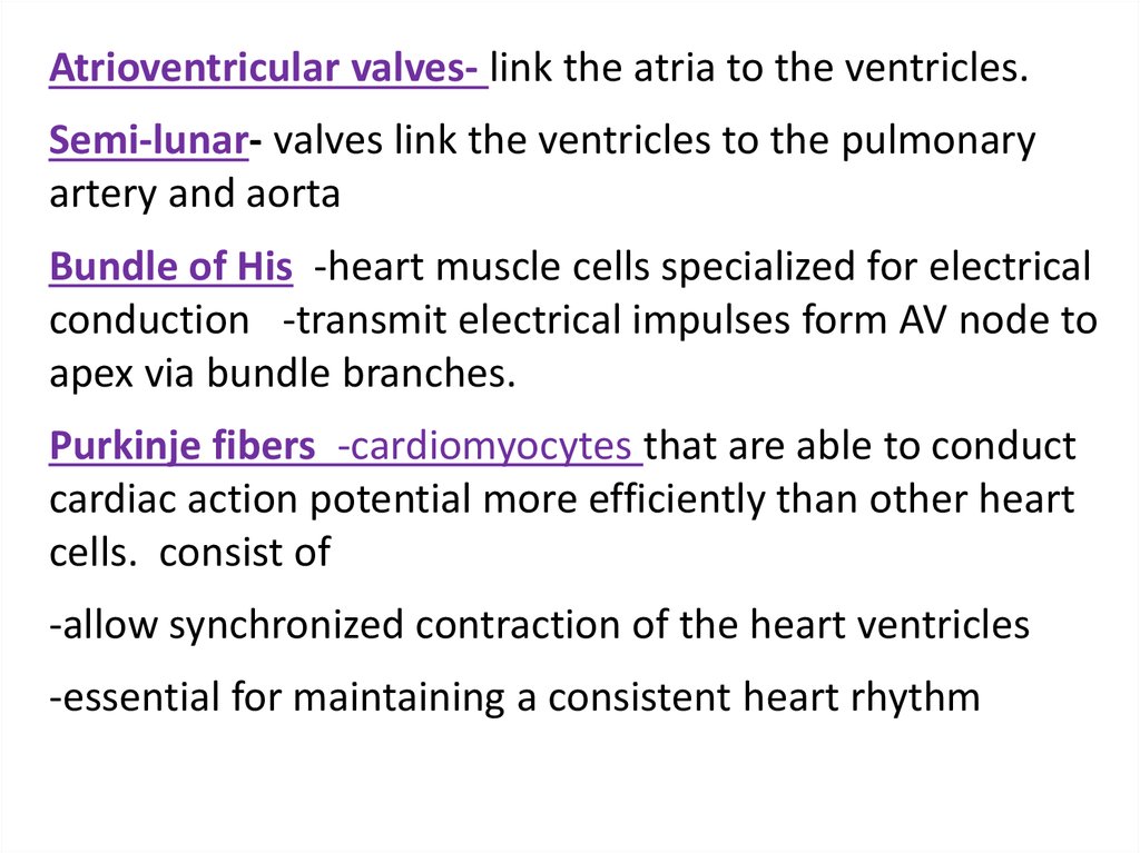
Atrioventricular valves- link the atria to the ventricles.Semi-lunar- valves link the ventricles to the pulmonary
artery and aorta
Bundle of His -heart muscle cells specialized for electrical
conduction -transmit electrical impulses form AV node to
apex via bundle branches.
Purkinje fibers -cardiomyocytes that are able to conduct
cardiac action potential more efficiently than other heart
cells. consist of
-allow synchronized contraction of the heart ventricles
-essential for maintaining a consistent heart rhythm
28.
Rest:(=)+ outside - inside
Depolarization:
(=) - outside + inside
Repolarization:
Returns to:
+ outside - inside
29.
30.
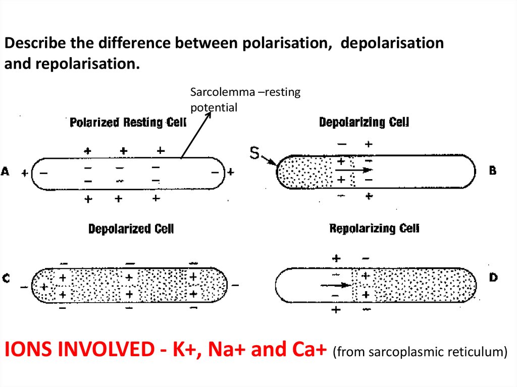
Describe the difference between polarisation, depolarisationand repolarisation.
Sarcolemma –resting
potential
IONS INVOLVED - K+, Na+ and Ca+ (from sarcoplasmic reticulum)
31. depolarization…..
• Depolarization is when a cell membrane's chargebecomes positive to generate an action potential. This is
usually caused by positive sodium and calcium ions going
into the cell
32. repolarization…..
• Repolarization is when a cell membrane's charge returnsto negative after depolarization. This is caused by positive
potassium ions moving out of the cell.
Depolarization and repolarization video
http://www.youtube.com/watch?v=4vkbywows-o
33. The QRS complex the combination of three of the graphical deflections seen on a typical deflections on an electrocardiogram =
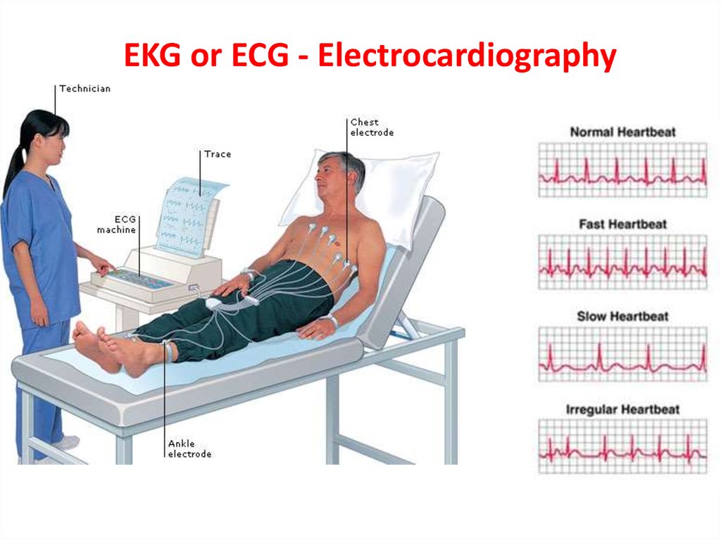
ECG orEKG
34.
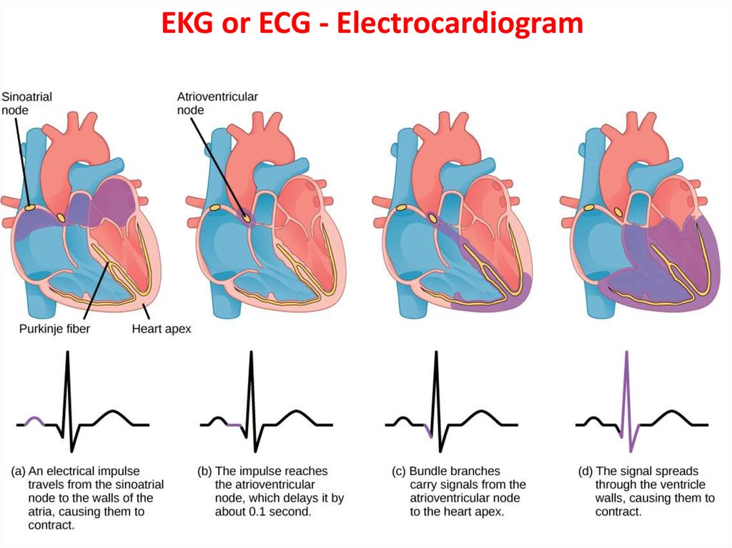
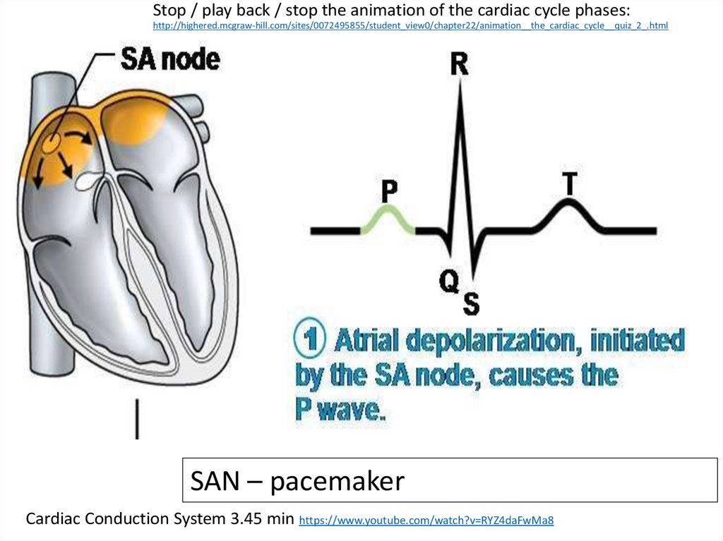
EKG or ECG - Electrocardiogram35.
Stop / play back / stop the animation of the cardiac cycle phases:http://highered.mcgraw-hill.com/sites/0072495855/student_view0/chapter22/animation__the_cardiac_cycle__quiz_2_.html
SAN – pacemaker
Cardiac Conduction System 3.45 min https://www.youtube.com/watch?v=RYZ4daFwMa8
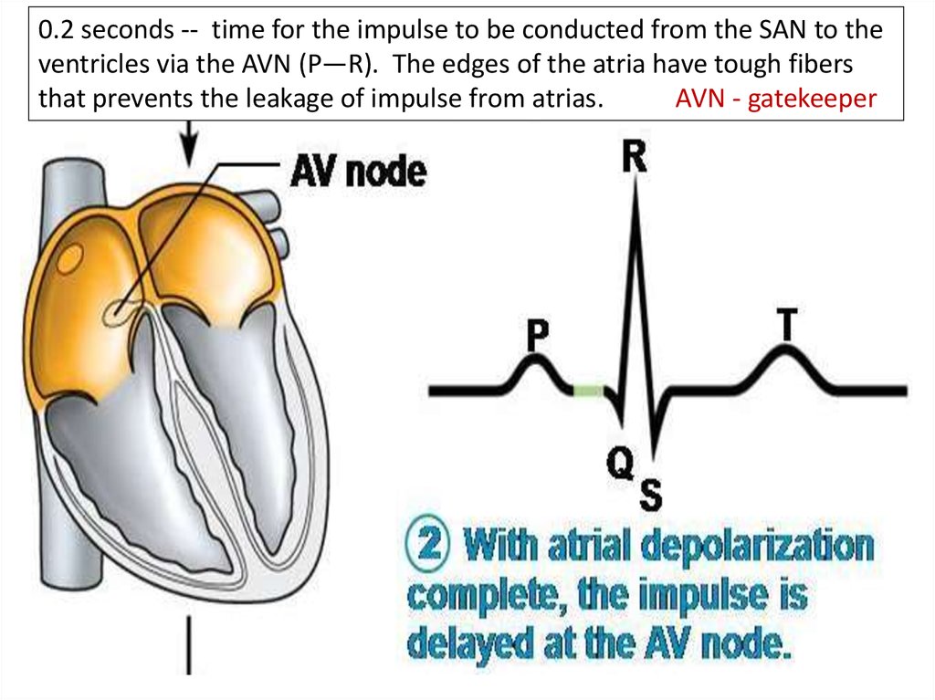
36.
0.2 seconds -- time for the impulse to be conducted from the SAN to theventricles via the AVN (P—R). The edges of the atria have tough fibers
that prevents the leakage of impulse from atrias.
AVN - gatekeeper
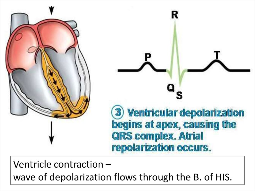
37.
Ventricle contraction –wave of depolarization flows through the B. of HIS.
38.
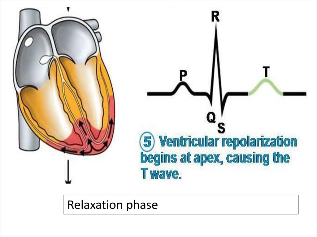
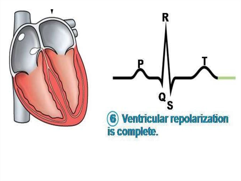
39.
Relaxation phase40.
41. What are some ways that SAN and AVN control the heart beat?
42. -SAN initiates heartbeat -Beat of heart is myogenic – spontaneous not started by nervous system stimulus -Rate of heartbeat is
FACTS-SAN initiates heartbeat
-Beat of heart is myogenic – spontaneous not started by nervous system stimulus
-Rate of heartbeat is influenced by nervous system
-Wave of electrical activity, impulses over atria triggers contraction of atrium
-Electrical activity may only pass to the ventricles via AVN and bundle of HIS (septum)
-Fibrous tissue prevents passage beyond atria
-Delay at AVN allows ventricles to fill completely from atria
43. EKG wave animation..
• http://en.wikipedia.org/wiki/Electrocardiography#mediaviewer/File:ECG_principle_slow.gif
44.
EKG or ECG - Electrocardiography45.
During the cardiac cycle (one contraction of the heartplus the relaxation period that follows), electrical
changes take place in the heart. These changes can be
visualized and recorded.
1) Detection of electrical forces in the heart.
Electrical forces in the heart can be detected on the
body's surface. Therefore, electrodes attached to
the patient's skin can detect electrical forces in the
heart.
2) Recording of electrical forces in the heart.
The recording of the electrical changes during the
cardiac cycle is called an electrocardiogram (ECG
or EKG). The instrument used to record these
changes is an electrocardiograph.
46.
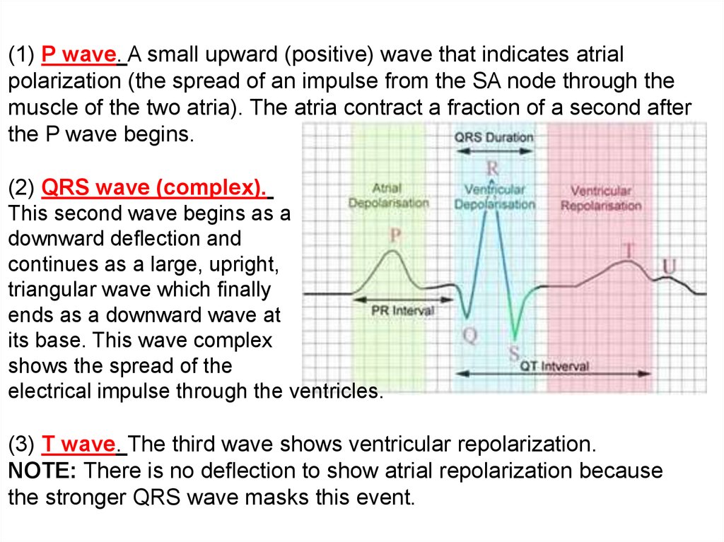
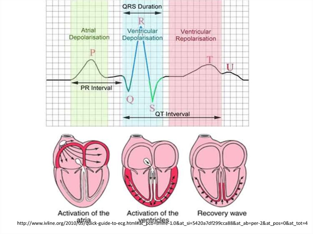
(1) P wave. A small upward (positive) wave that indicates atrialpolarization (the spread of an impulse from the SA node through the
muscle of the two atria). The atria contract a fraction of a second after
the P wave begins.
(2) QRS wave (complex).
This second wave begins as a
downward deflection and
continues as a large, upright,
triangular wave which finally
ends as a downward wave at
its base. This wave complex
shows the spread of the
electrical impulse through the ventricles.
(3) T wave. The third wave shows ventricular repolarization.
NOTE: There is no deflection to show atrial repolarization because
the stronger QRS wave masks this event.
47.
http://www.ivline.org/2010/05/quick-guide-to-ecg.html#at_pco=smlre-1.0&at_si=5420a7df299cca88&at_ab=per-2&at_pos=0&at_tot=448. Ventricular fibrillation
Heart BlockWhat do you think is happening in the ECG?
49. Ventricular fibrillation
Uncontrolledcontraction of the
ventricles causes
little blood to be
pumped
Heart Block
Ventricles are
not always
stimulated
What do you think is happening in the ECG?
50. Match the beat with the ECG.
Irregular TachycardiaBradycardia Normal
51.
NormalBradycardia - Slow
Tachycardia - Fast
Irregular
52. Wolff-Parkinson-White (WPW) Syndrome
An extra electrical pathwaybetween your heart's upper
and lower chambers causes
a rapid heartbeat. The extra
pathway is present at birth
and fairly rare.
WPW syndrome is defined
as a congenital condition
involving abnormal
conductive cardiac tissue
between the atria and the
ventricles that provides a
pathway for a reentrant
tachycardia circuit
The accessory bridge is
called the bundle of Kent.
It runs along the wall of the
the left ventricle.
53. Wolff Parkinson Wright Syndrome (WPW)
https://www.youtube.com/watch?v=9MDRKId2d0Q-Write information from the video and label
the 2nd heart on your worksheet.
-Record the information of the electrical
circuit of WPW
Accessory pathway
Bundle of Kent
Normal
Wolff Parkinson White Syndrome
54.
The Nanostim has no leads and is ten times smaller than the traditionalpacemaker. The nanostim sits in the right ventricle of the heart. It takes
under ten minutes to fit. The traditional pacemaker took 45 minutes to fit.
The AAA pacemaker: It's the size of a
small battery and takes just seven
minutes to fit... so could this revolutionary
device give new heart to millions?
The new pacemaker is put into a vein in the leg and is then pushed up to
the heart by the surgeon. When the new pacemaker is inside the right
ventricle, it is screwed into the wall. This keeps it in the same place.
By ALICE SMELLIE
25 January 2014
The leads on traditional pacemakers sometimes caused clots and
infections. This could be very dangerous. The new pacemaker has no
leads and this makes it much safer.
The new pacemaker is called the ‘Nanostim’.
Six Britons have been fit with the new pacemaker.
The new pacemaker is ten times smaller than the traditional pacemaker
It takes 10 minutes to fit. The traditional pacemaker takes 45 minutes to fit.
The first six British patients have been fitted with a wireless pacemaker –
smaller than an AAA battery – that is expected to change the treatment of
heart disease.
Traditional pacemakers have not changed much since 1958. Traditional p
acemakers control the heart beat by sending electrical impulses into the
heart using leads from a battery pack. The battery pack is put under the
patients’ collarbone.
Maureen McCleave, 77, has already had a new pacemaker fitted. She was
surprised because it only took 10 minutes to fit. As soon as it had been
fitted she felt much better.
Last year, more than 40,000
Britons had a pacemaker fitted.
They are used to treat people
who have problems with their
heartbeat. A person might
need a pacemaker if their heart
beats too fast or too slow.
They might also need a
pacemaker is their heart does
not beat in a normal rhythm.
In traditional pacemakers, three electrical leads run into the heart. The
pacemaker worked by sending an electrical impulse to the heart down the
leads.
The new pacemaker works by checking the electrical impulses in the heart.
If it notices that the heart has stopped beating it releases an electrical
impulse. The battery lasts up to 13 years. When the battery runs out, the
surgeon puts in another new pacemaker or changes the battery.
Pacemaker
Article
If the new pacemaker fell off the wall of the right ventricle it would not be
dangerous. If it fell off it would go to the lung and surgeons could take it
out.
55.
56. Pacemaker
57.
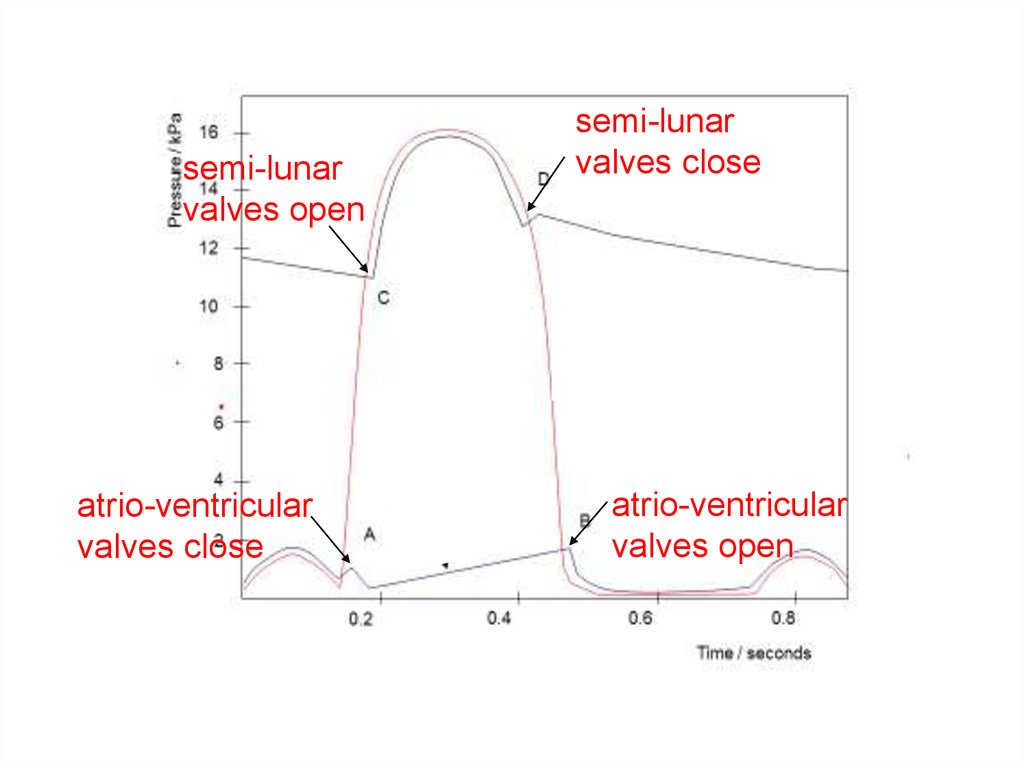
semi-lunarvalves open
atrio-ventricular
valves close
semi-lunar
valves close
atrio-ventricular
valves open
58.
59.
Match the letter on the graph to the following events______Semi-lunar valves open
______Atrio-ventricular valves close,
______Semi-lunar valves close
______Atrio-ventricular valves open
60.
semi-lunarvalves open
atrio-ventricular
valves close
semi-lunar
valves close
atrio-ventricular
valves open
61.
AtrialSystole
Ventricular
Systole
Diastole
D
“DUB”
C
“LUB”
B
How to calculate
the cardiac cycle
Beats per minute
(bpm).
1 minute (60s)
Length one cycle
A
Atrioventricular (bicuspid / mitral) valve(s) closes (“snaps shut”– makes 1st
louder heart sound “LUB”
B
Semilunar valve(s) (aortic valve) opens
C
Semilunar valve(s) closes – makes second softer heart sound “DUB”- shut
due to blood accumulating in their pockets
D
Atriioventricular (bicuspid) valve(s) opens
62.
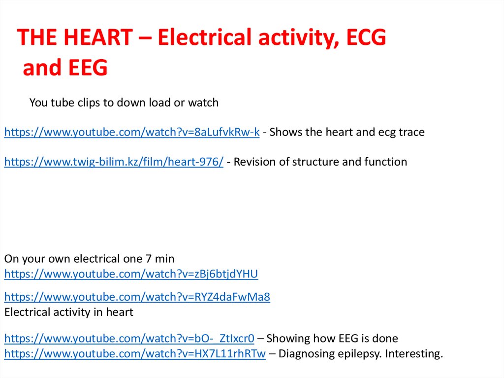
THE HEART – Electrical activity, ECGand EEG
You tube clips to down load or watch
https://www.youtube.com/watch?v=8aLufvkRw-k - Shows the heart and ecg trace
https://www.twig-bilim.kz/film/heart-976/ - Revision of structure and function
On your own electrical one 7 min
https://www.youtube.com/watch?v=zBj6btjdYHU
https://www.youtube.com/watch?v=RYZ4daFwMa8
Electrical activity in heart
https://www.youtube.com/watch?v=bO-_ZtIxcr0 – Showing how EEG is done
https://www.youtube.com/watch?v=HX7L11rhRTw – Diagnosing epilepsy. Interesting.
63. Extra Information
64. Tachycardia
• Increased heart rate is anormal response to:
Normal – for reference
6 beats per 5 seconds
=
72 bpm
Tachycardic
exercise
excitement
stress
drugs e.g. caffeine, nicotine,
amphetamine (speed).
• Tachycardia is elevated heart
rate for no reason.
• Sometimes heart rate is so
high that little blood is
actually pumped:
• filling time too short.
• Treatment might involve:
9 beats per 5 seconds =108 bpm
• relaxation therapy
• β-blocker.
65. Bradycardia
Normal – for reference6 beats per 5 seconds = 72 bpm
Bradycardic
• Pattern of electrical activity
is normal but slow.
• Reduced heart rate could
indicate:
• good aerobic fitness (elite
athletes like Steve Redgrave
have resting heart of ca. 45
bpm).
• Alternatively might be
caused by drugs:
• tranquilisers
• β-blocker.
• Cause may need
investigation:
4 beats per 5 seconds = 48 bpm
• stagnation
• risk of blood clots.
66. Heart block
Normal – for reference6 beats per 5 seconds = 72 bpm
Heart block
Dissociated P and QRS complex
• There is separation of the P
wave and the QRS complex.
• Pacemaker activity and atrial
contraction is normal.
• Delay in conduction
between atria and
ventricles.
67. Fibrillation
Normal – for reference6 beats per 5 seconds = 72 bpm
• Contraction of cardiac
muscle is normally
coordinated.
• In VF the ventricles contract,
but it is not coordinated:
• fluttering
• little blood is pumped.
• Defibrillation may work:
Ventricular fibrillation (VF)
Uncoordinated and weak contraction
• heart is shocked
• heart stops
• when it restarts, it may do
so with a normal rhythm.
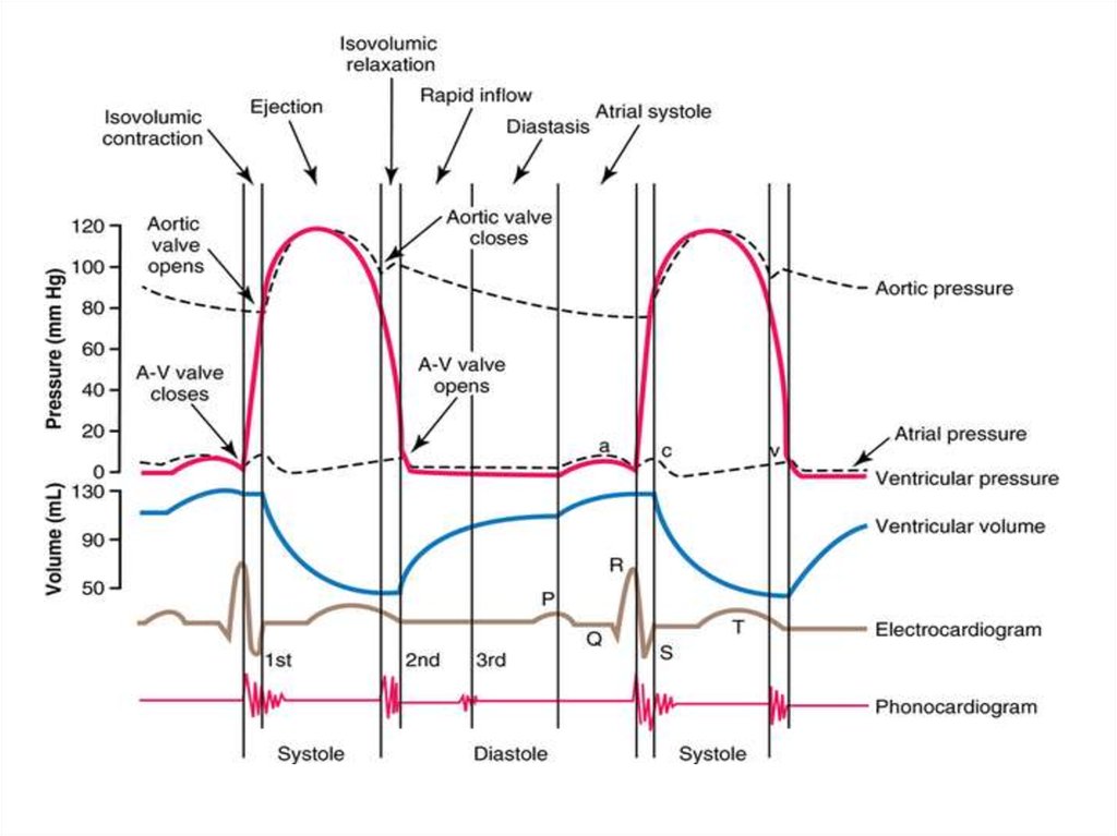
68. Cardiac Cycle
• General Principles.• Contraction of the myocardium generates pressure
changes which result in the orderly movement of blood.
• Blood flows from an area of high pressure to an area of
low pressure, unless flow is blocked by a valve.
• Events on the right and left sides of the heart are the
same, but pressures are lower on the right.
69. Atrial systole
• The heart is full of bloodand the ventricles are
relaxed
• Both the atria contract
and blood passes down to
the ventricles
• The atrio-ventricular
valves open due to blood
pressure
70% of the blood flows passively down to the ventricles so the
atria do not have to contract a great amount.
70. Ventricular systole
• The atria relax.• The ventricle walls
contract, forcing the
blood out
• The pressure of the
blood forces the atrioventricular valves shut
(producing the heart
sound ‘lub’)
71. Ventricular systole
• The pressure ofblood opens the
semi-lunar valves.
• Blood passes into
the aorta and
pulmonary arteries.
72. Diastole
• The ventricles relax• Pressure in the ventricles
falls below that in the
arteries
• Blood under high pressure in
the arteries causes the semi
lunar valves to shut. This
produces the second heart
sound, ‘dub’.
• During diastole, all the
muscle in the heart relaxes.
73.
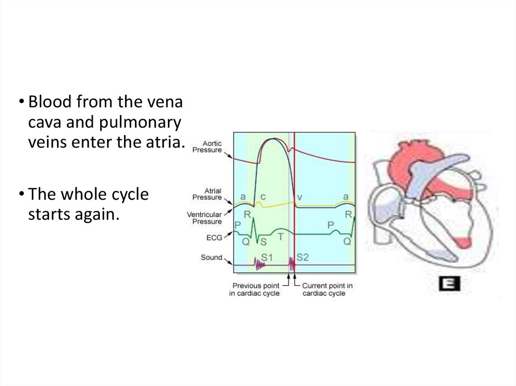
• Blood from the venacava and pulmonary
veins enter the atria.
• The whole cycle
starts again.
74.
Match the letter on the graph to the following events______Semi-lunar valves open
______Atrio-ventricular valves close,
______Semi-lunar valves close
______Atrio-ventricular valves open
75.
semi-lunarvalves open
atrio-ventricular
valves close
semi-lunar
valves close
atrio-ventricular
valves open
76.
AtrialSystole
Ventricular
Systole
Diastole
D
“DUB”
C
“LUB”
B
How to calculate
the cardiac cycle
Beats per minute
(bpm).
1 minute (60s)
Length one cycle
A
Atrioventricular (bicuspid / mitral) valve(s) closes (“snaps shut”– makes 1st
louder heart sound “LUB”
B
Semilunar valve(s) (aortic valve) opens
C
Semilunar valve(s) closes – makes second softer heart sound “DUB”- shut
due to blood accumulating in their pockets
D
Atriioventricular (bicuspid) valve(s) opens
77.
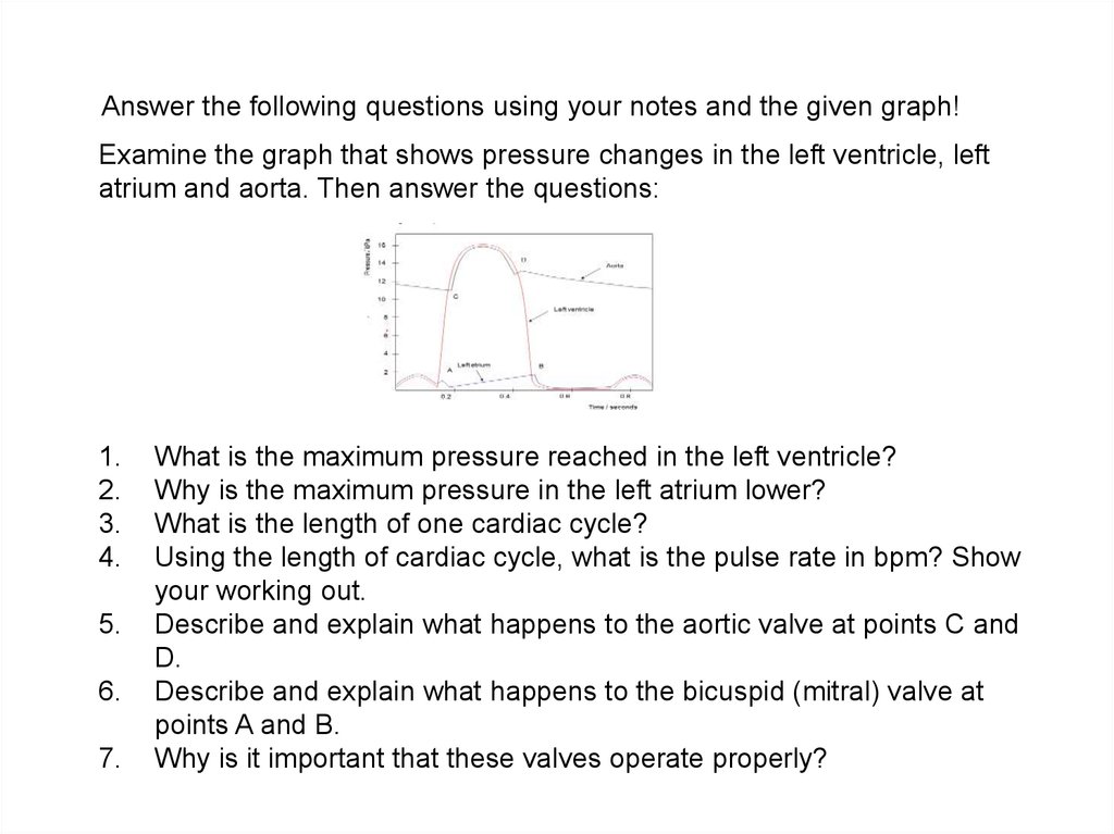
Answer the following questions using your notes and the given graph!Examine the graph that shows pressure changes in the left ventricle, left
atrium and aorta. Then answer the questions:
1.
2.
3.
4.
5.
6.
7.
What is the maximum pressure reached in the left ventricle?
Why is the maximum pressure in the left atrium lower?
What is the length of one cardiac cycle?
Using the length of cardiac cycle, what is the pulse rate in bpm? Show
your working out.
Describe and explain what happens to the aortic valve at points C and
D.
Describe and explain what happens to the bicuspid (mitral) valve at
points A and B.
Why is it important that these valves operate properly?
78.
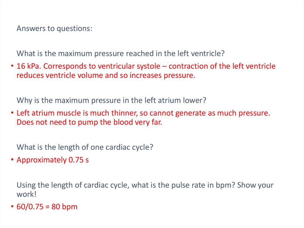
Answers to questions:What is the maximum pressure reached in the left ventricle?
• 16 kPa. Corresponds to ventricular systole – contraction of the left ventricle
reduces ventricle volume and so increases pressure.
Why is the maximum pressure in the left atrium lower?
• Left atrium muscle is much thinner, so cannot generate as much pressure.
Does not need to pump the blood very far.
What is the length of one cardiac cycle?
• Approximately 0.75 s
Using the length of cardiac cycle, what is the pulse rate in bpm? Show your
work!
• 60/0.75 = 80 bpm
79.
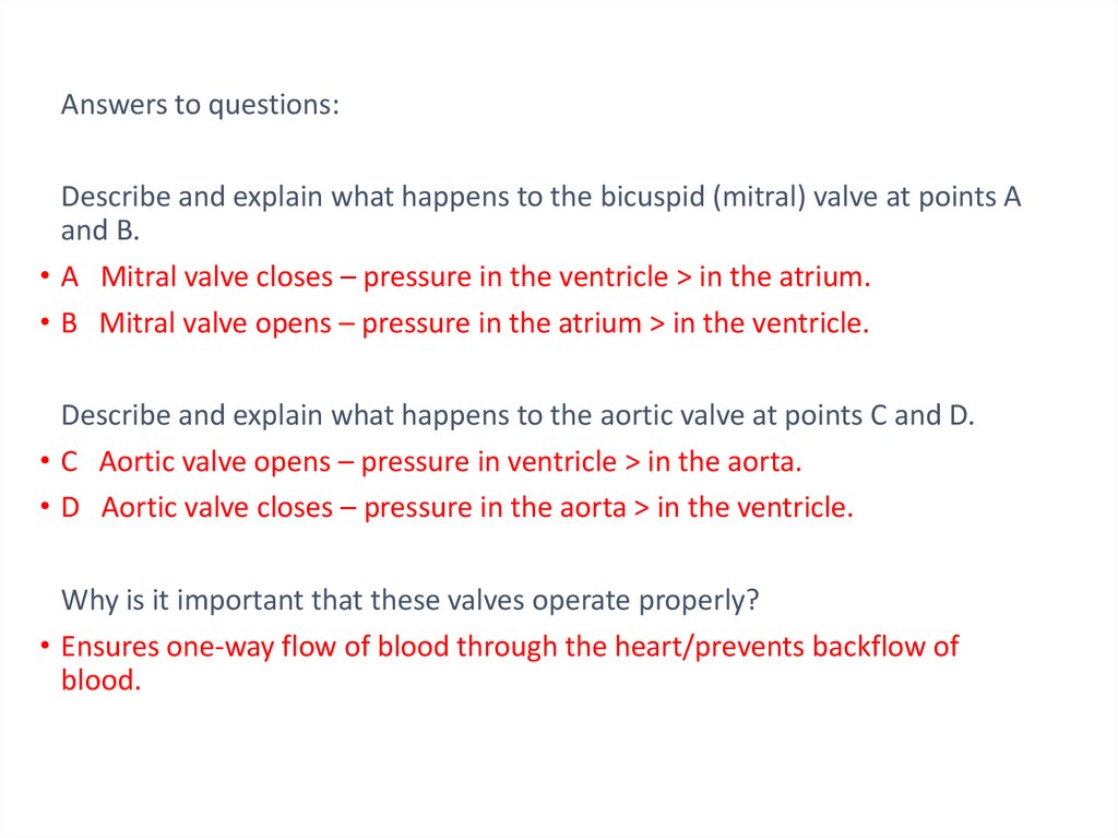
Answers to questions:Describe and explain what happens to the bicuspid (mitral) valve at points A
and B.
• A Mitral valve closes – pressure in the ventricle > in the atrium.
• B Mitral valve opens – pressure in the atrium > in the ventricle.
Describe and explain what happens to the aortic valve at points C and D.
• C Aortic valve opens – pressure in ventricle > in the aorta.
• D Aortic valve closes – pressure in the aorta > in the ventricle.
Why is it important that these valves operate properly?
• Ensures one-way flow of blood through the heart/prevents backflow of
blood.
80.
81.
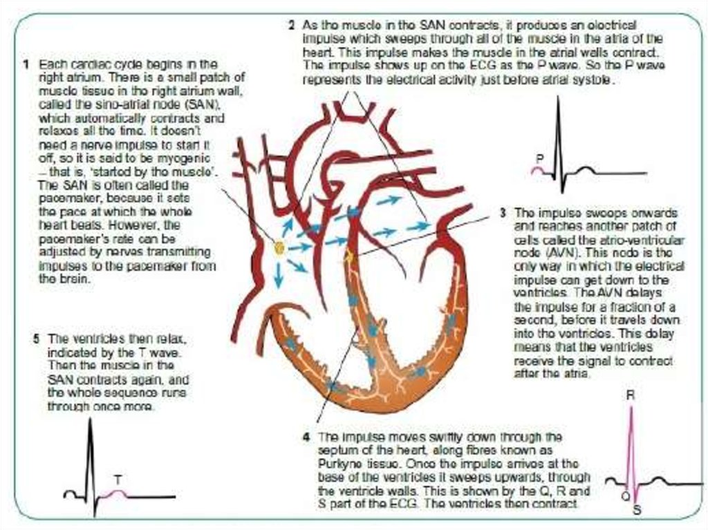
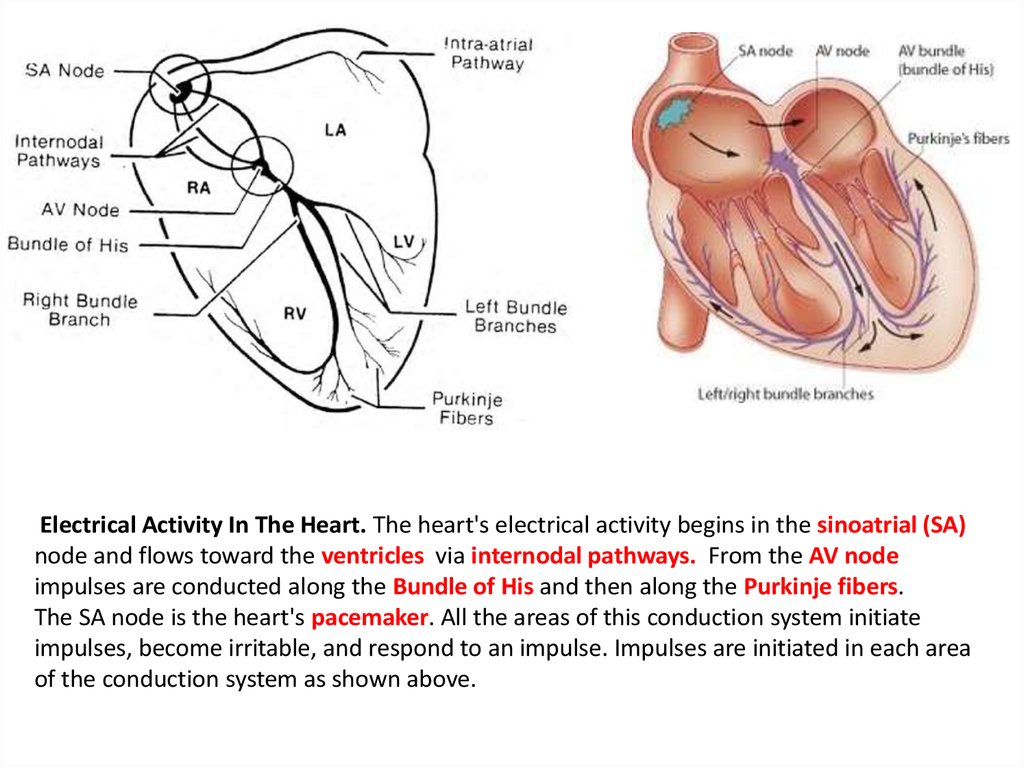
Electrical Activity In The Heart. The heart's electrical activity begins in the sinoatrial (SA)node and flows toward the ventricles via internodal pathways. From the AV node
impulses are conducted along the Bundle of His and then along the Purkinje fibers.
The SA node is the heart's pacemaker. All the areas of this conduction system initiate
impulses, become irritable, and respond to an impulse. Impulses are initiated in each area
of the conduction system as shown above.
82.
Only know (a)How does the structure of cardiac muscle differ from cardiac muscle?
83.
For interest – Dataresponse
1) Name the ions involved in cardiac muscle contraction.
2) Where does the action potential originate from?
3) How do you think the contraction of cardiac muscle differ from
that of striated muscle?