


 Медицина
Похожие презентации:

")


Hemoglobinopathies. Hb structure
1.
HemoglobinopathiesHemoglobinopathies
Thalassemia genetics
Hb synthesis
Hb A, A2, F
Hb ELP
Hb Constant-Spring
Hb Bart’s
Hb H
Hb Lepore
Hb E
Hb S
Hb C
Hb SC disease
HPFH
2.
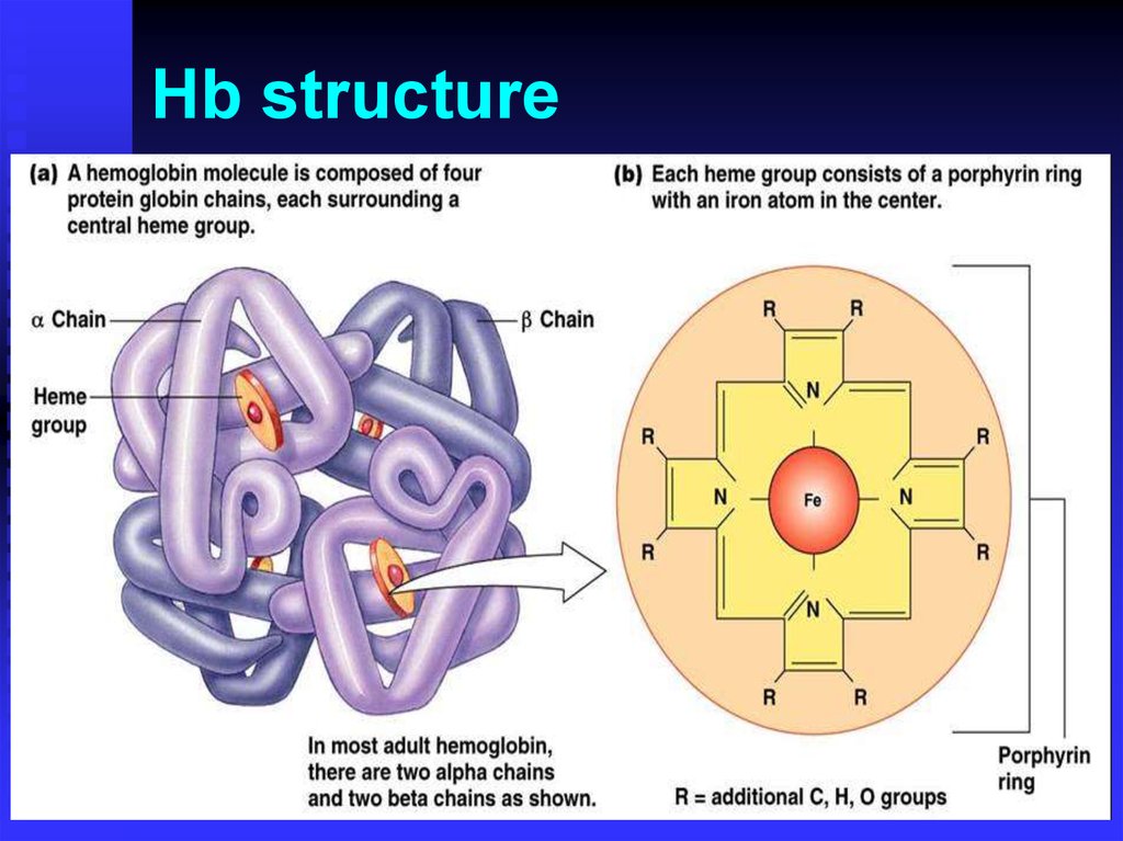
Hb structure3.
4.
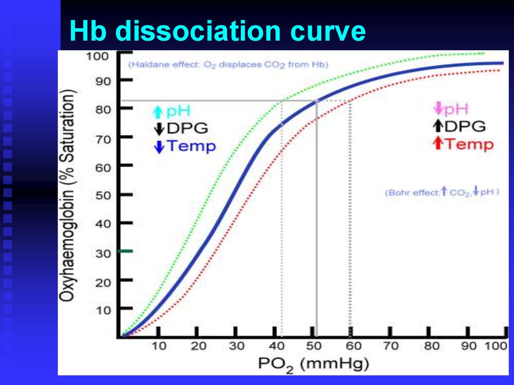
Hb dissociation curve5.
Anemia?
Production?
Survival/Destruction?
The key test is the …..
6.
The reticulocyte count(kinetic approach)
Increased reticulocytes (greater than 2-3% or
100,000/mm3 total) are seen in blood loss and
hemolytic processes, although up to 25% of
hemolytic anemias will present with a normal
reticulocyte count due to immune destruction of red
cell precursors.
Retic counts are most helpful if extremely low
(<0.1%) or greater than 3% (100,000/mm3 total).
7.
Causes of AnemiaDecreased erythrocyte production
Decreased erythropoietin production
Inadequate marrow response to erythropoietin
Erythrocyte loss
Hemorrhage
Hemolysis
8.
Morphological Approach(big versus little)
First, measure the size of the RBCs:
• Use of volume-sensitive automated blood cell
counters, such as the Coulter counter. The RBC’s pass
through a small aperture and generate a signal directly
proportional to their volume.
• Other automated counters measure red blood cell
volume by means of techniques that measure refracted,
diffracted, or scattered light
• By calculation
9.
Underproductionmacrocytic
MCV>115
B12, Folate
Drugs that impair
DNA synthesis
(AZT, chemo)
MDS
MCV 100 - 115
Endocrinopathy
(hypothyroidism)
Erythropoetin
Reticulocytosis
10.
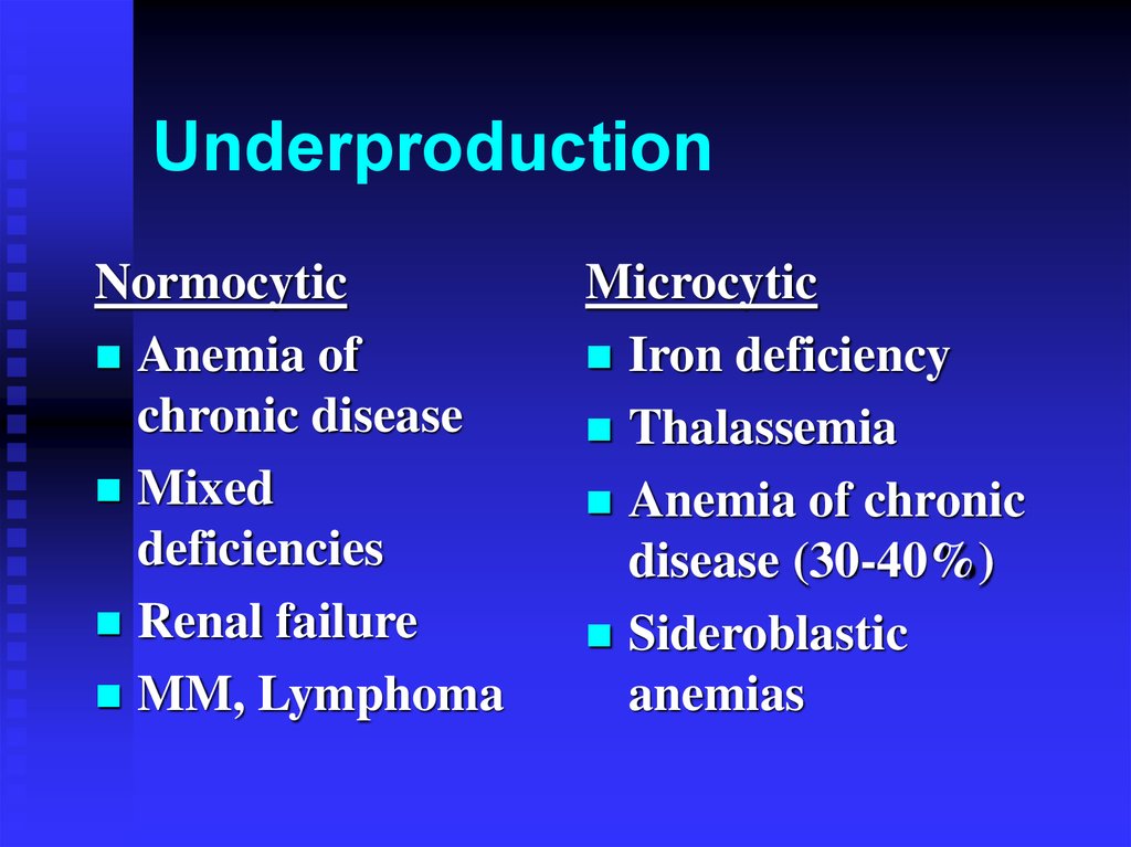
UnderproductionNormocytic
Anemia of
chronic disease
Mixed
deficiencies
Renal failure
MM, Lymphoma
Microcytic
Iron deficiency
Thalassemia
Anemia of chronic
disease (30-40%)
Sideroblastic
anemias
11.
Review red blood celldisorders
Marrow production
Thalassemias
Myelodysplasia
Myelophthisic
Aplastic anemia
Nutritional
deficiencies
Red cell destruction
Hemoglobinopathies
Enzymopathies
Membrane
disorders
Autoimmune
12.
Review red blood cell disordersMarrow Production - Aplastic Anemia
Acquired
Immunological
Toxins – Benzene
Drugs – methotrexate, chloramphenicol
Viruses – EBV, hepatitis
Hereditary
Fanconi,
Diamond-Shwachman
13.
Review red blood cell disordersMarrow Production - Myelodysplasia
Preleukemia, most commonly in the elderly.
Supportive care that involves transfusion
therapy is an option.
Poor response to growth factors
14.
Review red blood celldisorders
Marrow Production - Myelophthisic
Anemia associated with marrow infiltration
“teardrops”
Cancer, infections
Myelofibrosis
Treatment is aimed at the underlying
disease
Supportive transfusions as needed.
15.
Review red blood cell disordersRed cell destruction
Elevated reticulocyte count
Mechanical
Autoimmune
Drug
Congenital
16.
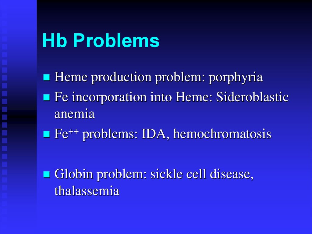
Hb ProblemsHeme production problem: porphyria
Fe incorporation into Heme: Sideroblastic
anemia
Fe++ problems: IDA, hemochromatosis
Globin problem: sickle cell disease,
thalassemia
17.
18.
HemoglobinopathiesDecrease, lack of, or abnormal globin
May be severe hemolytic anemia
Abnormal Hb with low functionality
Mutation may be deletion, substitution,
elongation
Hb electrophoresis may be helpful
19.
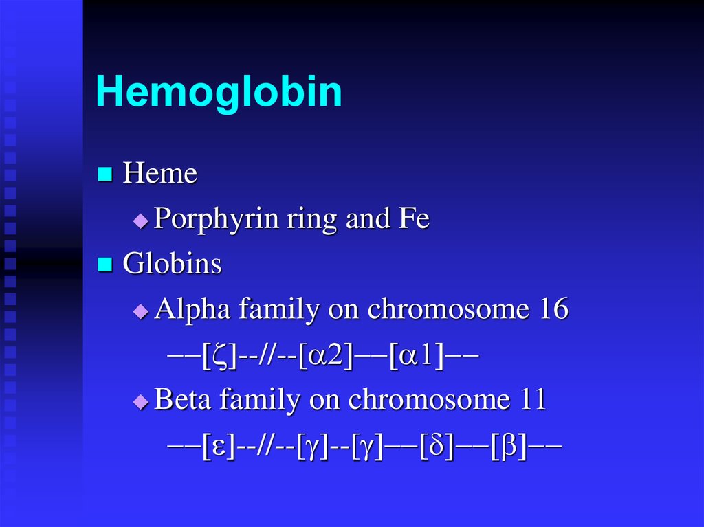
HemoglobinHeme
Porphyrin ring and Fe
Globins
Alpha family on chromosome 16
]--//--[
Beta family on chromosome 11
]--//--[ ]--[ [
20.
21.
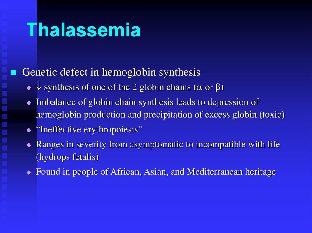
ThalassemiaGenetic defect in hemoglobin synthesis
synthesis of one of the 2 globin chains ( or )
Imbalance of globin chain synthesis leads to depression of
hemoglobin production and precipitation of excess globin (toxic)
“Ineffective erythropoiesis”
Ranges in severity from asymptomatic to incompatible with life
(hydrops fetalis)
Found in people of African, Asian, and Mediterranean heritage
22.
Thalassemia1925: Described by Dr. Thomas Cooley and Dr. Pearl
Lee of Detroit
1920’s: Osmotic fragility test
1932: Dr. George Whipple of Rochester coined the
name “thalassa anemia” from Greek story about
Xenophon’s army returning from Persia
1930’s: Familial pattern recognized
1950’s: Alkali denaturation test for Hb F, Hb ELP
1956: Coulter model A
1960’s: RBC indices
1980’s: Histogram, DNA analysis, PCR
23.
ThalassemiaGenetic defect in hemoglobin synthesis
synthesis of one of the 2 globin chains ( or )
Imbalance of globin chain synthesis leads to depression of
hemoglobin production and precipitation of excess globin (toxic)
“Ineffective erythropoiesis”
Ranges in severity from asymptomatic to incompatible with life
(hydrops fetalis)
Found in people of African, Asian, and Mediterranean heritage
24.
Signs and SymptomsHemolytic
Bone changes (hair on end)
Ethnicity: Mediterranean, Africa, Southeast
Asia
Hypo-Micro, Poikilocytosis
NRBC’s, reticulocytosis, basophilic
stippling
Siderocytes (with repeated transfusions)
25.
ThalassemiaBlood Smears
26.
X-ray of scullin Thalassemia:
“Hair-on-end”
27.
Perl’s iron stain (Prussianblue)
with potassium ferrocyanide
Siderocyte
Sideroblasts
28.
ThalassemiaDeletion of one or more alpha genes from
chromosome 16
- / : silent career with little signs
--/ : cis double deletion more common in SEA
- /- : trans double deletion
--/- : Hb H disease
--/--: Hb Bart’s hydrops fetalis
Hb Constant-Spring: elongation (discovered in
Kingston, Jamaica; 2% of Thai have it)
29.
Thalassemia Lab ChangesHigh RBC
Low H&H and indices
High RDW
May need to rule out IDA
Hb ELP not useful except in Hb H
BCB prep for Hb H
30.
31.
Hb H Prep withBrilliant cresyl blue
thalassemia
Hydrops fetalis
32.
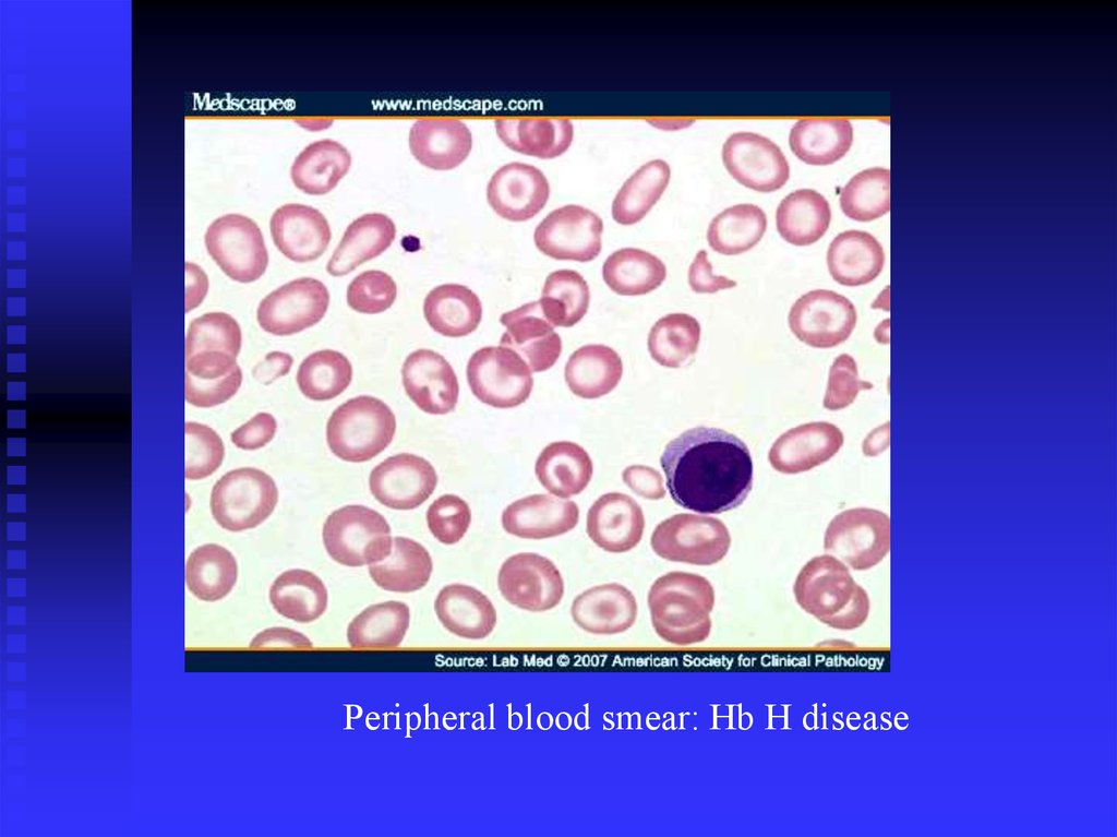
Peripheral blood smear: Hb H disease33.
ThalassemiaUsually point mutation in the control region chr 11
+ has minimal production
o has no production
+/ + or o/ o is thal major or Cooley’s anemia
Often not apparent at birth until chain takes over
chain production
High Hb A2, Hb F
Related: Hb Lepore ( fusion), HPFH
34.
Hb F preparation with Kleihauer-BetkeFetal Hb resists acid elution
35.
36.
37.
38.
ThalassemiaThe only treatments are stem cell transplant
and simple transfusion.
Chelation therapy to avoid iron overload
has to be started early.
39.
Sickle Cell AnemiaSingle base pair mutation results in a single
amino acid change.
Under low oxygen, Hgb becomes insoluble
forming long polymers
This leads to membrane changes
(“sickling”) and vasoocclusion
40.
41.
Red Blood Cells from Sickle Cell AnemiaDeoxygenation of SS erythrocytes leads to
intracellular hemoglobin polymerization, loss of
deformability and changes in cell morphology.
OXY-STATE
DEOXY-STATE
42.
Hb SSickling Hb
Autosomal
Sickle crisis in low oxygen condition
6 glutamate to valine substitution
Prevalent in Eastern Africa
Solubility test
Sickling test (meta-bisulfite)
43.
Other HemoglobinopathiesHb C ( 6 Glu-Lys) in Western Africa
Cigar-like crystals
Billiard ball cells
Folded cells
Hb SC disease
Washington monument cells
Mitten shape
Hb E ( 26 Glu-Lys) in SEA
Moves with Hb A2 in Hb ELP and A2 column
(ie, false elevated Hb A2)
44.
Hb C diseaseHb SC disease
45.
Unusual Hemoglobins in the World46.
Review red blood celldisorders
Red cell destruction – membrane disorders
Hereditary spherocytosis
Hereditary elliptocytosis
Hereditary pyropoikilocytosis
Southeast Asian ovalocytosis
47.
Review red blood cell disordersRed cell destruction – membrane disorders
48.
Review red blood cell disordersRed cell destruction – enzymopathies
G6PD deficiency
Pyruvate kinase deficiency
Other very rare deficiencies
49.
Thank youתודה רבה