


 Медицина
Похожие презентации:


Pheochromocytomas
1. PHEOCHROMOCYTOMA
Marina Nodelman, MDThe Diabetes, Endocrinology and
Metabolism Department
2. Pheochromocytomas
rare, catecholamine-secreting,vascular, neuroendocrine tumors
arising from chromaffin cells of the
adrenal medulla ~80%
extra-adrenal pheochromocytoma
or paraganglioma (PGL) ~15–20%
3. Pheochromocytoma localization
4. Epidemiology
rare cause of secondary hypertension
less than 0.2% of patients with HTN
incidence is approximately 0.8/100,000 p-y
0.05% in the autopsy (report from China)
occur at any age, most common in 40-50 y
male and female equally
5. Tumor characteristics
• ~ 95% of catecholamine-secreting tumorsare in the abdomen
• 85-90% of which are intraadrenal (PHEO)
• 10-15% of catecholamine-secreting
tumors are extra-adrenal (paraganglioma(
• 5-10% multiple PHEO
• ~ 10% malignant PHEO: local invasion
into surrounding tissues and organs
(kidney, liver) or distant metastases
6.
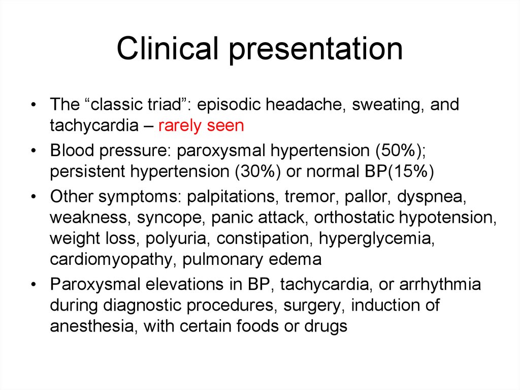
Clinical presentation• The “classic triad”: episodic headache, sweating, and
tachycardia – rarely seen
• Blood pressure: paroxysmal hypertension (50%);
persistent hypertension (30%) or normal BP(15%)
• Other symptoms: palpitations, tremor, pallor, dyspnea,
weakness, syncope, panic attack, orthostatic hypotension,
weight loss, polyuria, constipation, hyperglycemia,
cardiomyopathy, pulmonary edema
• Paroxysmal elevations in BP, tachycardia, or arrhythmia
during diagnostic procedures, surgery, induction of
anesthesia, with certain foods or drugs
7.
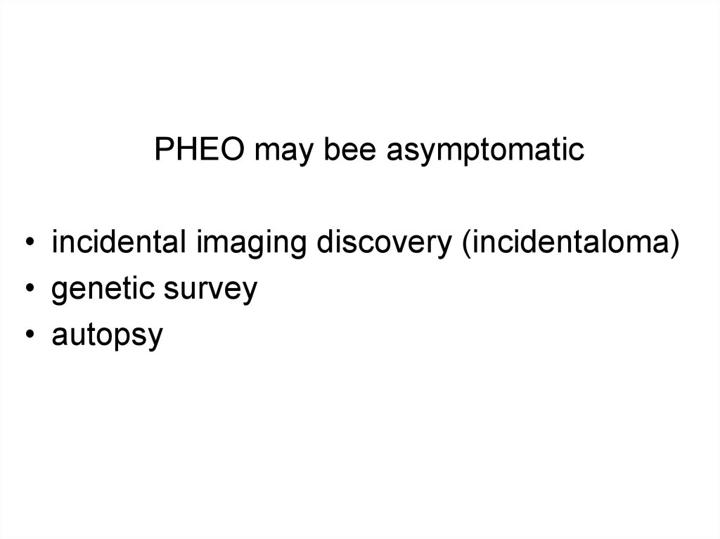
PHEO may bee asymptomatic• incidental imaging discovery (incidentaloma)
• genetic survey
• autopsy
8. Familial pheochromocytoma
MEN 2 syndrome• 95% autosomal dominant RET proto-oncogene
mutation
• prevalence ~1/ 35,000 individuals
• ~ 50% of patients with MEN 2 syndrome develop
PHEO in the adrenal glands
• rarely malignant
• younger age (30-40 years)
9. Familial pheochromocytoma
MEN 2A (Sipple's syndrome)• medullary thyroid cancer (MTC) in all patients,
PHEO in 50%, primary hyperparathyroidism in
20%, and cutaneous lichen amyloidosis in 5%
10. Familial pheochromocytoma
MEN 2B (mucosal neuroma syndrome)• MTC in all patients, PHEO in 50%,
mucocutaneous neuromas,
skeletal deformities, marfanoid
habitus and intestinal
ganglioneuromas (Hirschsprung's
disease)
11. Familial pheochromocytoma
Neurofibromatosis type 1• prevalence ~ 1/ 3,000 individuals
• neurofibromas, multiple café-au-lait
spots, axillary and inguinal freckling,
iris hamartomas (Lisch nodules),
macrocephaly and cognitive deficits
• ~ 2% solitary, benign adrenal PHEO
12. Familial pheochromocytoma
von Hippel–Lindau disease (VHL)• prevalence ~2–3/ 100,000 persons
• hemangioblastoma (cerebellum, spinal cord or
brainstem), retinal angioma, clear cell renal
carcinoma, pancreatic tumors, endolymphatic
sac tumors of the middle ear
• bilateral or malignant PHEO, paraganglioma in
the mediastinum, abdomen and pelvis
13. Familial pheochromocytoma
Familial paraganglioma syndromesParaganglioma syndrome type 1-4
• usually nonfunctional parasympathetic
paragangliomas at skull base and neck
• sometimes adrenal pheochromocytoma
• type 4 may be malignant PHEO
14. Genetic vs. sporadic PHEO
15. When to suspect PHEO?
• Hyperadrenergic spells• Resistant hypertension
• A familial syndrome that predisposes to PHEO
(MEN2, NF1, VHL(
• A family history of pheochromocytoma
• An incidentally discovered adrenal mass
• Hypertension and new onset or atypical DM
• Pressor response to anesthesia, surgery, or
angiography
• Onset of hypertension at a young age (<20 years)
16. Catecholemine metabolism
NorepinephrineCOMT
COMT
Metanephrine
Normetanephrine
MAO
MAO
Vanillylmandelic acid
17. Pheochromocytoma diagnosis
24-hour urine collection for fractionatedmetanephrines and catecholamines
Norepinephrine >170 mcg/24 h
Epinephrine >35 mcg/24 h
Dopamine >700 mcg/24 h
Normetanephrine >900 mcg/24 h
Metanephrine >400 mcg/24 h
measurement of
urinary creatinine to
verify an adequate
collection
may be false-positive
have to be used if clinical probability is low
18. Pheochromocytoma diagnosis
Plasma fractionated metanephrines• metanephrine <0.3 nmol\l (fast), <0.5 nmo\l (non-fast)
• normetanephrine <0.66 nmol\l (fast), <0.9 nmo\l (non-fast)
high predictive value of a negative test
high rate of false-positive tests
have to be used if clinical probability is high
19.
20. Pheochromocytoma diagnosis
24-hour urinary vanillylmandelic acid(VMA) excretion
poor diagnostic sensitivity and specificity
Chromogranine A in serum
increased in 80% of patients with PHEO
not specific for PHEO and may be seen with
other neuroendocrine tumors (carcinoid), and
in a variety of other conditions (atrophic
gastritis, cirrhosis, CRF, PPI treatment …)
21. Pheochromocytoma imaging
CT or MRI of the abdomen and pelvisPheo Imaging characteristics
• Usually large size (>3 sm)
May be bilateral
Cystic and hemorrhagic changes
Increased mass vascularity
Increased attenuation on non-enhanced CT (>20HU)
Additional imaging: MIBG, FDG-PET, DOTATATE-Scan
Biopsy of suspected Pheo should be avoided!
22. Pheochromocytoma imaging
a- FDG-PETb- abdominal CT
c- DOTATATE-Scan
23. Pheochromocytoma treatment
• all patients should undergo a resection of the Pheo(laparascopic or open adrenalectomy)
• preoperative medical therapy
hypertension and tachycardia control:
target BP 120/80 mmHg
combined α-adrenergic blockade (Phenoxybenzamine,
Prazocine, Doxazocine) and β-blockade (Deralin)
volume expansion (high sodium diet, IV 0.9% NS)
• prevention of the hypertensive crisis during surgery
(Nitroprusside, Phentolamine, Nicardipine)