


")


Похожие презентации:


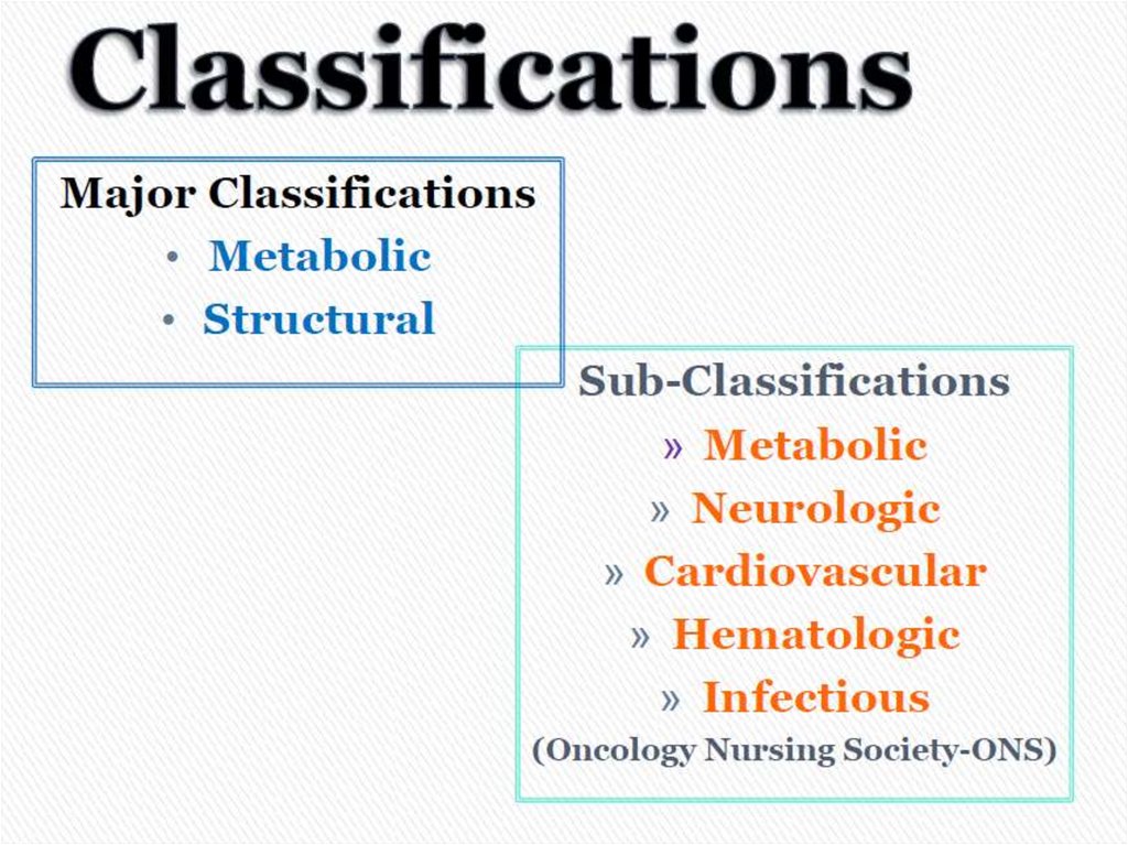
Oncological Emergencies
1. Oncological Emergencies
2. What is Oncologic Emergency?
A clinical condition resulting from a metabolic,neurologic, cardiovascular, hematologic,
and/or infectious change caused by cancer or
its treatment that requires immediate
intervention to prevent loss of life or quality of
life.
3.
4.
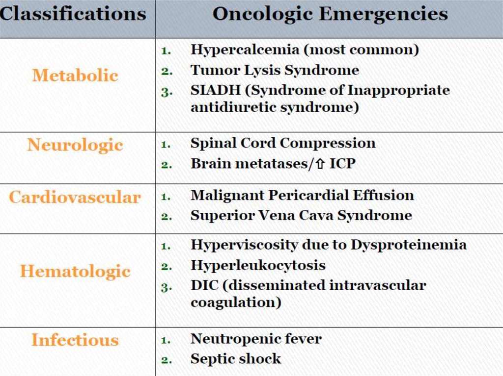
5. METABOLIC
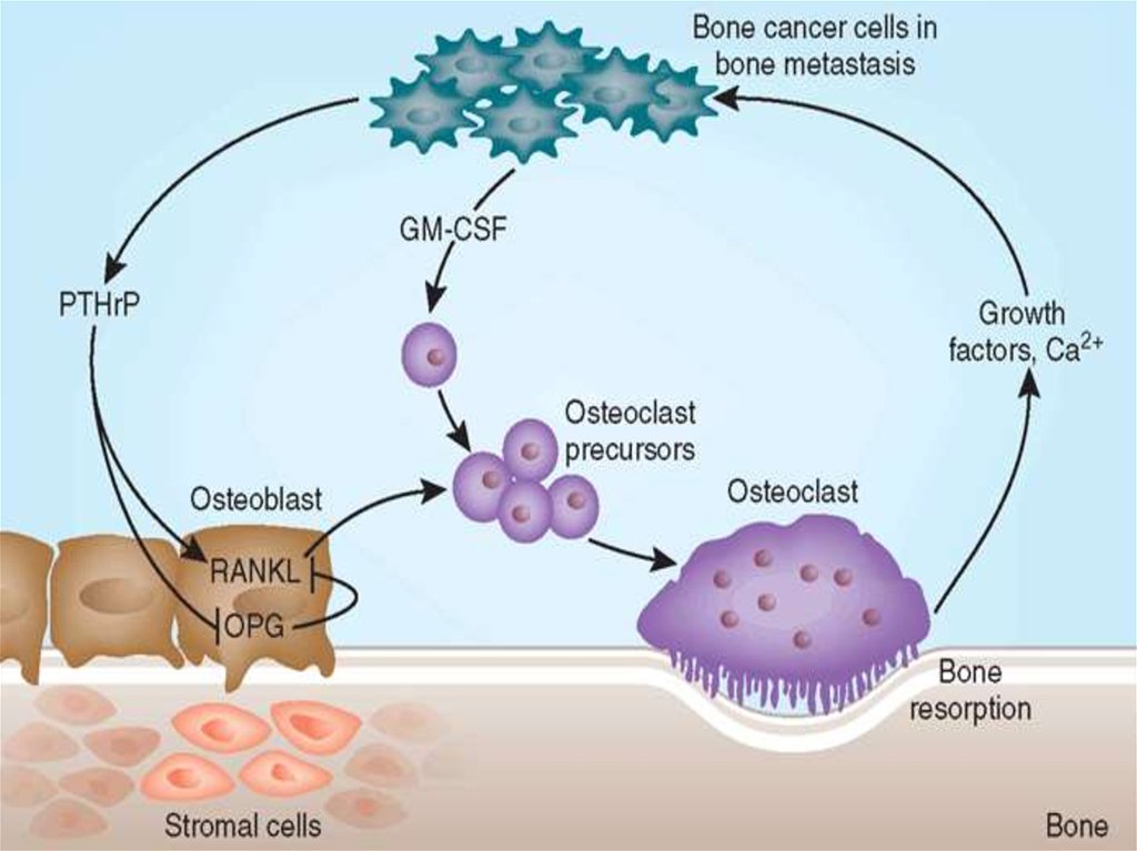
6. Hypercalcemia of Malignancy. Major Mechanisms:
1)Local osteolytic hypercalcemiaOsteoclastic bone resorbing cytokines
In Extensive bone metastases - 20%
2) Humoral hypercalcemia of malignancy
Parathyroid hormone related peptide (PTHrP)
secreted systemically - 80%
7.
8. Symptoms
• GI :Nausea, vomiting, Anorexia,Constipation
• Renal
Polyuria due to interference with ADH- Diabetes
insipidus-like syndrome, Polydipsia
• Neurologic
Lethargy and fatigue ,Cognitive and behavioural
changes ,Altered mental status to coma
Muscle weakness
9. Lab
• Total calcium & albumin or ionized calcium– Medical emergency above 10.5 mg/dL
• Phosphorus
• Creatinine, urea
– Electrolytes
• 50% are hypokalemic
– PTH level
• If elevated may be primary hyperparathyroidism (or
rarely ectopic PTH production)
10.
11.
12.
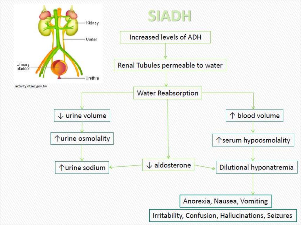
13. Cиндром неадекватной секреции антидиуретического гормона (SIADH)
14.
15. Osmotic Demyelination Syndrome
• Recall that during chronichyponatremia, osmolytes
are shifted out of brain cells
to avoid shift of water into
cells and brain edema
• With rapid correction of
[Na], brain cells not able to
reaccumulate these
osmolytes quickly enough
resulting in water shift out of
cells hence cell shrinkage
and concentrated ion
damage1
16.
17.
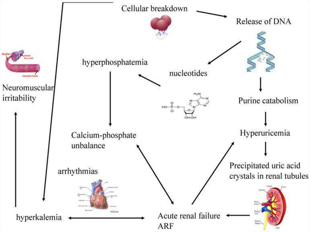
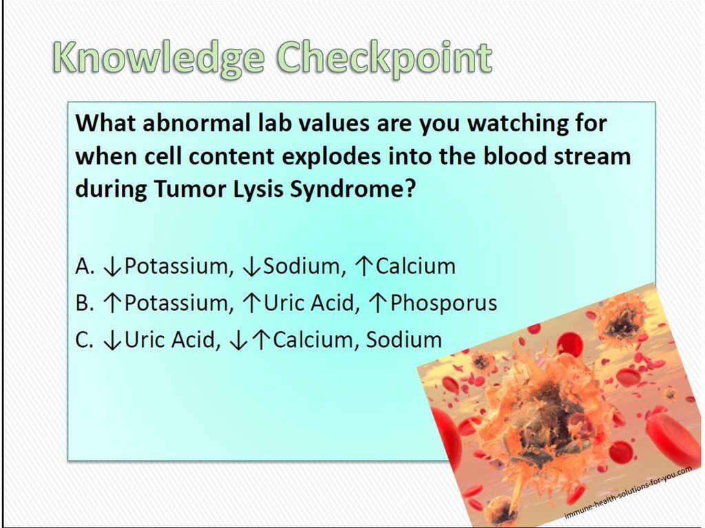
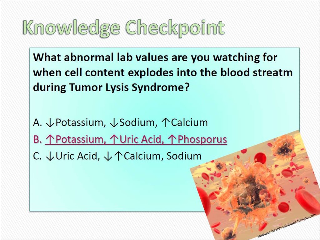
18. Acute Tumor Lysis Syndrome
• Usually starts 6-72 h from initiation of chemoor radiotherapy
• Due to rapid release of cell contents into
blood stream
• Most common tumor cause:
Leukemias
Lymphomas
Small cell ca
19. Etiologic Factors
Large Tumor burden
High growth fraction
High pre treatment serum LDH or Uric Acid
Preexisting renal insufficiency
20.
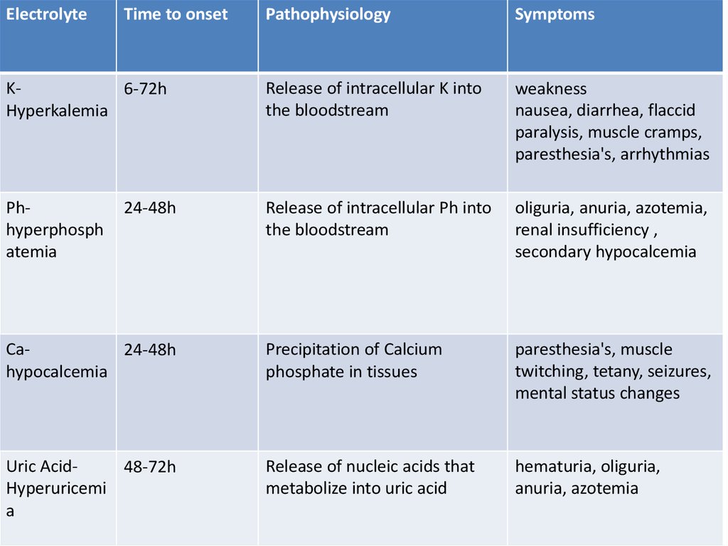
ElectrolyteTime to onset
Pathophysiology
Symptoms
K6-72h
Hyperkalemia
Release of intracellular K into
the bloodstream
weakness
nausea, diarrhea, flaccid
paralysis, muscle cramps,
paresthesia's, arrhythmias
Phhyperphosph
atemia
Release of intracellular Ph into
the bloodstream
oliguria, anuria, azotemia,
renal insufficiency ,
secondary hypocalcemia
Ca24-48h
hypocalcemia
Precipitation of Calcium
phosphate in tissues
paresthesia's, muscle
twitching, tetany, seizures,
mental status changes
Uric Acid48-72h
Hyperuricemi
a
Release of nucleic acids that
metabolize into uric acid
hematuria, oliguria,
anuria, azotemia
24-48h
21.
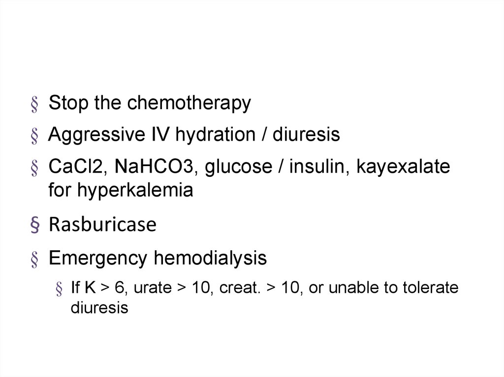
22. Treatment
Best treatment – prevention• Hydration – 3L\24h, better started 24-48 h
before treatment initiation
• Stop nephrotoxic drugs
• Monitoring of electrolyte levels
• Urine alkalinization Ph >7.5
• Allopurinol
23.
§ Stop the chemotherapy§ Aggressive IV hydration / diuresis
§ CaCl2, NaHCO3, glucose / insulin, kayexalate
for hyperkalemia
§ Rasburicase
§ Emergency hemodialysis
§ If K > 6, urate > 10, creat. > 10, or unable to tolerate
diuresis
24.
25.
26.
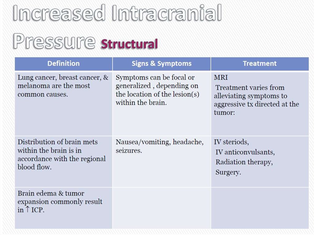
27. STRUCTURAL:
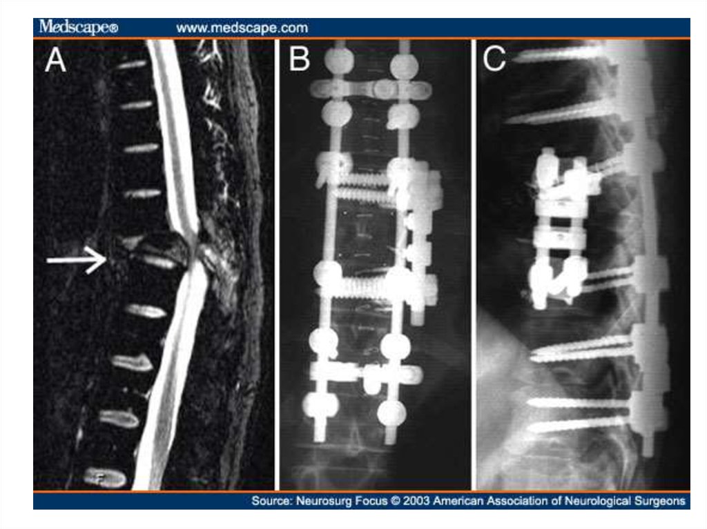
Neurologic emergencies28. Spinal Cord Compression
29. What is malignant spinal cord compression?
• Occurs when cancer cells growin/near to spine and press on the
spinal cord & nerves
• Results in swelling & reduction in
the blood supply to the spinal
cord & nerve roots
• The symptoms are caused by the
increasing pressure
(compression) on the spinal cord
& nerves
30. What types of cancer cause it?
Most commonly seen inBreast
Lung
Prostate
Lymphoma
Myeloma
– About 10% of patients with cancer overall
31. Method of spread
85%From vertebral body orpedicle
10% Through intervertebral
foramina (from
paravertebral nodes or
mass)
4% Intramedullary spread
1%(Low) Direct spread to
epidural space (Batson’s
plexus)
32. Location
Thoracic spine60-70%
Lumbosacral spine 20-30%
Cervical and sacral spine
less then 10% each
33.
34. First Symptoms
PainWeakness
Ataxia
Sensory loss
RED FLAGS…..
95%
5%
1%
1%
35. First Red Flag: Pain
• Usually first and most common symptom(80-90%)
• Usually precedes other neurologic symptoms
by weeks to month
• Severe local back pain
• Aggravated by lying down
• Pain may feel like a 'band' around the chest
or abdomen (radicular)
36. Second Red Flag: Motor
• Weakness: 60-85%• At or above conus medularis
– Extensors of the upper extremities
• Above the thoracic spine
– Weakness from corticospinal dysfunction
– Affects flexors in the lower extremities
• Patients may be hyper reflexic below the
lesion and have extensor plantars
37. Third Red Flag: Bladder & Bowel Function
Third Red Flag: Bladder & Bowel Function• Loss is late finding
• Problems passing urine
– may include difficulty controlling bladder function
– passing very little urine
– or passing none at all
• Constipation or problems controlling bowels
• Autonomic neuropathy presents usually as urinary retention
– Rarely sole finding
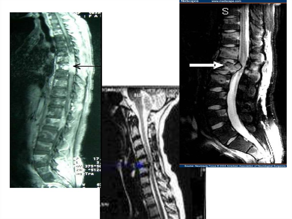
38. Investigations & information needed prior to therapy
Investigations & information needed priorto therapy
1.
MRI scan of the whole spine
2.
3.
4.
Knowledge of cancer type &
stage
Knowledge of patient fitness
Current neurological function
5.
Can get compression at multiple
levels
Have they lost power in their
legs?
Can they walk?
Do they need a catheter?
Do they have pain?
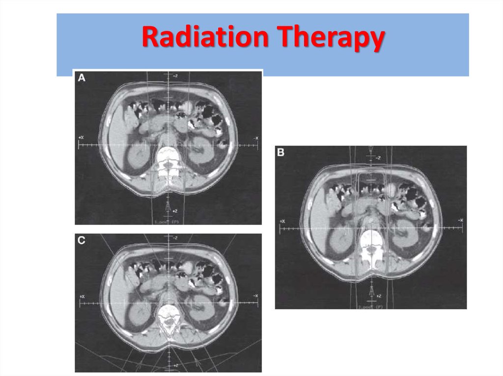
39. Treatment options include:
1.2.
3.
4.
Immobilisation
Steroids & gastric protection
Analgesia
Surgery – decompression & stabilisation of
the spine
5. Radiotherapy
6. Chemotherapy e.g. lymphoma
7. Hormonal manipulation e.g. prostate Ca
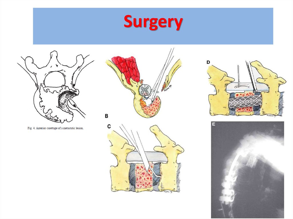
40. Indications for Surgery
• Unknown primary tumour• Relapse post RT
• Progression while on RT
• Intractable pain
• Instability of spine
• Patients with a single level of cord
compression who have not been totally
paraplegic for longer than 48 hours
• Prognosis >3 months
41.
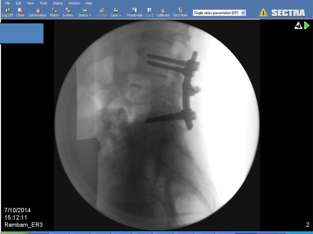
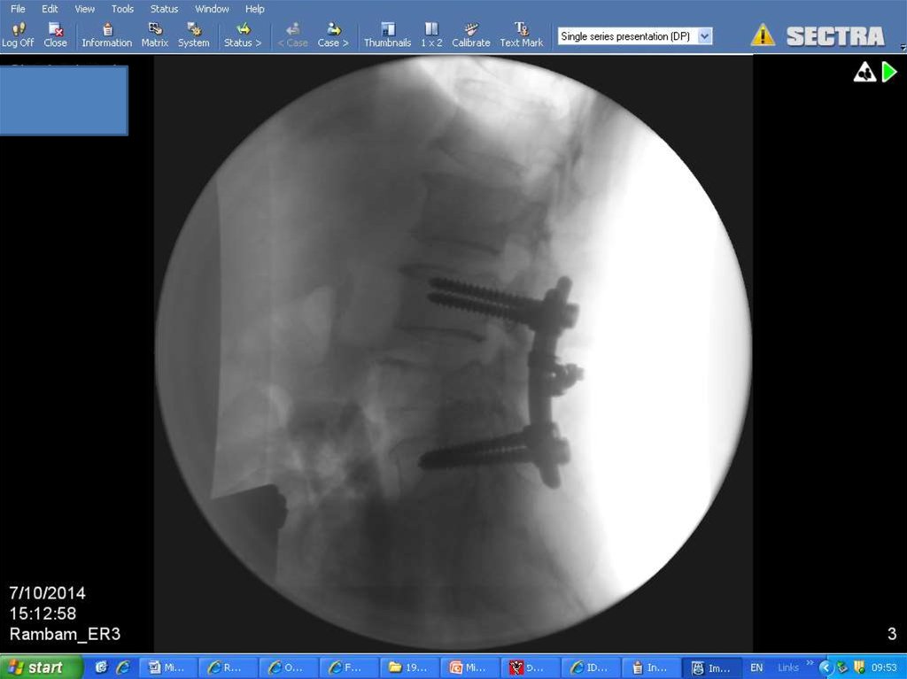
Surgery42.
43.
44.
45. RCT comparing surgery followed by RT vs. RT alone
• Improvement in surgery + RT– Days remained ambulatory (126 vs. 35)
– Percent that regained ambulation after therapy (56% vs.
19%)
– Days remained continent (142 vs. 12)
– Less steroid dose, less narcotics
– Trend to increase survival
Patchell, R, Tibbs, PA, Regine, WF, et al. A randomized trial of
direct decompressive surgical resection in the treatment of spinal
cord compression caused by metastasis (abstract). proc Am Soc
Clin Oncol 2003; 22:1.
46.
Radiation Therapy47.
48. Prognosis
• Median survival with MSCC is 6 months• Ambulatory patients with radiosensitive
tumours have the best prognosis
– Likely to remain mobile
MSCC is a poor prognostic indicator in cancer patients
Need better detection rates
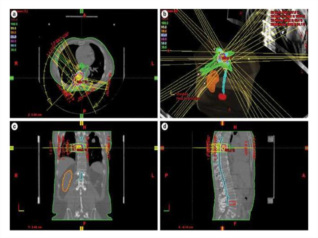
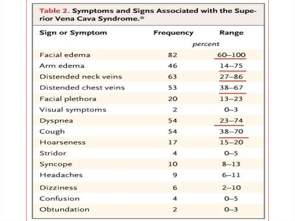
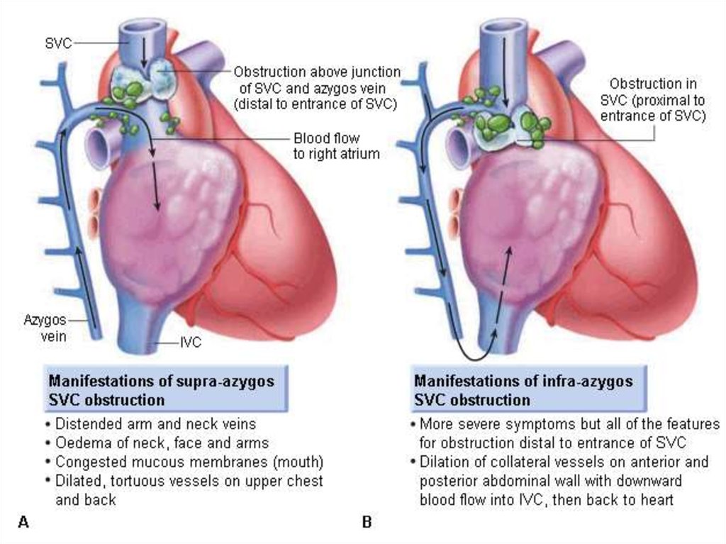
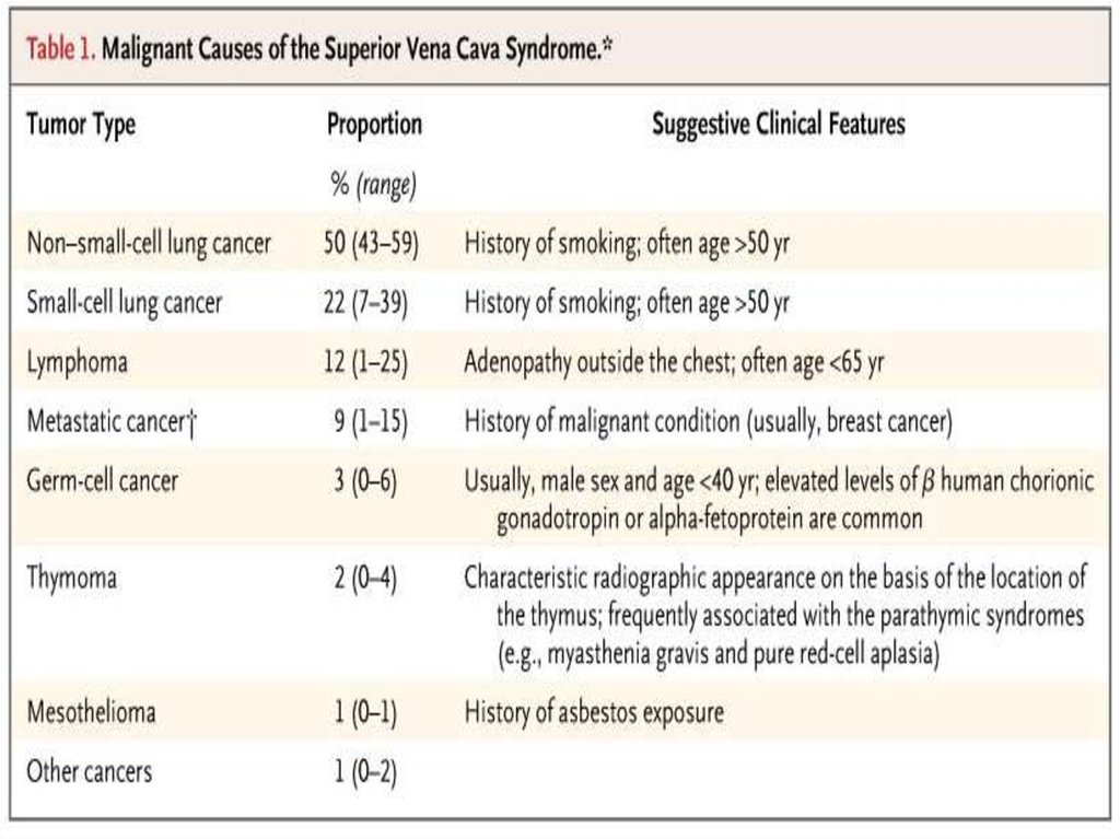
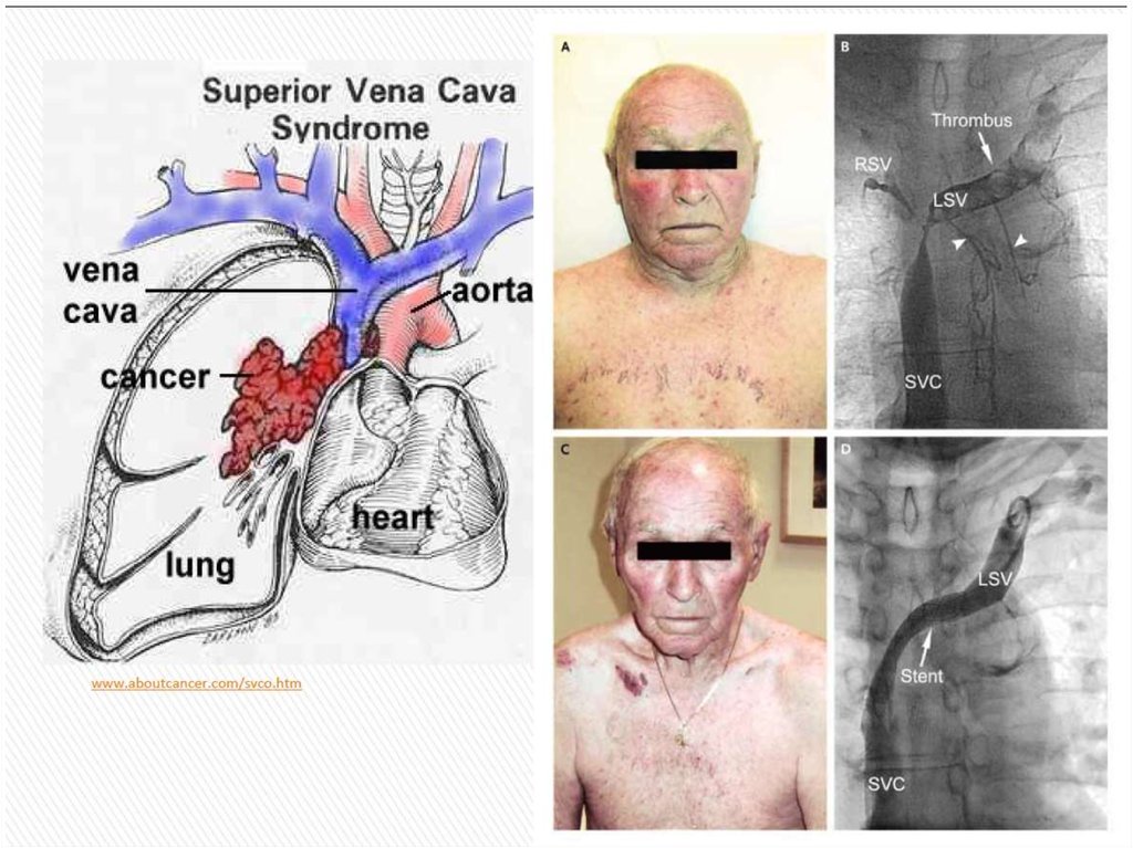
49. Superior Vena Cava Syndrome
50.
51. Superior Vena Cava Syndrome
52. Superior Vena Cava Syndrome
53.
54.
Superior Vena Cava Syndrome55.
56.
57. Superior Vena Cava Syndrome
In rare cases can be disease presentation• No time for pathology
• Urgent treatment without tissue diagnosis
Median survival – 6 month
2 year survivale – 15%
58. Exeption: Treatment Sensitive Tumors
• NHLs, germ cells, and limited-stage small celllung cancers usually respond to chemotherapy
and or radiation
• Can achieve long term remission with tumor
specific directed therapy
• Symptomatic improvement usually takes 1-2
weeks after start of therapy
59. Superior Vena Cava Syndrome
60. Superior Vena Cava Syndrome
61. Superior Vena Cava Syndrome
62. Treatment Options
• Radiation therapy• Chemotherapy
• Intraluminal Stent
+supportive care
63. Supportive Care:
Rest
Head elevation
Oxygen
Diuretics
Anticoagulation
Steroids
Avoid high volume fluid infusion through
upper extremities
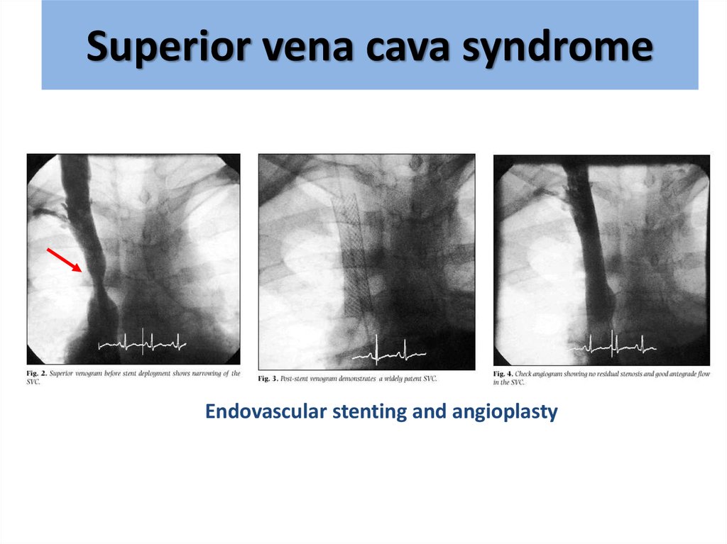
64. Intraluminal Stents
• Endovascular placement under fluoroscopy• Patients who have recurrent disease in
previously irradiated fields
• Tumors refractory chemotherapy
• Patient too ill to tolerate radiation or
chemotherapy
65.
66.
Superior vena cava syndromeEndovascular stenting and angioplasty
67.
68.
69.
70.
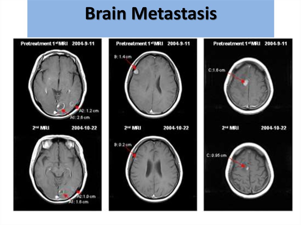
Brain Metastasis• Most Common type of CNS malignancy
• 20-40% of cancer patients will develop brain mets
• Most common types: Breast, Lung, Melanoma,
Colorectal Ca
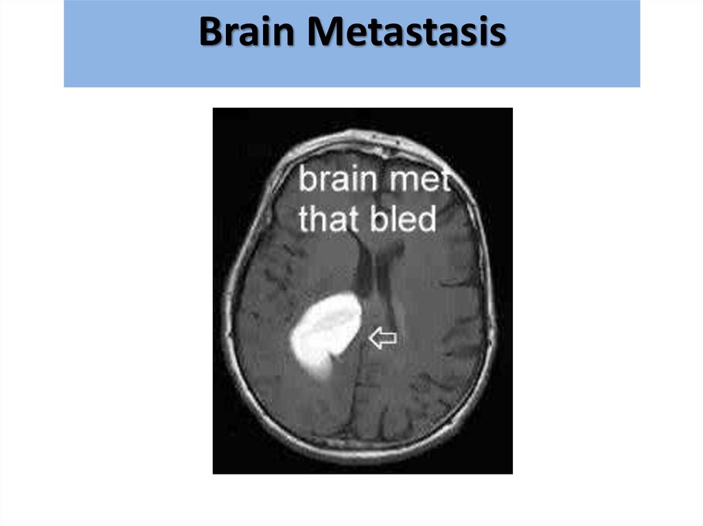
• Highest risk for bleeding
–
–
–
–
–
RCC
Melanoma
Choriocarcinoma
Papillary thyroid
Lung Cancer
71.
BrainMetastasis
מוחיות
גרורות
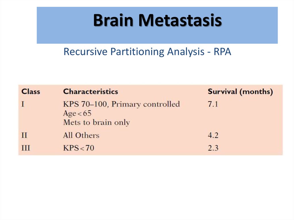
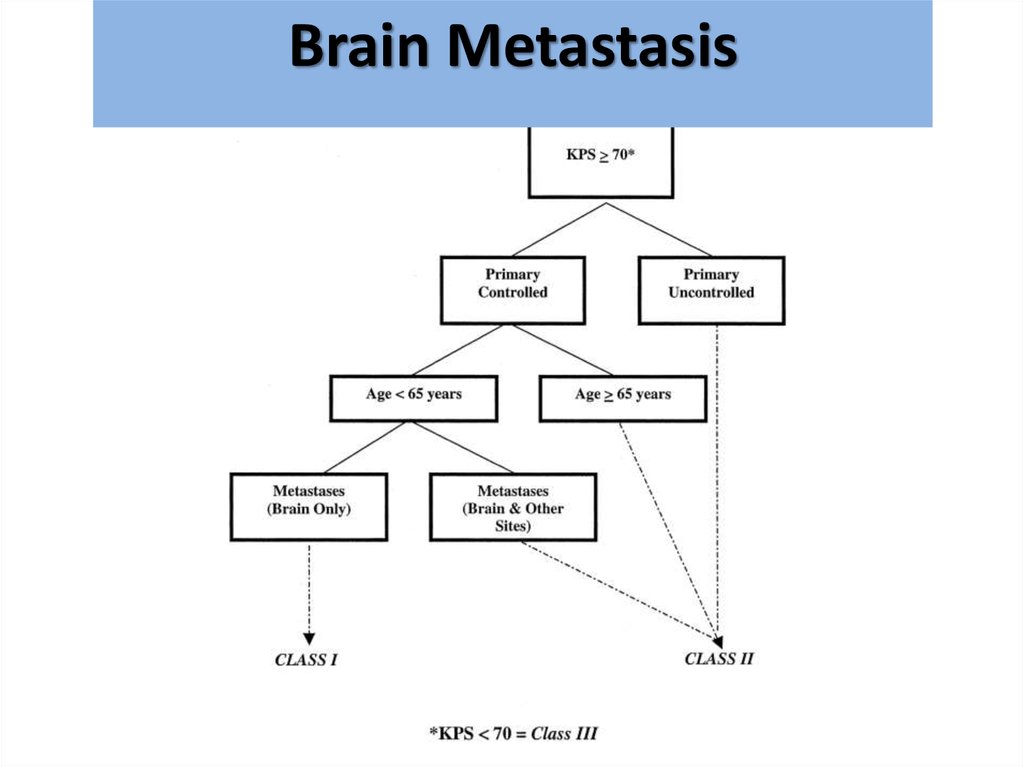
Recursive Partitioning Analysis - RPA
72.
BrainMetastasis
גרורות מוחיות
73.
BrainMetastasis
גרורות מוחיות
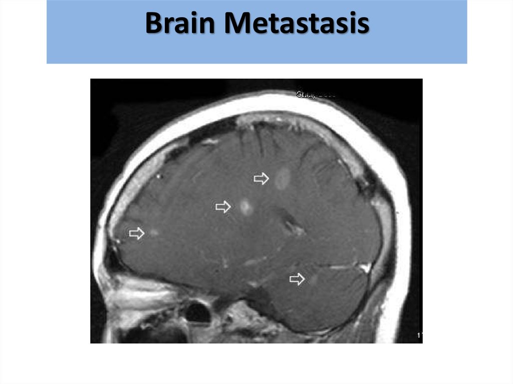
Diagnosis:
• CT with and without contrast
• MRI – modality of choice for small lesions including
leptomeningial spread
• If no previous history of malignancy - consider total
body imaging
74.
BrainMetastasis
גרורות מוחיות
75.
BrainMetastasis
גרורות מוחיות
76.
BrainMetastasis
גרורות מוחיות
77.
BrainMetastasis
גרורות מוחיות
Treatment:
Steroids – Dexamethasone 16mg*2
Anticonvulsant
Surgery?
Radiation therapy
78.
BrainMetastasis
גרורות מוחיות
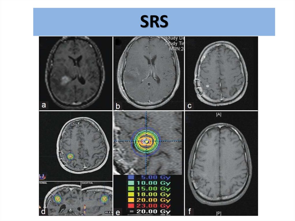
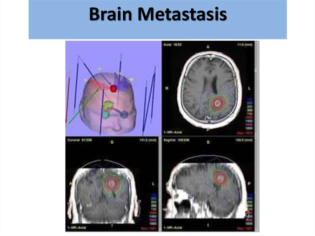
• Radiation therapy
– WBRT=Whole Brain RT
– SRS=Stereotactic Radio Surgery
79.
BrainMetastasis
גרורות מוחיות
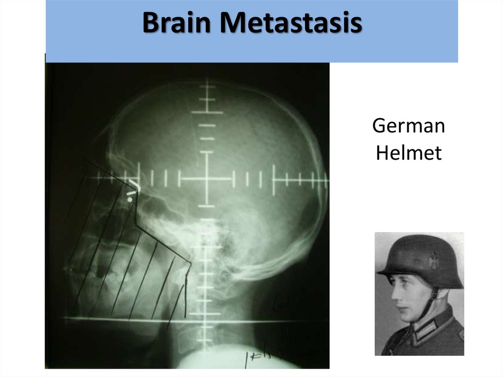
German
Helmet
80.
BrainMetastasis
גרורות מוחיות
81.
BrainMetastasis
גרורות מוחיות
82.
SRS83.
BrainMetastasis
גרורות מוחיות